Dr Željko Kojadinović — NEUROHIRURGIJA I LEČENJE BOLA
Dr Zeljko Kojadinovic — Pain Treatment & Neurosurgery


Author:
Dr. Zeljko Kojadinovic, MD, PhD
— Consultant Neurosurgeon
Specialized Experience:
30 years of clinical expertise in neurosurgery.
Last medically reviewed:
October 16, 2026
Who This Arachnoiditis Page Is For
This page is intended for patients diagnosed with or suspected of having spinal arachnoiditis, as well as their families seeking a clear explanation of the condition, its symptoms, diagnosis, progression, and treatment options.
If the diagnosis remains uncertain, symptoms persist despite treatment, different specialists have recommended conflicting approaches, or there is a possibility that another treatable pain generator may be contributing to the symptoms, an individualized online neurosurgical consultation may help clarify the findings, identify the dominant source of pain, and assess realistic treatment options.
Arachnoiditis — Quick Summary (Read This First)
- Arachnoiditis is inflammation involving the arachnoid membrane and the subarachnoid space surrounding the spinal cord and nerve roots. In some patients, inflammation leads to fibrosis and adhesions, producing chronic adhesive arachnoiditis. The lumbar and thoracic regions are commonly affected, although arachnoiditis can also involve the cervical and sacral spine. It may also occur around the brain (intracranial arachnoiditis).
- Arachnoiditis may develop after spinal surgery, infection, hemorrhage, trauma, or certain invasive spinal procedures. In some cases, no identifiable cause can be established.
- Adhesive arachnoiditis can damage nerve roots and the spinal cord through several mechanisms. Adhesions may distort neural structures, impair local circulation, and obstruct cerebrospinal fluid (CSF) flow. More extensive disease may be associated with arachnoid cysts, spinal cord compression, or secondary syringomyelia.
- Persistent neuropathic pain is a common clinical manifestation. Lumbosacral involvement often causes lower back pain radiating into the buttocks and legs. Cervical disease may affect the neck, shoulders, arms, and hands, while thoracic disease can produce pain around the chest or abdomen. Sensory disturbances, muscle weakness, walking difficulties, and bladder or bowel dysfunction may also occur.
- Arachnoiditis does not always progress. Some patients remain stable for years, while others experience fluctuating symptoms or gradual neurological deterioration. The severity of pain does not necessarily correspond to the extent of abnormalities visible on MRI.
- MRI is the principal diagnostic method for chronic adhesive arachnoiditis. Characteristic findings may include nerve root clumping, adhesion of nerve roots to the dural sac, distortion of CSF spaces, and associated spinal cord abnormalities. No blood or CSF test can independently confirm the diagnosis, and lumbar puncture is not routinely required.
- There is currently no established treatment that reliably reverses mature arachnoid adhesions. Management focuses on neuropathic pain control, preservation of neurological function, rehabilitation, and treatment of complications. Surgery is considered selectively, particularly when spinal cord compression, significant CSF flow obstruction, or progressive syringomyelia is present.
- Not every pain symptom in a patient with arachnoiditis is caused by arachnoiditis itself. Disc abnormalities, foraminal stenosis, sacroiliac or facet joint disorders, and peripheral nerve conditions may coexist. Identifying a separate, treatable pain generator can provide meaningful relief even when the underlying arachnoid fibrosis remains unchanged.
Most readers benefit from the Quick Summary and the sections on Symptoms of Adhesive Arachnoiditis, How Is Adhesive Arachnoiditis Diagnosed?, Can Other Pain Generators Coexist With Arachnoiditis?, and Treatment Options for Adhesive Arachnoiditis. The remaining sections provide more detailed clinical explanations.
Contents
- Who this is for
- Quick summary
- What is arachnoiditis?
- Inflammatory vs. adhesive
- How arachnoiditis develops
- How nerve damage occurs
- Causes of arachnoiditis
- Surgery and medical procedures
- Infections, hemorrhage and trauma
- Spinal locations
- Intracranial arachnoiditis
- Symptoms of arachnoiditis
- Does arachnoiditis always progress?
- Diagnosis
- MRI findings and limitations
- Normal MRI?
- Differential diagnosis
- Other pain generators
- Finding the dominant pain generator
- Treatment options
- Anti-inflammatory treatment
- Neuropathic pain medication
- Rehabilitation and functional support
- Psychological and multidisciplinary support
- Neuromodulation and interventions
- When is surgery considered?
- Prognosis and long-term management
- When to seek second opinion
- Frequently asked questions
What Is Arachnoiditis?
The spinal cord and nerve roots are surrounded by three protective membranes: the dura mater, arachnoid mater, and pia mater. The subarachnoid space, located between the arachnoid and pia, contains cerebrospinal fluid (CSF) and provides a protective environment for neural structures.
Arachnoiditis refers to inflammation involving the arachnoid membrane and subarachnoid space. In persistent or severe cases, the inflammatory response may result in the formation of fibrous tissue and adhesions. These adhesions can alter the normal arrangement of the nerve roots, attach them to surrounding structures, and disturb CSF circulation.
In the lumbar spine, where the spinal cord usually ends around the L1–L2 vertebral level, adhesive arachnoiditis primarily affects the nerve roots forming a bundle (cauda equina). In the cervical and thoracic regions, the inflammatory and adhesive process may also involve the spinal cord and cause myelopathy.
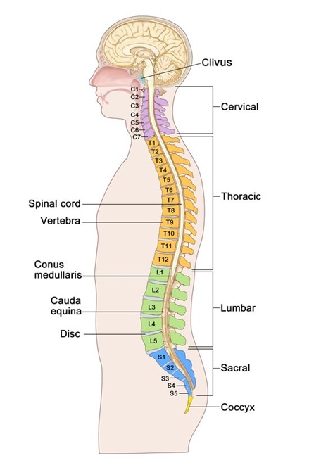
Image: Shows the anatomy of the vertebral column and spinal canal. The spinal cord ends at the L2 level, and below it, within the spinal canal, lie the spinal nerve roots forming a bundle (cauda equina). The nerve roots of the cauda equina will give rise to the nerves for the legs and pelvic region.
Inflammatory and Adhesive Arachnoiditis — Understanding the Terminology
Arachnoiditis can involve different degrees of inflammation and scarring within the arachnoid membrane and subarachnoid space. Two descriptive terms are sometimes used: non-adhesive arachnoiditis and adhesive arachnoiditis.
Non-adhesive arachnoiditis describes inflammatory involvement without clearly visible adhesions or scarring on MRI. Patients may experience neuropathic pain and neurological symptoms even when structural changes are not readily demonstrated on imaging.
Adhesive arachnoiditis develops when inflammation is associated with fibrosis and adhesions involving the nerve roots or surrounding meningeal structures. These adhesions may cause nerve root clumping, mechanical distortion, and disturbances of cerebrospinal fluid circulation.
The distinction primarily describes the presence or absence of demonstrable adhesive changes rather than two universally established, separate diseases. MRI findings must always be interpreted together with the clinical history and neurological examination.
How Does Arachnoiditis Develop?
Arachnoiditis usually begins when an inflammatory or traumatic event affects the arachnoid membrane and subarachnoid space. The initiating process may involve direct tissue injury, infection, blood products, chemical irritation, or another inflammatory mechanism.
Inflammation can disrupt normal meningeal tissue architecture and stimulate fibroblast activity, collagen deposition, and scar formation. If fibrosis develops, the resulting adhesions may connect structures that normally remain separated by cerebrospinal fluid.
Inflammation and established fibrosis should not be considered identical processes. Inflammatory activity may be more prominent during earlier disease phases, whereas mature fibrous adhesions represent structural changes that are much more difficult to reverse. Chronic inflammation and arachnoid adhesions may disrupt normal cerebrospinal fluid (CSF) circulation, creating localized fluid compartments and contributing to the formation of arachnoid cysts. In longstanding cases, the affected arachnoid membrane may also develop calcification or, more rarely, ossification. These changes can further distort or compress neural structures and interfere with CSF flow, depending on their location and extent.
The process may remain localized or extend over several spinal segments. Depending on its location, it can interfere with the mobility and function of nerve roots, distort the spinal cord, or obstruct the circulation of CSF.
Not every case of arachnoid inflammation progresses to adhesive arachnoiditis. The factors determining whether inflammation resolves or develops into persistent fibrosis and adhesions remain incompletely understood.
How Does Arachnoiditis Damage Nerve Roots and the Spinal Cord?
Arachnoiditis can damage nerve roots and the spinal cord through several interconnected mechanisms. The process begins with inflammation, which may subsequently progress to fibrosis and the formation of adhesions. These adhesions can cause nerve roots to clump together, become attached to the dural sac, and lose their normal mobility. This may result in mechanical distortion, traction, and persistent irritation of neural structures.
Inflammation and fibrous adhesions may also impair local microcirculation, causing venous congestion, reduced tissue perfusion, and potentially ischemic injury to the nerve roots or spinal cord. Extensive adhesions can obstruct cerebrospinal fluid (CSF) circulation, altering CSF pressure dynamics and contributing to spinal cord edema or secondary syringomyelia, in which a fluid-filled cavity develops within the spinal cord and damages neural tissue from within. Disturbed CSF circulation and adhesions may also contribute to the formation of arachnoid cysts, which can compress the spinal cord or the nerve roots of the cauda equina, depending on their location.
These mechanisms, individually or in combination, can cause persistent neuropathic pain, sensory and motor deficits, and progressive neurological dysfunction. Prolonged abnormal nerve signaling may also contribute to central sensitization, amplifying pain beyond the effects of the initial structural injury.
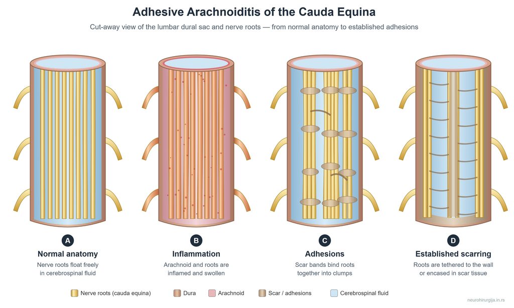
Image: Stages of arachnoiditis involving the cauda equina.
What Causes Spinal Arachnoiditis?
Several mechanisms can initiate inflammation or injury within the spinal subarachnoid space.
Spinal Surgery and Medical Procedures
Arachnoiditis is a recognized potential complication of spinal surgery, particularly when procedures involve dural opening, intradural manipulation, hemorrhage, infection, or other complications affecting the meninges. It has also been reported following certain spinal injections, intrathecal drug administration, spinal anesthesia, and historical myelographic procedures. Nevertheless, most patients undergoing spinal surgery or injections do not develop adhesive arachnoiditis. A previous procedure alone does not establish causation, particularly when the diagnosis is based only on persistent pain.
Infection, Hemorrhage and Trauma
Meningitis and other infections affecting the meninges can initiate inflammation that subsequently produces adhesions. Tuberculous meningitis is a well-recognized association, although bacterial, fungal, and other infections may also be involved.
Subarachnoid hemorrhage exposes the meninges to blood products and inflammatory reactions that may lead to fibrosis. Significant spinal trauma can similarly produce direct tissue injury, bleeding, and secondary inflammatory changes.
Other Causes and Idiopathic Arachnoiditis
Less common causes of arachnoiditis include autoimmune and immune-mediated inflammatory disorders, such as neurosarcoidosis and certain systemic autoimmune diseases that can affect the spinal meninges. These conditions may cause chronic meningeal inflammation and, in some cases, lead to fibrosis and adhesions. However, autoimmune mechanisms account for only a small proportion of reported cases, and their role in adhesive arachnoiditis remains incompletely understood.
Idiopathic arachnoiditis refers to cases in which no identifiable cause can be established despite appropriate clinical investigation.
A detailed medical history is important, but the absence of a known risk factor does not automatically exclude arachnoiditis.
Where in the Spine Does Arachnoiditis Occur?
Arachnoiditis can affect the cervical, thoracic, lumbar, or sacral regions of the spine. Its neurological consequences depend on whether the inflammatory and adhesive process involves the spinal cord, the nerve roots of the cauda equina, or both.
In a 2024 systematic review of 510 published cases of spinal adhesive arachnoidopathy, the thoracic region was the most frequently reported location (49%), followed by the lumbar (28.7%), cervical (15.8%), and sacrococcygeal regions (6.4%). These figures come largely from selected case reports and series, frequently involving surgically treated patients with complex spinal pathology, and should not be interpreted as the distribution of arachnoiditis in the general patient population. Lumbosacral involvement is also clinically important, particularly in patients with a history of lumbar spine surgery.
Lumbosacral arachnoiditis primarily affects the nerve roots of the cauda equina and may cause radicular pain, sensory and motor deficits, or bladder and bowel dysfunction. Cervical and thoracic arachnoiditis may directly affect the spinal cord, potentially causing myelopathy, CSF flow obstruction, and secondary syringomyelia.
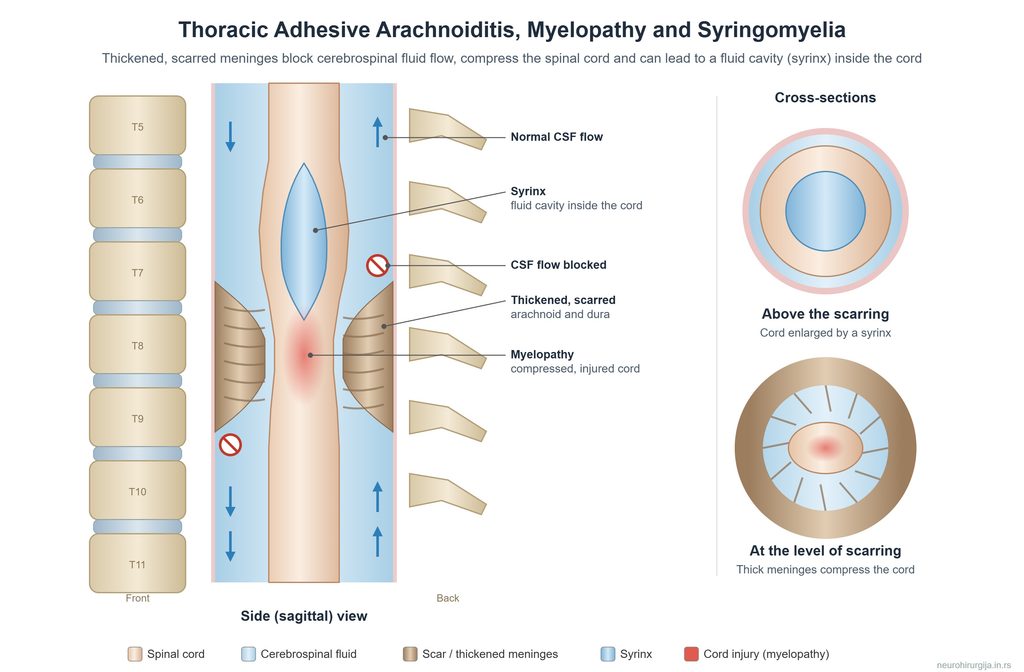
Image: Arachnoiditis causing myelopathy and syringomyelia.
Can Arachnoiditis Also Affect the Brain?
Yes. Although arachnoiditis is most commonly discussed as a spinal disorder, inflammation and adhesions can also affect the arachnoid membrane surrounding the brain, a condition known as intracranial (cranial) arachnoiditis.
Intracranial arachnoiditis may develop following meningitis, particularly tuberculous meningitis, subarachnoid hemorrhage, head trauma, or intracranial surgery. Inflammation and adhesions may involve cranial nerves, particularly in the basal cisterns, causing visual disturbances, eye movement abnormalities, hearing impairment, or other cranial nerve deficits. Adhesions may also obstruct cerebrospinal fluid (CSF) circulation, potentially leading to hydrocephalus and increased intracranial pressure.
The rest of the text will focus solely on spinal arachnoiditis.
Symptoms of Adhesive Arachnoiditis
The clinical presentation varies considerably depending on the affected spinal level, extent of neural involvement, and presence of additional neurological or musculoskeletal disorders.
Neuropathic Pain and Sensory Disturbances
One of the most characteristic pain presentations of spinal arachnoiditis is persistent lower back pain radiating into one or both legs, often accompanied by burning, shooting, electric, or stabbing sensations. Pain may extend into the buttocks, pelvis, and feet, particularly when the lumbosacral nerve roots of the cauda equina are affected.
Cervical arachnoiditis may cause pain in the neck, shoulders, arms, and hands, while thoracic arachnoiditis can produce upper or middle back pain radiating around the chest or abdomen.
Patients may also experience numbness, tingling, hypersensitivity to touch, or unusual sensations, such as feeling water droplets or insects crawling on the skin. These symptoms are not specific to arachnoiditis and must be interpreted together with neurological findings and imaging.
Muscle Weakness and Walking Difficulties
Neurological impairment may include weakness, altered reflexes, muscle wasting, cramps, reduced walking endurance, and balance difficulties. When the spinal cord is involved, patients may develop spasticity, increased reflexes, or other signs of myelopathy. The pattern of deficits should correspond as closely as possible to the anatomical structures involved.
Bladder, Bowel and Sexual Dysfunction
Extensive involvement of the lumbosacral nerve roots may affect bladder, bowel, and sexual function. Symptoms can include urinary retention, impaired bladder emptying, incontinence, altered perineal sensation, or bowel dysfunction.
New urinary retention, saddle anesthesia, rapidly progressive weakness, or significant new bladder or bowel dysfunction requires urgent medical assessment. These symptoms must not automatically be attributed to previously diagnosed chronic arachnoiditis because an acute compressive lesion may require emergency treatment.
Does Arachnoiditis Always Progress?
No. Arachnoiditis does not always progress. Some patients remain clinically stable for years, while others experience fluctuating symptoms, episodic exacerbations, or gradual neurological deterioration. The course of the disease is highly variable and cannot be reliably predicted from the initial symptoms or MRI findings alone.
When arachnoiditis progresses, neurological deterioration may manifest as increasing sensory disturbances, muscle weakness, impaired walking, and reduced mobility. In more severe cases, patients may develop bladder and bowel dysfunction, significant motor deficits, or loss of independent mobility. Progression may be associated with extensive adhesions, spinal cord compression, obstruction of cerebrospinal fluid (CSF) circulation, or secondary syringomyelia.
The severity of the disease can range from mild symptoms with preserved neurological function to severe disability requiring walking aids, a wheelchair, or assistance with daily activities.
Importantly, new or worsening pain does not always indicate progression of arachnoiditis. Other spinal, musculoskeletal, or peripheral nerve disorders may coexist and should be considered when symptoms change.
How Is Adhesive Arachnoiditis Diagnosed?
Diagnosis requires careful consideration of the patient’s history, neurological findings, imaging abnormalities, and possible alternative explanations. A history of spinal surgery, meningitis, hemorrhage, or another relevant event may raise suspicion. Persistent neuropathic pain, sensory abnormalities, weakness, or bladder dysfunction can further support the need for investigation. However, clinical symptoms are often nonspecific and overlap with lumbar stenosis, radiculopathy, peripheral neuropathy, and other disorders.
The diagnosis should not be made solely because a patient has severe pain after spinal surgery. Objective imaging and anatomical correlation are especially important when establishing chronic adhesive disease.
MRI and Additional Investigations
MRI is the principal imaging method for evaluating suspected spinal adhesive arachnoiditis. It allows assessment of nerve root configuration, the subarachnoid space, spinal cord abnormalities, and associated degenerative or postoperative conditions.
CT myelography or specialized MRI sequences may occasionally provide additional information, particularly when CSF flow obstruction or complex anatomy requires further clarification.
Cerebrospinal fluid (CSF) analysis may reveal elevated protein levels, particularly when CSF circulation is obstructed. Increased number of white blood cells in the CSF (pleocytosis) may occur in active inflammatory or infectious conditions, while CSF findings can remain normal in chronic adhesive arachnoiditis. Blood tests, including inflammatory markers and selected infectious or autoimmune investigations, may help identify an underlying cause. However, no specific blood or CSF biomarker reliably confirms adhesive arachnoiditis. Lumbar puncture is not routinely required to diagnose chronic adhesive arachnoiditis.
Electrophysiological studies may help assess coexisting peripheral neuropathy or radiculopathy, but they cannot independently confirm adhesive arachnoiditis. Because lumbar puncture and CT myelography are invasive procedures, they should be performed only when clinically indicated.
MRI Findings and Their Limitations
The characteristic MRI abnormalities of adhesive arachnoiditis include nerve root clumping, abnormal distribution of the cauda equina roots, peripheral adherence to the dural sac, and distortion or obliteration of normal CSF spaces.
Additional findings may include arachnoid cysts, localized adhesions, spinal cord deformity, or secondary syringomyelia.
The Three Classical MRI Patterns
Three characteristic MRI patterns of adhesive arachnoiditis involving the cauda equina nerve roots have been described in the lumbar spine. These patterns reflect the distribution and adhesion of nerve roots within the thecal sac, rather than successive stages of disease progression.
Type I — Central nerve root clumping
The nerve roots are adherent to one another and form abnormal central groups within the dural sac.
Type II — Empty thecal sac sign
The nerve roots become adherent to the peripheral wall of the dural sac, leaving the central CSF space relatively empty in appearance.
Type III — Soft-tissue mass replacing the normal root configuration
Extensive adhesive changes produce a conglomerated soft-tissue appearance in which individual nerve roots become difficult to distinguish.
These patterns describe radiological morphology rather than separate clinical stages. They do not reliably predict pain severity, functional disability, or the response to treatment.
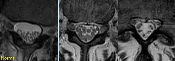
Image: Three classical MRI patterns of adhesive arachnoiditis involving the cauda equina.
Can Arachnoiditis Be Present With a Normal MRI?
MRI findings and symptoms do not always correlate. Some patients with established abnormalities have relatively limited symptoms, while others experience substantial pain despite less extensive imaging changes.
However, a normal MRI does not establish a diagnosis of early or non-adhesive arachnoiditis. When characteristic abnormalities are absent, the diagnosis remains uncertain, and alternative causes of symptoms should be carefully considered. In selected complex cases, review of the original MRI images by a specialist and additional targeted imaging may help clarify the findings.
Arachnoiditis, Epidural Fibrosis and Other Conditions
Adhesive arachnoiditis must be distinguished from several disorders that can cause similar symptoms, particularly after spinal surgery.
Epidural fibrosis is scar formation outside the dura mater, usually at the site of a previous operation. It may surround or tether a nerve root externally but is anatomically different from intradural arachnoid adhesions.
Arachnoid cysts and arachnoid webs may produce localized spinal cord compression or CSF flow obstruction. These conditions can coexist with adhesive arachnoiditis but may have different indications for surgical treatment.
Tarlov cysts are CSF-containing perineural cysts, usually located in the sacral region. They may cause pain and neurological symptoms in selected patients, but their presence alone does not establish arachnoiditis or demonstrate generalized CSF dysfunction.
A careful distinction between these conditions is important because the treatment options and prognosis can differ substantially.
Can Other Pain Generators Coexist With Arachnoiditis?
Yes. A confirmed diagnosis of arachnoiditis does not exclude other causes of pain. This is particularly important when symptoms persist despite treatment, change their anatomical distribution, or become more localized. A patient may have genuine neuropathic pain caused by arachnoiditis while simultaneously experiencing pain from a degenerative spinal lesion, joint, muscle, ligament, or peripheral nerve. If every symptom is automatically attributed to arachnoiditis, a separate and potentially treatable condition may remain undiagnosed. Identifying the responsible lesion requires correlation between the pain distribution, neurological examination, and imaging findings. A visible MRI abnormality is not automatically the source of pain.
Spinal and Foraminal Pain Generators
Degenerative disc disease, recurrent disc herniation, foraminal stenosis, lateral recess stenosis, and segmental instability may coexist with arachnoiditis. For example, a patient with established lumbosacral arachnoiditis may subsequently develop foraminal stenosis compressing a specific nerve root. The resulting new unilateral radicular pain may have a different mechanism from the patient’s pre-existing diffuse neuropathic symptoms.
Facet Joints, Sacroiliac Joints and Musculoskeletal Pain
Facet joints, sacroiliac joints, muscles, and ligaments can generate pain independently of arachnoiditis. These disorders may cause localized lower back, buttock, pelvic, or referred leg pain that overlaps with neuropathic symptoms. In some patients, pain from an additional musculoskeletal source becomes the dominant complaint. Clinical examination is particularly important because these pain generators may not be clearly demonstrated on routine spinal MRI.
When clinically appropriate, targeted diagnostic injections can help assess whether a particular structure contributes substantially to the patient’s pain. Their results must nevertheless be interpreted cautiously because diagnostic blocks are not perfectly specific.
Peripheral Neuropathy and Nerve Entrapment
Peripheral nerve entrapment, polyneuropathy, and other neurological disorders may produce burning, tingling, numbness, or radiating pain similar to symptoms attributed to arachnoiditis. The distribution of sensory changes, muscle weakness, reflex abnormalities, and electrophysiological findings may help distinguish peripheral nerve disorders from spinal nerve root disease.
Why Small Additional Lesions May Cause Substantial Pain
In patients with adhesive arachnoiditis, chronic nerve root injury and persistent abnormal pain signaling may increase the sensitivity of peripheral and central pain pathways. As a result, even a relatively small additional lesion, such as a disc protrusion, foraminal narrowing, or local inflammation, may trigger or substantially aggravate pain that is disproportionate to the size of the lesion on MRI.
This does not mean that every small imaging abnormality is clinically significant. The additional lesion on MRI must still correspond to the patient’s symptoms, pain distribution, and neurological examination.
Why Identifying the Dominant Pain Generator Matters
When several abnormalities coexist, the central diagnostic question is not simply which conditions are visible on MRI, but which structure or mechanism is primarily responsible for the patient’s current symptoms.
Identifying a separate pain generator is important because it may be treatable without attempting to remove or surgically release the existing arachnoid adhesions. Treatment can then target the additional source of pain, potentially providing meaningful symptom relief or functional improvement even though the underlying arachnoid fibrosis remains unchanged.
Treatment Options for Adhesive Arachnoiditis
Treatment must be individualized according to the severity of symptoms, neurological deficits, imaging findings, disease complications, and associated conditions. There is currently no established treatment that reliably reverses extensive, mature arachnoid adhesions. Consequently, the principal goals are pain reduction, preservation of neurological function, improvement of daily activities, and treatment of identifiable complications.
Coordinated care involving neurology, neurosurgery, pain medicine, rehabilitation specialists, and primary care may be necessary in complex cases.
Anti-Inflammatory Treatment
Corticosteroids and other anti-inflammatory treatments have been proposed for selected patients, particularly when an active inflammatory process is suspected. However, evidence supporting corticosteroid treatment specifically for established adhesive arachnoiditis remains limited. There is no universally accepted corticosteroid regimen proven to prevent progression or reverse mature fibrosis.
Some proposed protocols recommend early methylprednisolone treatment, but these recommendations should not be presented as established standards of care without stronger clinical evidence. The potential benefits must be weighed against risks such as infection, hyperglycemia, osteoporosis, gastrointestinal complications, and other adverse effects.
Neuropathic Pain Medication
Gabapentin, pregabalin, and certain antidepressants used for neuropathic pain may be considered according to the patient’s symptoms, comorbidities, tolerance, and previous treatment response. These medications aim to reduce abnormal pain signaling rather than remove arachnoid adhesions. In selected patients with severe refractory pain, other analgesic strategies may be required under specialist supervision. Long-term opioid treatment requires particular caution because of tolerance, dependence, adverse effects, and uncertain sustained benefit.
Rehabilitation and Functional Support
Rehabilitation should focus on maintaining mobility, muscle function, endurance, and independence without provoking substantial or prolonged symptom exacerbations. Individualized physiotherapy, pacing of activities, appropriate assistive devices, and modifications to daily activities may improve function and reduce secondary disability.
Exercise has not been demonstrated to dissolve established arachnoid adhesions. Its role is primarily functional rehabilitation, prevention of deconditioning, and support of overall health. Aggressive stretching or exercises that repeatedly aggravate neurological symptoms should be avoided.
Psychological and Multidisciplinary Support
Chronic neuropathic pain and functional limitations can profoundly affect sleep, mood, relationships, and quality of life. Psychological support, pain education, and multidisciplinary rehabilitation may help patients manage the consequences of chronic illness. Such interventions do not imply that the pain is psychological in origin.
Neuromodulation and Other Interventional Treatments
Spinal cord stimulation or other neuromodulation techniques may be considered in selected patients with refractory neuropathic pain after careful multidisciplinary assessment. However, the evidence specifically supporting neuromodulation in adhesive arachnoiditis is limited, and the presence of extensive adhesions or altered spinal anatomy may complicate implantation.
Intrathecal drug delivery and other invasive interventions also require careful selection because previous procedures, meningeal injury, and complex spinal anatomy may increase procedural risks.
No interventional technique should be presented as universally effective for arachnoiditis.
When Is Surgery Considered?
Surgery is generally not recommended solely to remove diffuse arachnoid fibrosis in a patient with chronic pain. Extensive adhesions may involve multiple nerve roots or spinal segments, and surgical manipulation can produce additional tissue injury or recurrent scarring.
Nevertheless, surgery may be beneficial in selected patients when arachnoiditis causes spinal cord compression, significant obstruction of cerebrospinal fluid (CSF) circulation, or progressive syringomyelia. In such cases, the goal is to relieve compression or restore CSF flow rather than attempt to eliminate all arachnoid adhesions.
The available procedures may involve microsurgical adhesiolysis, cyst fenestration, decompression, restoration of CSF pathways, or selected shunting techniques.
Why Are Surgical Results So Variable?
The outcome depends on the anatomical extent of disease, duration of neurological injury, presence of syringomyelia, and ability to restore CSF circulation without causing further damage. Patients with localized abnormalities may have more favorable surgical options than those with diffuse, extensive adhesions.
The 2024 systematic review by Zhang and colleagues reported postoperative improvement in many published cases of spinal adhesive arachnoidopathy. However, most included clinical studies were case reports or small case series, often involving surgically selected patients with identifiable structural abnormalities. These reported improvement rates should not be interpreted as evidence that surgery is generally superior to conservative treatment for all patients with adhesive arachnoiditis. Selection bias, differences in pathological anatomy, and inconsistent outcome definitions limit such conclusions.
When surgery is considered, the expected neurological benefit, possibility of recurrent adhesions, and risk of worsening symptoms should be discussed carefully.
Prognosis and Long-Term Management
The prognosis of arachnoiditis is highly variable. Some patients retain useful neurological function for many years, while others experience persistent severe pain, restricted mobility, or progressive neurological impairment. Factors that may influence the clinical course include the extent and location of arachnoid pathology, severity of pre-existing neural injury, presence of CSF obstruction or syringomyelia, and coexisting spinal disorders.
Long-term care should include reassessment when symptoms change, attention to neurological function, individualized pain management, and appropriate rehabilitation. New weakness, progressive walking difficulties, or changes in bladder and bowel function require renewed evaluation rather than automatic attribution to the established diagnosis.
Equally important is the recognition that worsening pain does not always indicate progression of arachnoid fibrosis. A separate musculoskeletal or neurological condition may be responsible and may offer an additional opportunity for treatment.
When Should Patients Seek a Specialist Second Opinion?
A specialist second opinion may be useful when the diagnosis remains uncertain, MRI findings are difficult to interpret, or symptoms are not adequately explained by the existing clinical assessment.
It may also be appropriate when pain persists despite several treatments, neurological function changes, or another invasive procedure has been recommended without a clearly identified anatomical target.
For patients with confirmed arachnoiditis, a second opinion should address three central questions:
1. What abnormalities are objectively demonstrated? The evaluation should distinguish established arachnoid adhesions, nerve root involvement, CSF flow disturbances, and associated spinal abnormalities.
2. Which mechanisms best explain the current symptoms? The clinical assessment should determine whether the dominant complaint arises from arachnoiditis itself, another identifiable pain generator, or a combination of mechanisms.
3. Which treatment options are realistic and appropriate? Recommendations should distinguish potentially correctable structural conditions from chronic neural injury requiring long-term symptom management.
A remote neurosurgical second opinion may help review previous investigations, interpret available MRI studies, assess the history and distribution of symptoms, and identify questions that require further examination or testing. However, online assessment cannot replace emergency evaluation when progressive neurological deficits are present.
Frequently Asked Questions About Arachnoiditis
What is adhesive arachnoiditis, and how is it different from inflammation alone?
Arachnoiditis refers to inflammation involving the arachnoid membrane and subarachnoid space surrounding the spinal cord and nerve roots. In some patients, inflammation leads to fibrosis and the formation of adhesions that attach nerve roots to one another or to surrounding meningeal structures. This established scarring is called adhesive arachnoiditis. The adhesions may distort neural structures, impair local circulation, and interfere with cerebrospinal fluid (CSF) movement. However, not every episode of arachnoid inflammation progresses to permanent fibrosis. The term non-adhesive arachnoiditis is sometimes used when inflammation is suspected without demonstrable adhesions, but it is not a universally validated diagnostic category. Distinguishing active inflammation from established fibrosis is important because treatment possibilities differ.
What are the most common symptoms of spinal arachnoiditis?
Persistent neuropathic pain is a prominent symptom of spinal arachnoiditis. In lumbosacral disease, patients commonly describe lower back pain radiating into the buttocks, pelvis, legs, or feet. The pain may feel burning, electric, stabbing, or shooting and can be accompanied by numbness, tingling, or sensitivity to touch. Cervical involvement may cause neck, shoulder, arm, or hand symptoms, while thoracic disease may produce pain around the chest or abdomen. Depending on the affected neural structures, patients can also develop muscle weakness, walking difficulties, or bladder and bowel dysfunction. These symptoms are not specific to arachnoiditis, so diagnosis requires correlation with neurological findings and imaging rather than symptoms alone.
Can spinal surgery or injections cause arachnoiditis?
Yes. Arachnoiditis is a recognized potential complication of spinal procedures, particularly when there is dural injury, intradural manipulation, bleeding, infection, or another event affecting the meninges. Adhesive arachnoiditis has also been reported following epidural steroid injections, intrathecal drug administration, spinal anesthesia, and historical contrast-based myelographic procedures. However, it appears to be an uncommon complication, and most patients undergoing these procedures do not develop adhesive arachnoiditis. Published case reports describe an association, but persistent pain after a procedure does not automatically establish the diagnosis or prove causation. Other postoperative conditions, including recurrent nerve root compression, epidural fibrosis, or musculoskeletal pain, may explain the symptoms. Clinical history, MRI findings, and neurological examination must be considered together.
Is adhesive arachnoiditis always progressive?
No. Progressive neurological deterioration is not inevitable. Some patients remain stable for many years, although chronic neuropathic pain may continue to interfere with everyday activities. Others experience fluctuating symptoms or a gradual decline in neurological function. When the disease progresses, symptoms may include increasing sensory loss, muscle weakness, impaired walking, and, in severe cases, bladder or bowel dysfunction. Extensive adhesions associated with spinal cord compression, CSF flow obstruction, or secondary syringomyelia may increase concern about neurological deterioration. However, worsening pain alone does not prove that arachnoid fibrosis has progressed. A new disc lesion, peripheral nerve problem, or another pain generator may be responsible and should be evaluated.
Can arachnoiditis cause permanent nerve damage or paralysis?
Yes, severe adhesive arachnoiditis can produce lasting neurological deficits, but permanent paralysis is not an inevitable outcome. Adhesions may distort or tether nerve roots, impair local microcirculation, and obstruct CSF circulation. When the spinal cord is affected, progressive compression or secondary syringomyelia may cause additional tissue injury. Clinical consequences depend on the location and extent of disease and may include sensory loss, muscle weakness, difficulty walking, and bladder or bowel dysfunction. Some deficits may persist even when the underlying structural complication is treated, particularly if nerve tissue has already sustained substantial damage. New or rapidly worsening weakness requires prompt assessment to exclude potentially reversible causes.
How is adhesive arachnoiditis diagnosed on MRI?
MRI is the principal imaging examination for suspected spinal adhesive arachnoiditis. In the lumbar region, characteristic findings include clumping of cauda equina nerve roots, abnormal central grouping, or adhesion of the roots to the dural sac, producing an empty thecal sac appearance. Severe cases may show a conglomerated soft-tissue configuration in which individual roots are difficult to distinguish. In cervical and thoracic disease, MRI may demonstrate arachnoid adhesions, distortion of the spinal cord, abnormal CSF spaces, arachnoid cysts, or syringomyelia. These findings must be interpreted in the context of symptoms and neurological examination. Importantly, the extent of abnormalities seen on MRI does not always correspond to pain severity.
Can I have arachnoiditis if my MRI is normal?
If MRI shows none of the characteristic changes, established adhesive arachnoiditis is less likely, although subtle abnormalities may sometimes be difficult to detect. Inflammation may occasionally be suspected clinically before obvious adhesions develop, but persistent pain with a normal MRI is not sufficient by itself to establish the diagnosis. Symptoms such as burning pain, tingling, weakness, or sensory disturbances can arise from many neurological and musculoskeletal conditions. When characteristic MRI abnormalities are absent, the diagnosis may remain uncertain. If clinical suspicion persists, a specialist may review the original MRI images, check whether the appropriate spinal regions and sequences were examined, and consider additional investigations when justified. At the same time, other potentially treatable causes of the symptoms should be investigated so that opportunities for pain relief are not overlooked.
Is lumbar puncture necessary to diagnose adhesive arachnoiditis?
No. Lumbar puncture is not routinely required when chronic adhesive arachnoiditis is being evaluated and the clinical and MRI findings provide sufficient information. Cerebrospinal fluid analysis may reveal increased protein levels, especially when CSF circulation is obstructed. An increased number of white blood cells, called pleocytosis, may occur with active infection or inflammation. However, CSF findings may be normal in chronic adhesive disease, and no specific CSF biomarker reliably confirms the diagnosis. Lumbar puncture is mainly considered when another condition, such as infectious meningitis or an active inflammatory disorder, needs investigation. Its benefits and procedural risks must be assessed individually.
What is the difference between adhesive arachnoiditis and epidural fibrosis?
Both conditions involve scar tissue, but they occur in different anatomical spaces. Epidural fibrosis develops outside the dura mater, usually following spinal surgery, and may surround or tether a nerve root externally. Adhesive arachnoiditis occurs within the intradural subarachnoid space, where inflammation and fibrosis can cause nerve roots to adhere to one another or surrounding structures. These disorders may coexist, particularly after complicated spinal procedures, but they should not be treated as interchangeable diagnoses. MRI can help identify the location and extent of postoperative changes. This distinction matters because the mechanisms of pain, potential neurological consequences, and available treatment options differ.
Can other spinal problems cause pain even when I already have arachnoiditis?
Yes. A confirmed diagnosis of arachnoiditis does not mean that every pain symptom comes from arachnoid adhesions. Patients may also develop disc herniation, foraminal stenosis, facet or sacroiliac joint pain, muscular disorders, or peripheral nerve entrapment. A new condition can cause pain in the same region as established arachnoiditis, making the source difficult to recognize. Chronic neural injury may also increase sensitivity within pain pathways, so a relatively small additional lesion can sometimes produce substantial symptoms. Nevertheless, an MRI abnormality should not automatically be considered responsible. Its location must correspond to the pain distribution, clinical examination, and other relevant findings.
Can my pain improve if another pain source is treated, even though the arachnoiditis remains?
Yes. Meaningful pain relief may be possible without reversing the underlying arachnoid fibrosis. For example, a patient with established arachnoiditis may also have symptomatic foraminal stenosis, sacroiliac joint dysfunction, or peripheral nerve entrapment. If careful examination and appropriate investigations show that this separate condition contributes significantly to the current pain, targeted treatment may improve symptoms and daily function. This does not mean that the arachnoid adhesions have disappeared or that chronic nerve injury has been cured. The key is to identify the dominant pain generator and avoid invasive treatment directed at established adhesions when another, more accessible source of pain is responsible.
Can adhesive arachnoiditis be treated without surgery?
Yes. Chronic adhesive arachnoiditis is usually managed without surgery when there is no progressive neurological deficit or surgically correctable structural complication. Treatment may include medications for neuropathic pain, individualized rehabilitation, management of sleep and daily activity limitations, and assessment of additional pain generators. Gabapentin, pregabalin, or certain antidepressants may help reduce neuropathic pain in selected patients, but they do not remove established adhesions. Treatment should be adjusted to symptoms, neurological function, medication tolerance, and coexisting conditions. Long-term management focuses on preserving mobility and independence, improving quality of life, and recognizing complications that require renewed specialist assessment.
Can corticosteroids stop arachnoiditis from progressing or reverse adhesions?
Corticosteroids may be considered in selected patients when there is evidence of an active inflammatory process, particularly if a treatable inflammatory disease is identified. However, there is no established corticosteroid regimen proven to reverse mature arachnoid fibrosis or reliably prevent progression of chronic adhesive arachnoiditis. The distinction between active inflammation and established scarring is important because anti-inflammatory medication cannot be assumed to dissolve adhesions that have already formed. Steroid treatment also carries risks, including infection, elevated blood glucose, osteoporosis, and gastrointestinal complications. Decisions about corticosteroids should therefore be based on the suspected underlying mechanism, objective clinical findings, and an individualized assessment of benefits and risks.
When is surgery considered for adhesive arachnoiditis?
Surgery is generally not recommended simply to remove diffuse arachnoid adhesions in a patient with chronic pain. Such procedures can injure already compromised neural structures and may lead to recurrent scarring. Surgery may be considered when there is a defined structural complication, such as spinal cord compression, significant CSF flow obstruction, a symptomatic arachnoid cyst, or progressive syringomyelia. Depending on the anatomy, procedures may involve decompression, microsurgical adhesiolysis, cyst fenestration, reconstruction of CSF pathways, or selected shunting techniques. The goal is usually to relieve compression or improve CSF circulation, not to eliminate every adhesion. Expected benefit must be balanced against neurological injury, recurrence, and the possibility of limited improvement.
Why do some patients improve after arachnoiditis surgery while others do not?
Surgical outcomes depend heavily on the type and extent of arachnoid pathology. A localized adhesion, arachnoid cyst, or focal CSF flow obstruction may offer a more clearly defined surgical target than extensive fibrosis involving multiple spinal segments and nerve roots. Outcomes also depend on the duration of neurological injury, the presence of syringomyelia, and whether CSF circulation can be improved without causing further damage. Even successful surgery may not reverse established neural injury or eliminate chronic neuropathic pain. Published surgical series often involve carefully selected patients with structural complications, so their results should not be generalized to everyone with adhesive arachnoiditis. Recurrent adhesions and further neurological deterioration remain important concerns.
Which symptoms of arachnoiditis require urgent medical assessment?
New urinary retention, loss of bladder or bowel control, saddle anesthesia, rapidly progressive leg weakness, or sudden loss of walking ability require urgent medical assessment. These symptoms may indicate significant neurological deterioration, but they must not automatically be attributed to previously diagnosed chronic arachnoiditis. An acute disc herniation, spinal cord compression, cauda equina syndrome, infection, hemorrhage, or another potentially treatable condition may be responsible. Prompt neurological examination and urgent imaging may be necessary, depending on the presentation. Patients should not wait for a routine follow-up appointment or online consultation when new major neurological deficits develop.
Can adhesive arachnoiditis be cured?
There is currently no established treatment that reliably removes mature arachnoid adhesions or completely reverses established nerve damage. However, this does not mean that symptoms cannot improve. Medications for neuropathic pain, individualized rehabilitation, and treatment of additional pain generators may provide meaningful relief in selected patients. When a specific complication such as spinal cord compression, CSF flow obstruction, or progressive syringomyelia is present, surgery may offer neurological benefit, although it cannot guarantee recovery or prevent recurrent scarring. Treatment should therefore focus on preserving neurological function, reducing pain, improving daily activities, and identifying complications or coexisting disorders that may be amenable to treatment.
Why is sitting often painful with arachnoiditis?
Prolonged sitting can aggravate pain in some patients with spinal arachnoiditis, particularly when lumbosacral nerve roots are affected. Chronic neuropathic pain, altered nerve sensitivity, and intolerance of sustained spinal or pelvic positions may contribute to discomfort. However, pain during sitting is not specific to arachnoiditis. Sacroiliac joint disorders, coccygeal pain, lumbar disc abnormalities, deep gluteal pain, pudendal nerve entrapment, and symptomatic Tarlov cysts may cause similar symptoms or coexist with arachnoiditis. If sitting has become the dominant trigger, these additional pain generators should be assessed rather than automatically attributing all symptoms to arachnoid adhesions. A more detailed explanation is available in our
guide to pain when sitting
.