Dr Željko Kojadinović — NEUROHIRURGIJA I LEČENJE BOLA
Dr Zeljko Kojadinovic — Pain Treatment & Neurosurgery


Author:
Dr. Zeljko Kojadinovic, MD, PhD
— Neurosurgeon
Specialized Experience:
30 years of clinical expertise in neurosurgery.
Last medically reviewed:
September 18, 2026
Who This Spontaneous Spinal CSF Leak Page Is For
This page is intended for patients with suspected or confirmed spontaneous spinal cerebrospinal fluid (CSF) leak and spontaneous intracranial hypotension.
It is especially relevant if you have orthostatic headache, symptoms that worsen when sitting or standing and improve when lying down, neck pain, dizziness, tinnitus, hearing changes, visual symptoms, fatigue, cognitive fog, brain sagging or other signs of intracranial hypotension on MRI.
It may also be useful if brain or spine MRI is normal or inconclusive despite persistent suspicion, if the leak cannot be localized, if a CSF-venous fistula, meningeal diverticulum, or Tarlov cyst has been suspected, or if previous blood patching or other treatment has not resolved the symptoms.
If the diagnosis remains unclear, imaging reports are contradictory, specialized myelography has been proposed, treatment has failed, or surgery or another targeted procedure is being considered, an individualized neurosurgical second opinion can help clarify the most reasonable next step.
When patients seek a second opinion for a suspected spontaneous spinal CSF leak
• Orthostatic headache, symptoms that worsen when upright, or brain MRI findings suggest spontaneous intracranial hypotension, but the diagnosis remains uncertain
• Brain or spine MRI is normal or inconclusive despite a clinical pattern that remains suspicious for spinal CSF leak
• CT myelography, dynamic CT myelography, or digital subtraction myelography has been performed, but the leak site or mechanism has not been clearly identified
• A CSF-venous fistula, meningeal diverticulum, perineural cyst, or Tarlov cyst has been reported, but it is unclear whether the finding actually explains the symptoms
• One or more epidural blood patches have failed, symptoms have returned, or the response to treatment has been incomplete
• A new or different headache develops after treatment and it is unclear whether this represents persistent leakage, recurrence, or rebound intracranial hypertension
• Targeted treatment, endovascular treatment, or surgery has been proposed, but the patient is unsure whether the leak mechanism and location have been defined clearly enough
Spontaneous spinal CSF leak can be difficult to diagnose because the leak may be small, intermittent, rapid, or caused by a CSF-venous fistula that does not produce a large extradural fluid collection.
If your situation involves uncertainty about the diagnosis, imaging interpretation, leak localization, treatment failure, or the choice of a targeted procedure, you may request an individualized neurosurgical review here:
Request Second Opinion
Spontaneous Spinal CSF Leak — Quick Summary (Read This First)
- A spontaneous spinal CSF leak occurs when cerebrospinal fluid escapes through a defect in the spinal dura. It most often involves the thoracic or cervicothoracic spine, although it can occur at other spinal levels.
- Unlike cranial CSF leaks, a spinal CSF leak usually remains internal. The patient usually does not see clear fluid leaving the nose, ear, or body.
- The main consequence is loss of CSF volume, which can lead to spontaneous intracranial hypotension (SIH). Reduced CSF volume changes the normal support of the brain and may cause characteristic symptoms and MRI findings.
- The most typical symptom is orthostatic headache. Headache and other symptoms often worsen when standing or sitting and improve when lying down, although this positional pattern may become less obvious in chronic cases.
- Other symptoms may include neck pain, nausea, dizziness, imbalance, tinnitus, hearing changes, visual symptoms, fatigue, and cognitive fog. The clinical picture may overlap with migraine, cervical spine disorders, vestibular disorders, and other neurological conditions.
- Several different mechanisms can cause a spontaneous spinal CSF leak. These include a ventral dural tear, sometimes related to a calcified disc or small bony spur; a leak near a nerve root sleeve; a meningeal diverticulum or perineural cyst; and a CSF-venous fistula, in which CSF drains directly into a vein without forming a large extradural fluid collection.
- Brain MRI may show indirect signs of spontaneous intracranial hypotension, but it cannot by itself reliably confirm that a spinal CSF leak is present. Findings may include diffuse pachymeningeal enhancement, brain sagging, venous engorgement, subdural fluid collections, pituitary enlargement, or low cerebellar tonsils that can mimic Chiari I malformation.
- A normal or inconclusive brain or spine MRI does not completely exclude a spinal CSF leak. Some leaks are small, intermittent, rapid, slow, or difficult to demonstrate, while CSF-venous fistulas may not produce an obvious extradural fluid collection.
- Different imaging techniques are used for different leak mechanisms. Spine MRI and non-contrast MR myelography may provide structural clues, while CT myelography, dynamic CT myelography, or digital subtraction myelography may be needed to localize difficult leaks or CSF-venous fistulas.
- Treatment depends on the leak mechanism, location, symptom severity, and response to previous treatment. Options may include conservative measures, epidural blood patching, targeted patching, fibrin sealant in selected cases, endovascular treatment for some CSF-venous fistulas, or surgical repair.
- A new or different headache after successful treatment does not always mean that the leak remains open. In some patients, rebound intracranial hypertension may develop after the leak has been sealed, producing a different headache pattern that may be worse when lying down.
If you have orthostatic headache, MRI signs of spontaneous intracranial hypotension, suspected brain sagging, normal or inconclusive initial imaging, an unlocalized spinal CSF leak, or a possible CSF-venous fistula, the sections below explain how these findings are evaluated, which imaging techniques may be required, and how treatment is selected when the leak is difficult to identify or symptoms persist.
Contents
- Who This Is For
- Quick Summary
- Spinal CSF Leak Definition
- Causes of Spinal Leak
- Connective Tissue Disorders
- Symptoms
- Orthostatic Headache
- Brain MRI Signs
- Chiari I Mimic
- Diagnosis
- Spine MRI
- MR Myelography
- CT Myelography
- Digital Subtraction Myelography
- Normal MRI: Next Steps
- Negative Myelography: Next Steps
- CSF-Venous Fistula
- Tarlov Cysts and Diverticula
- Post-Lumbar Puncture Headache
- Treatment
- Epidural Blood Patch
- Fibrin Sealant
- Endovascular Treatment
- Surgical Repair
- Persistent or Recurrent Symptoms
- Rebound Intracranial Hypertension
- Recovery and Prognosis
- Second Opinion
- FAQ
- Email Contact
What Is a Spontaneous Spinal CSF Leak and Spontaneous Intracranial Hypotension?
A spontaneous spinal cerebrospinal fluid (CSF) leak occurs when CSF escapes through a defect in the spinal dura without a recent lumbar puncture, spinal procedure, surgery, or major trauma that would otherwise explain the leak. Unlike cranial CSF leaks, spinal leaks usually remain internal, so the patient does not see clear fluid leaving the nose or ear. Spontaneous spinal CSF leaks most often involve the thoracic or cervicothoracic spine, although they can occur at other spinal levels.
The main clinical consequence is loss of CSF volume, which can lead to spontaneous intracranial hypotension (SIH). This condition is often associated with headache that worsens when upright and improves when lying down, but the clinical picture can be broader and may include neck pain, dizziness, tinnitus, hearing changes, visual symptoms, fatigue, imbalance, and cognitive fog.
Diagnosis can be challenging because the leak is not always visible on routine imaging. Brain MRI may show indirect signs of intracranial hypotension, while dedicated spinal imaging may be required to identify the actual leak site and mechanism. The key clinical task is therefore not only to recognize spontaneous intracranial hypotension, but to determine how and where CSF is being lost. More about other kinds of spontaneous CSF leaks read on this page.
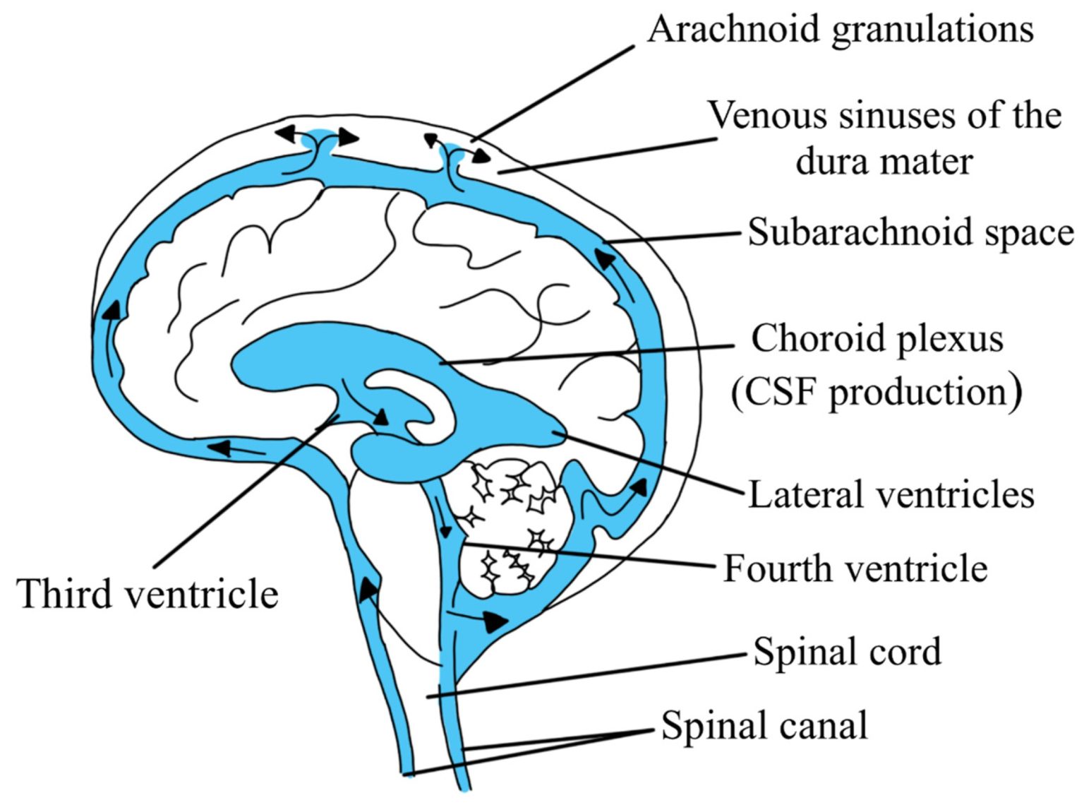
Image: Cerebrospinal fluid (CSF) flow: from production within the ventricles, circulation through the ventricular system, and exit into the subarachnoid space (cranial and spinal), where it flows before being resorbed into the venous system (specifically the dural venous sinuses).
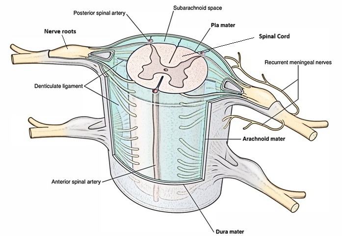
Image: The subarachnoid space is shown between the spinal cord, the spinal nerves, and the arachnoid membrane. Cerebrospinal fluid (CSF) flows through this space, and spinal nerves pass through it before they receive their individual nerve coverings.
What Causes a Spontaneous Spinal CSF Leak?
Spontaneous spinal CSF leaks can arise from several different mechanisms.
A leak may originate from the ventral dura, meaning the front side of the dural sac. In some patients, a calcified disc or small bony spur may irritate or tear the adjacent dura. The leak may also arise laterally near the nerve root sleeve, where the dura extends around the exiting spinal nerve. Another possible mechanism involves a spinal meningeal diverticulum or perineural cyst. In some patients, the problem is not a conventional dural tear but a CSF-venous fistula, where cerebrospinal fluid drains directly into a vein instead of forming an obvious extradural fluid collection.
Because these mechanisms are different, the imaging method and treatment strategy may also need to be different.
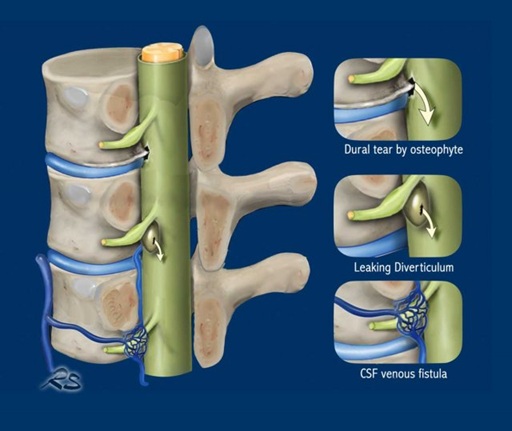
Image: Types of spinal CSF leak
Connective Tissue Disorders and Spontaneous CSF Leak
Some spontaneous CSF leaks are associated with underlying weakness of connective tissue.
This may be seen in patients with:
- hypermobility spectrum disorders
- Ehlers-Danlos syndrome
- Marfan syndrome
- dural ectasia
- other conditions that may make the dura more fragile
In these patients, leaks may be multiple, recurrent, or more difficult to localize. The clinical history may include joint hypermobility, unusual flexibility, easy bruising, poor wound healing, scoliosis, tall slender body habitus, or a family history of vascular or connective tissue disease. However, a connective tissue disorder does not prove that a CSF leak is present. Its presence only increases suspicion when the symptoms, imaging findings, recurrence pattern, and other clinical features also support the diagnosis.
Symptoms of Spinal CSF Leak and Intracranial Hypotension
Spinal CSF leak may produce a wide range of symptoms.
The most characteristic feature is headache that changes with body position.
Other symptoms may include:
- neck pain
- nausea
- dizziness
- imbalance
- tinnitus
- hearing changes
- visual blurring or other visual symptoms
- fatigue
- cognitive fog
- sensitivity to upright posture
Some patients feel significantly better when lying flat. However, not all patients have a textbook presentation. Headache may become less clearly positional over time, especially in chronic cases. Symptoms may fluctuate, and some patients may experience more neck pain, dizziness, ear symptoms, or cognitive fatigue than classic headache.
Imaging may also be incomplete or interpreted as nonspecific.
For this reason, the absence of a perfectly positional headache does not always exclude a spinal CSF leak.
Orthostatic Headache: Why Body Position Matters
An orthostatic headache becomes worse when the patient is standing or sitting and improves after lying down. It is one of the most important clinical clues in suspected spinal CSF leak and spontaneous intracranial hypotension. Improvement after lying down may occur quickly or gradually.
Some patients describe the headache as:
- pressure
- pulling
- heaviness
- pain in the head
- pain extending into the neck or upper spine
The positional relationship is important because reduced CSF volume changes the mechanical support of the brain. When the patient is upright, the brain and its supporting structures are more affected by the loss of normal CSF support. Lying down may reduce traction and temporarily improve symptoms.
However, not every patient develops a perfect orthostatic pattern. In chronic cases, the headache may become less clearly positional, while dizziness, neck pain, tinnitus, hearing symptoms, or cognitive fatigue become more prominent.
Brain MRI Signs of Spontaneous Intracranial Hypotension
Brain MRI can provide important indirect evidence of spontaneous intracranial hypotension.
Typical findings may include:
- diffuse pachymeningeal enhancement
- downward displacement or sagging of the brain
- enlarged or engorged venous structures
- subdural fluid collections
- changes around the pituitary gland
- low cerebellar tonsils
These findings reflect the intracranial response to reduced CSF volume. However, brain MRI does not always show the full classical pattern. Some patients with real spinal CSF leaks may have only subtle or incomplete findings. Others may have imaging that appears normal or nonspecific. Therefore, brain MRI can support the diagnosis of spontaneous intracranial hypotension, but it cannot by itself reliably confirm the presence of a spinal CSF leak.
Spinal CSF Leak Can Mimic Chiari I Malformation
Spontaneous intracranial hypotension may cause downward displacement of the cerebellar tonsils. This can mimic Chiari I malformation on MRI. The distinction is important. In primary Chiari I malformation, the problem is usually related to the anatomy of the posterior fossa and crowding around the foramen magnum. In spontaneous intracranial hypotension, the tonsillar descent is secondary to loss of CSF volume and downward sagging of the brain.
If acquired Chiari-like tonsillar descent is mistaken for primary Chiari I malformation, posterior fossa decompression may be performed without treating the actual cause. In that situation, symptoms may persist because the underlying problem is the CSF leak.
For this reason, low cerebellar tonsils should be interpreted together with the full MRI pattern, including:
- pachymeningeal enhancement
- venous engorgement
- brain sagging
- subdural collections
- pituitary enlargement
- the clinical history of orthostatic headache
Low-lying tonsils should not be interpreted in isolation when spontaneous intracranial hypotension is possible.
How Is Spontaneous Spinal CSF Leak Diagnosed?
Diagnosis begins with the clinical history.
The doctor should determine:
- how the symptoms started
- whether they worsen when the patient is upright
- whether lying down improves them
- whether the pattern has changed over time
- whether there has been previous surgery, trauma, lumbar puncture, or spinal anesthesia
- whether imaging already shows signs of intracranial hypotension
Brain MRI may provide evidence of spontaneous intracranial hypotension. Spine MRI may show extradural fluid collections, meningeal diverticula, perineural cysts, or other structural clues. If the leak must be localized, more specialized imaging may be required.
The diagnostic process therefore has two main goals:
1. Determine whether the clinical and imaging pattern supports spontaneous intracranial hypotension.
2. Identify the actual spinal leak mechanism and location whenever possible.
The second task can be considerably more difficult.
ICHD-3 Criteria and Spontaneous Intracranial Hypotension
According to ICHD-3, headache attributed to spontaneous intracranial hypotension requires:
- headache associated with low CSF pressure or evidence of CSF leakage
- no recent procedure or trauma that would explain the CSF loss
- a temporal relationship between the headache and the low CSF pressure or leakage
- no other headache disorder that better explains the symptoms
Evidence may include low CSF pressure or imaging findings consistent with CSF leakage. These criteria should be interpreted together with the clinical pattern and imaging findings rather than in isolation.
Spine MRI, MR Myelography, CT Myelography and Digital Subtraction Myelography in Spinal CSF Leak
Different spinal imaging techniques answer different questions.
Spine MRI in Spinal CSF Leak
Spine MRI can show indirect signs of spinal CSF leak.
It may demonstrate:
- extradural fluid collections
- meningeal diverticula
- perineural cysts
- other structural abnormalities
However, routine spine MRI does not detect every leak.
Non-Contrast MR Myelography in Spinal CSF Leak
Non-contrast MR myelography uses heavily T2-weighted MRI sequences to make cerebrospinal fluid more conspicuous. It can help demonstrate CSF collections or the general leak pathway without intrathecal contrast.
CT Myelography in Spinal CSF Leak
CT myelography uses iodinated contrast injected into the intrathecal CSF space through a lumbar puncture, followed by CT imaging of the spine to detect abnormal escape of contrast from the dural sac. It can help confirm that a spinal CSF leak is present and, more importantly, localize the level and anatomical pathway of leakage.
The lumbar puncture is usually performed under fluoroscopic guidance. After correct intrathecal needle position has been confirmed, contrast is injected into the CSF space. The patient is then positioned so that the contrast can travel through the region of the spine being examined, and CT imaging is performed according to the suspected type of leak.
Patient position is not the same in every case. Prone imaging may be useful for some ventral dural leaks, while lateral decubitus imaging can improve detection of subtle lateral leaks or CSF-venous fistulas. In decubitus CT myelography, contrast can be injected while the patient remains on the side being investigated, and the patient may be kept in that position so that gravity concentrates contrast along the dependent nerve root sleeves.
Timing is also important. With a fast spinal CSF leak, contrast may escape and spread through the extradural space so rapidly that delayed conventional CT no longer shows the exact point where the leak began. In such cases, CT imaging may need to start immediately or very soon after intrathecal contrast injection, using a dynamic CT myelographic technique.
When no definite leak is seen on the initial scan, selected protocols may include delayed CT imaging to look for slower leakage. Additional targeted or differently positioned myelographic examinations may also be considered when the first study is nondiagnostic and clinical suspicion remains high.
Contrast administration is therefore planned around the suspected leak mechanism rather than simply repeated routinely. Additional myelographic examinations, including imaging in a different position or on another occasion, may be required when searching for an occult CSF-venous fistula or a leak that was not demonstrated by the first study. Repeated intrathecal contrast administration is possible when clinically justified, but each examination is invasive and should be planned by an experienced neuroradiology team.
The main value of CT myelography is therefore not simply that contrast is present in the spinal canal, but that contrast administration, patient position, and CT timing are coordinated to match the suspected type and speed of the CSF leak.
Digital Subtraction Myelography in Spinal CSF Leak
Digital subtraction myelography (DSM) is a dynamic X-ray examination used to detect and localize spinal CSF leaks, particularly when routine MRI or conventional CT myelography does not clearly show the source. The technique uses the same basic subtraction principle known from digital subtraction angiography: a background image is acquired first, and subsequent images obtained during contrast passage are digitally subtracted from it so that moving contrast becomes much easier to see. The difference is that, in DSM for CSF leak, contrast is followed inside the intrathecal CSF space rather than inside blood vessels.
The examination usually begins with a lumbar puncture, most often under fluoroscopic guidance. The needle is advanced into the thecal sac, and a small test injection can be used to confirm that the needle tip is intrathecal. In published DSM protocols, intrathecal contrast is then injected while rapid sequential X-ray images are acquired. The exact contrast volume and injection protocol vary between centers and should not be presented as one universal standard.
Patient positioning is crucial. DSM may be performed prone or in the lateral decubitus position, depending on the suspected leak mechanism. Prone DSM has traditionally been useful for some ventral leaks, while lateral decubitus DSM has become particularly important for detecting CSF-venous fistulas. In the decubitus position, gravity helps concentrate intrathecal contrast along the dependent nerve root sleeves, increasing the chance of seeing contrast enter an abnormal paraspinal vein. Studies have shown a substantially higher detection rate for CSF-venous fistulas with lateral decubitus DSM than with prone DSM in appropriately selected patients.
The study is highly time-dependent because the radiologist is trying to capture the actual movement of contrast at the moment it leaves the normal CSF space. This is why DSM can reveal rapid or subtle leaks that may be missed on delayed static imaging. It is especially useful when conventional spinal imaging shows no obvious extradural CSF collection but the clinical and brain MRI pattern still strongly suggests spontaneous intracranial hypotension.
When a CSF-venous fistula is suspected, one side of the spine may be examined first in lateral decubitus position. If the study is negative and suspicion remains high, the opposite side may need to be examined separately, either during another acquisition sequence or in a subsequent examination, depending on the protocol. Some centers also combine lateral decubitus DSM with CT performed shortly afterward to improve fistula localization.
DSM is therefore not simply “another myelogram.” Its value comes from combining intrathecal contrast, carefully chosen patient position, rapid image acquisition, and digital subtraction to make very small or rapidly appearing leaks visible. It is particularly useful for difficult-to-localize spinal CSF leaks and CSF-venous fistulas when standard MRI or CT myelography has been inconclusive.
What to Do When Spinal CSF Leak Is Suspected but MRI Is Normal
A normal or unclear MRI does not always exclude spinal CSF leak.
A leak may be:
- small
- intermittent
- slow
- technically difficult to image
- not visible with the first technique used
CSF-venous fistulas are particularly challenging because CSF may drain directly into a vein without forming a large extradural fluid collection. Brain MRI may also show only subtle or incomplete evidence of spontaneous intracranial hypotension. In this situation, repeating the same test without a clear diagnostic strategy may not be useful. The more important question is whether the imaging method used was appropriate for the suspected leak mechanism. If the clinical pattern remains convincing, further evaluation with a different or more targeted myelographic technique may be required.
What to Do When Myelography Does Not Localize the Spinal CSF Leak
Some spinal CSF leaks are obvious. Others are extremely difficult to detect.
Failure to localize the leak may occur because it is:
- intermittent
- slow
- technically difficult to demonstrate
- very rapid
- associated with a mechanism requiring a specific imaging technique
CSF-venous fistulas are especially difficult because cerebrospinal fluid can drain directly into the venous system without producing a large visible extradural collection. When initial imaging is negative, the important question is whether the correct diagnostic technique was used for the suspected leak type. A negative or unclear first imaging study does not automatically end the diagnostic process when the clinical suspicion remains strong.
Spinal CSF-Venous Fistula: A Hidden Cause of Intracranial Hypotension
A CSF-venous fistula is a type of spinal CSF leak in which cerebrospinal fluid drains directly into a vein. Because the CSF does not necessarily accumulate in the extradural space, routine imaging may look unclear or even normal.
The patient can still develop typical symptoms of spontaneous intracranial hypotension, including:
- orthostatic headache
- neck pain
- dizziness
- tinnitus
- hearing symptoms
- fatigue
- cognitive fog
Brain MRI may also show signs such as brain sagging. CSF-venous fistulas often require specific myelographic techniques and experienced interpretation. Digital subtraction myelography or dynamic CT myelography may be needed in selected cases. Treatment may include targeted procedures, endovascular treatment in selected fistulas, or surgical repair depending on the anatomy.
Meningeal Diverticula and Tarlov Cysts: When Are They Relevant for Spinal CSF Leak?
Spinal meningeal diverticula, perineural cysts, and Tarlov cysts may be seen in patients evaluated for spontaneous intracranial hypotension. In some cases, a diverticulum or cyst may be related to the leak site. However, a Tarlov cyst on MRI is not the same as a proven active CSF leak.
These findings may also be present without causing CSF loss.
The important question is whether targeted imaging demonstrates:
- actual leakage
- abnormal filling
- a CSF-venous fistula
- another mechanism that explains the patient’s symptoms
A cyst or diverticulum must therefore be interpreted together with the headache pattern, brain MRI, spinal imaging, and treatment response. Meningeal diverticula and Tarlov cysts may be relevant, but they are not by themselves proof that they are the source of the leak.
Spontaneous Intracranial Hypotension and Post-Lumbar Puncture Headache Are Not the Same Condition
Post-lumbar puncture headache and spontaneous intracranial hypotension can feel similar. Both may involve loss of CSF volume and headache that worsens when upright. The difference is the cause.
A post-lumbar puncture headache occurs after a known dural puncture, such as:
- lumbar puncture
- spinal anesthesia
Spontaneous intracranial hypotension, in contrast, occurs without a recent procedure or trauma that explains the CSF loss. The symptoms may overlap, but spontaneous intracranial hypotension often requires a different diagnostic approach because the actual leak site may be hidden and difficult to localize.
Treatment of Spontaneous Spinal CSF Leak
Treatment depends on:
- severity of symptoms
- duration of symptoms
- imaging findings
- whether the leak can be localized
- the underlying leak mechanism
- response to previous treatment
Some mild and recent cases may initially be treated conservatively. However, conservative treatment should not continue indefinitely if significant symptoms persist. In suspected spinal CSF leak with spontaneous intracranial hypotension, early specialist evaluation and epidural blood patching are usually preferred over prolonged observation when symptoms do not improve.
Treatment may include:
- conservative measures
- epidural blood patch
- targeted patching
- fibrin sealant in selected cases
- endovascular treatment for some CSF-venous fistulas
- surgical repair
The choice depends on the actual leak mechanism and location.
Epidural Blood Patch for Spinal CSF Leak
An epidural blood patch is an important treatment for spinal CSF leak associated with spontaneous intracranial hypotension. During the procedure, at least 20 mL of the patient’s own venous blood is usually drawn under sterile conditions and injected into the epidural space near the suspected leak site. The blood is generally injected immediately after collection, before it clots in the syringe.
The injection is performed slowly and may be stopped earlier if the patient develops:
- significant back pressure
- leg pain
- neurological symptoms
The injected blood is not a medication or foreign substance. It works by forming a natural clot and seal around the dural defect, which may reduce or stop the CSF leak and help restore normal CSF volume. Doctors typically perform the procedure under fluoroscopy or CT guidance so that the needle can be positioned accurately in the epidural space. The blood patch may be non-targeted or targeted, depending on whether the leak site is known.
Non-Targeted Epidural Blood Patch
A non-targeted blood patch may be used when the clinical pattern strongly suggests spinal CSF leak but the exact location has not been identified. The goal is to allow blood to spread through the epidural space and potentially seal an unlocalized leak.
Targeted Epidural Blood Patch
A targeted blood patch is directed toward the suspected leak site when imaging has localized the abnormality. This may be particularly useful when a more specific anatomical source has been identified. A blood patch can help significantly, but it does not work in every patient. Some patients require repeated or targeted procedures. The result depends in part on whether the leak mechanism and location have been correctly understood.
When conservative treatment or blood patching does not adequately control the leak, more direct treatment may be required.
Fibrin Sealant in Spinal CSF Leak
Fibrin sealant may be used in selected cases as a targeted treatment around a known or suspected leak site.
Endovascular Treatment in Spinal CSF Leak
Some CSF-venous fistulas may be treated using an endovascular approach. This option is considered only in selected cases, depending on the anatomy and the leak mechanism.
Surgical Repair in Spinal CSF Leak
Surgery may be considered when:
- the leak is clearly localized
- conservative treatment and blood patching have failed
- there is a structural dural defect
- a calcified disc or bony spur is associated with the leak
- a CSF-venous fistula requires direct treatment
Why Symptoms May Persist or Return After Treatment
Symptoms may persist or return after treatment for several reasons.
Possible explanations include:
- the leak may not have been fully sealed
- the treated site may not have been the true source
- there may be more than one leak
- a CSF-venous fistula may have been missed
- another disorder may coexist with the CSF leak
Other conditions can produce overlapping symptoms.
These may include:
- migraine
- cervical spine disease
- vestibular disorders
- sinus disease
- neuralgia
- central sensitization
Persistent symptoms therefore do not automatically prove that the previous treatment was incorrect. They mean that the diagnosis, leak mechanism, treatment response, and remaining symptoms must be reassessed carefully.
Rebound Intracranial Hypertension After Spinal CSF Leak Treatment
A new or different headache after treatment does not always mean that the original leak remains open. One important possibility is rebound intracranial hypertension. This can occur after successful sealing of a spinal CSF leak, particularly after an epidural blood patch or targeted repair. The original low-pressure headache may improve, but the patient then develops a different headache pattern related to increased CSF pressure.
The new headache may:
- become less orthostatic
- become worse when lying down
- cause pressure behind the eyes
- be associated with nausea
- cause blurred vision
- be accompanied by pulsatile tinnitus
This distinction is important because treating rebound intracranial hypertension as though the original leak is still open may lead to the wrong next step. A change in headache pattern after treatment should therefore be interpreted carefully rather than automatically assumed to represent treatment failure.
Recovery and Prognosis After Spontaneous Spinal CSF Leak
The prognosis depends on:
- the type of spinal leak
- how long symptoms have been present
- whether the leak can be localized
- the severity of intracranial hypotension
- whether there are subdural collections or severe brain sagging
- whether the leak recurs
- how effectively the underlying mechanism is treated
Some mild leaks may improve with conservative measures, particularly when symptoms are recent and gradually improving.
Persistent spinal CSF leaks often require:
- epidural blood patch
- targeted patching
- embolization in selected CSF-venous fistulas
- surgical repair
Recovery is not always immediate. Headache may improve before other symptoms resolve. Fatigue, dizziness, hearing symptoms, cognitive fog, and neck pain may take longer to settle. In long-standing cases, symptoms may fluctuate for some time after the leak has been treated. The most useful prognostic question is not simply whether a CSF leak existed. It is whether the leak type, location, treatment response, and remaining symptoms all fit together.
Request Spontaneous Spinal CSF Leak Neurosurgical Second Opinion — 24-Hour Review (Priority Option Available)
Patients with a suspected spontaneous spinal CSF leak or spontaneous intracranial hypotension most often request a second opinion when symptoms strongly suggest a leak but brain or spine MRI is normal or inconclusive, the leak cannot be localized, CT myelography or digital subtraction myelography findings are unclear, a CSF-venous fistula is suspected, or symptoms persist after an epidural blood patch or other treatment.
An independent neurosurgical second opinion can help clarify whether the symptoms and imaging findings fit a spontaneous spinal CSF leak, which leak mechanism is most likely, whether additional targeted imaging may be needed, and whether observation, epidural blood patch, targeted treatment, endovascular treatment, or surgery is the most reasonable next step.
- ✔ Send a brief message describing your main symptoms, especially orthostatic headache, symptoms that worsen when upright and improve when lying down, neck pain, dizziness, tinnitus, hearing changes, cognitive fog, or MRI findings suggesting spontaneous intracranial hypotension.
- ✔ Mention whether brain MRI or spine MRI was normal or abnormal, whether a leak has been localized, and whether CT myelography, dynamic CT myelography, digital subtraction myelography, epidural blood patch, fibrin sealant, embolization, or surgery has already been performed.
- ✔ You’ll receive a reply within 24 hours explaining whether we can help with an online neurosurgical consultation.
- ✔ Priority cases: if there is severe disabling orthostatic headache, marked brain sagging, subdural collections, neurological deterioration, rapidly worsening symptoms, or an urgent recommendation for spinal surgery or another invasive procedure, consultations can often be arranged within a few hours — write PRIORITY in your first message.
- ✔ Radiology reports and/or original brain and spine MRI images, MR myelography, CT myelography, dynamic CT myelography, digital subtraction myelography, previous blood patch reports, operative reports, and other specialist evaluations can be reviewed once initial contact is established.
- ✔ During the consultation, we explain whether the clinical pattern supports a spontaneous spinal CSF leak, whether a ventral dural tear, nerve root sleeve leak, meningeal diverticulum, Tarlov cyst, or CSF-venous fistula may be relevant, whether further imaging is justified, and which treatment strategy is most appropriate — with up to 10 days of follow-up for brief clarification questions.
Consultation fees typically range from $180–250, depending on case complexity, previous treatment, and the amount of imaging that must be reviewed.
Secure payment by credit card, PayPal invoice (USD), or bank transfer.
This is within the usual range for international specialist telehealth neurosurgical second opinions.
Frequently Asked Questions About Spontaneous Spinal CSF Leak and Intracranial Hypotension
What is a spontaneous spinal CSF leak and how does it cause spontaneous intracranial hypotension?
A spontaneous spinal CSF leak occurs when cerebrospinal fluid escapes through a defect in the spinal dura without a recent lumbar puncture, spinal procedure, major trauma, or surgery that would otherwise explain the leak. CSF may escape into the spinal epidural space or, in a CSF-venous fistula, drain directly into a vein. The resulting loss of CSF volume can reduce the normal support of the brain and produce spontaneous intracranial hypotension. Patients may develop orthostatic headache, neck pain, nausea, dizziness, tinnitus, hearing changes, visual symptoms, imbalance, fatigue, or cognitive fog. Brain MRI may show indirect signs such as pachymeningeal enhancement, brain sagging, venous engorgement, subdural collections, pituitary enlargement, or low cerebellar tonsils.
What symptoms are most typical of a spontaneous spinal CSF leak?
The most characteristic symptom of a spontaneous spinal CSF leak is an orthostatic headache that becomes worse when sitting or standing and improves after lying down. Other symptoms may include neck pain, nausea, dizziness, imbalance, tinnitus, hearing changes, visual blurring, fatigue, and cognitive fog. The headache may feel like pressure, pulling, heaviness, or pain involving the head, neck, or upper spine. The pattern is not identical in every patient. In long-standing spontaneous spinal CSF leak, the positional component may become less obvious and other symptoms may become more prominent.
Can a spontaneous spinal CSF leak occur without a typical orthostatic headache?
Yes. A spontaneous spinal CSF leak does not always produce a perfectly positional headache. In chronic cases, the relationship between headache and upright posture may become less obvious, and some patients primarily report neck pain, dizziness, tinnitus, hearing symptoms, fatigue, imbalance, or cognitive difficulty. The diagnosis should therefore not depend on one symptom alone. The entire clinical pattern, brain MRI, spinal imaging, and response to previous treatment should be considered together.
Can brain MRI be normal in a patient with a spontaneous spinal CSF leak?
Yes. Brain MRI can show important indirect evidence of spontaneous intracranial hypotension, including pachymeningeal enhancement, brain sagging, venous enlargement, subdural collections, pituitary enlargement, or low cerebellar tonsils. However, some patients with a genuine spontaneous spinal CSF leak have only subtle or incomplete MRI findings, and occasionally the study may appear normal. Brain MRI therefore supports the diagnosis of spontaneous intracranial hypotension but cannot by itself reliably confirm or exclude the presence of a spinal CSF leak.
Can spine MRI be normal even when a spontaneous spinal CSF leak is present?
Yes. Routine spine MRI does not detect every spontaneous spinal CSF leak. A leak may be small, intermittent, rapid, slow, or difficult to demonstrate with the imaging technique used. A CSF-venous fistula can be particularly difficult because CSF drains directly into a vein and may not create a large extradural fluid collection. When clinical suspicion remains strong despite a normal or unclear MRI, the next step may involve a more targeted myelographic technique rather than simply repeating the same MRI.
When is spine MRI used for suspected spontaneous spinal CSF leak, how is it performed, and what are its limitations or risks?
Spine MRI is often one of the first spinal examinations used when a spontaneous spinal CSF leak is suspected. It can demonstrate extradural CSF collections, meningeal diverticula, perineural cysts, dural abnormalities, or other structural clues that help determine which type of leak may be present. The examination is non-invasive and is performed with the patient lying in the MRI scanner; depending on the protocol, different sequences may be obtained through the cervical, thoracic, and lumbar spine.
Spine MRI does not require lumbar puncture and may be performed without intrathecal contrast. Its main limitation is that it may show the consequences of a leak without precisely identifying the actual defect, and some spontaneous spinal CSF leaks or CSF-venous fistulas may remain invisible. MRI has no ionizing radiation, but usual MRI safety restrictions apply in patients with certain implants or metallic foreign bodies, and some patients may have difficulty with claustrophobia or remaining still during the examination.
When is non-contrast MR myelography used for spontaneous spinal CSF leak, how is it performed, and what are its limitations?
Non-contrast MR myelography may be used when evaluating a suspected spontaneous spinal CSF leak, particularly when the goal is to visualize CSF collections or the general pathway of leakage without performing a lumbar puncture. The technique uses heavily T2-weighted MRI sequences that make cerebrospinal fluid appear very bright and can therefore highlight abnormal fluid outside the normal thecal sac.
Because no intrathecal contrast or ionizing radiation is required, the examination is non-invasive. However, non-contrast MR myelography has limited temporal resolution and may not identify the exact origin of a rapid leak or a CSF-venous fistula. If the leak mechanism remains unclear, CT myelography, dynamic CT myelography, or digital subtraction myelography may be required.
When is CT myelography indicated for a spontaneous spinal CSF leak, how is it performed, and what complications can occur?
CT myelography is used when a spontaneous spinal CSF leak needs to be localized more precisely or when MRI does not adequately identify the leak mechanism. The examination begins with a lumbar puncture, usually performed under fluoroscopic guidance. After the needle tip is confirmed within the intrathecal CSF space, iodinated contrast is injected and the patient is positioned so that contrast reaches the spinal region being studied. CT images are then obtained to determine whether contrast escapes from the dural sac.
Patient position and timing depend on the suspected leak. Prone imaging may be useful for some ventral leaks, while lateral decubitus positioning can improve visualization of lateral nerve root abnormalities or CSF-venous fistulas. Complications are uncommon but may include post-lumbar-puncture headache, back discomfort, bleeding, infection, reaction to iodinated contrast, or, rarely, neurological complications. CT myelography also exposes the patient to ionizing radiation.
When is dynamic CT myelography used for a spontaneous spinal CSF leak, how does it differ from conventional CT myelography, and what are the risks?
Dynamic CT myelography is particularly useful when a spontaneous spinal CSF leak is suspected to be rapid. In a fast leak, contrast may leave the dural sac and spread through the epidural space so quickly that conventional delayed CT cannot show where the leak originally began. Dynamic CT myelography therefore coordinates intrathecal contrast injection with very rapid or immediate CT acquisition.
The examination requires lumbar puncture and intrathecal iodinated contrast, similar to conventional CT myelography, but timing is much more tightly controlled. The main risks are those associated with lumbar puncture and intrathecal contrast, including post-procedure headache, bleeding, infection, contrast-related reactions, and very rare neurological complications. Radiation exposure may be greater than with routine imaging because multiple rapid CT acquisitions may be required.
When is digital subtraction myelography used for a spontaneous spinal CSF leak, how is it performed, and what complications can occur?
Digital subtraction myelography may be used when a spontaneous spinal CSF leak remains difficult to localize, particularly when a rapid leak, subtle leak, or CSF-venous fistula is suspected. The examination begins with lumbar puncture and injection of iodinated contrast into the intrathecal CSF space. Rapid X-ray images are then obtained while contrast moves through the thecal sac.
The subtraction principle is similar to digital subtraction angiography: a background image is digitally removed from subsequent images so that moving contrast becomes easier to see. The difference is that DSM follows contrast within the CSF space rather than within blood vessels. The examination may be performed prone or in the lateral decubitus position depending on the suspected leak mechanism. Lateral decubitus positioning is particularly useful when a CSF-venous fistula is suspected because gravity helps concentrate intrathecal contrast along the dependent nerve root sleeves, increasing the chance of seeing contrast pass into an abnormal paraspinal vein.
In selected patients with spontaneous intracranial hypotension and no extradural CSF collection on conventional spinal imaging, lateral decubitus DSM has shown a substantially higher detection rate for CSF-venous fistulas than prone DSM. This does not mean that lateral decubitus DSM is always superior to every other myelographic technique; the choice between DSM, conventional CT myelography, and lateral decubitus CT myelography depends on the suspected leak mechanism, previous imaging, available expertise, and local protocol.
Because DSM is highly time-dependent, contrast injection and image acquisition must be closely coordinated so that the radiologist can capture the moment when contrast leaves the normal CSF space or enters an abnormal vein. In some cases, one side is examined first in lateral decubitus position, and the opposite side may require a separate acquisition if the initial study is negative and suspicion remains high.
Possible complications are generally related to lumbar puncture, intrathecal contrast, and radiation exposure. These may include post-lumbar-puncture headache, temporary back discomfort, bleeding, infection, contrast-related reactions, or, rarely, neurological complications.
Are connective tissue disorders such as Ehlers-Danlos syndrome, Marfan syndrome, or hypermobility associated with spontaneous spinal CSF leak?
Yes. Some patients with a spontaneous spinal CSF leak have an underlying connective tissue disorder or generalized tissue fragility. Associations have been described with hypermobility spectrum disorders, Ehlers-Danlos syndrome, Marfan syndrome, dural ectasia, and other conditions that may weaken the dura or supporting connective tissues.
Clinical clues may include joint hypermobility, unusually flexible joints, easy bruising, poor wound healing, scoliosis, a tall slender body habitus, or a family history of vascular or connective tissue disease. However, having Ehlers-Danlos syndrome, Marfan syndrome, or hypermobility does not by itself prove that a spinal CSF leak is present. These findings increase clinical suspicion only when the symptoms, imaging findings, recurrence pattern, and other features are compatible.
What is a CSF-venous fistula in spontaneous spinal CSF leak and why can it be missed on routine imaging?
A CSF-venous fistula is a form of spontaneous spinal CSF leak in which cerebrospinal fluid drains directly from the CSF space into a vein. Because the CSF is carried away through the venous system, there may be no large extradural fluid collection visible on routine spine MRI. This is why conventional imaging can appear normal even when the patient has clear clinical and brain MRI evidence of spontaneous intracranial hypotension.
Detecting a CSF-venous fistula often requires a technique designed to show rapid contrast passage from the CSF space into an adjacent vein. Lateral decubitus CT myelography or digital subtraction myelography may be required, and examination of both sides may sometimes be necessary.
What should be done when a spontaneous spinal CSF leak is strongly suspected but MRI and initial myelography are negative?
Negative imaging does not automatically exclude a spontaneous spinal CSF leak. The first question is whether the imaging method was appropriate for the suspected leak mechanism. A small, intermittent, very rapid, or technically difficult leak may require a different technique, and a CSF-venous fistula may not be visible on routine MRI or conventional myelography.
Previous brain and spine imaging should be reviewed carefully, preferably by a team experienced in spontaneous intracranial hypotension. Depending on the findings, further evaluation may include differently timed CT myelography, dynamic CT myelography, lateral decubitus imaging, or digital subtraction myelography. Repeating the same study without a clear diagnostic strategy may be less useful than selecting a method specifically suited to the suspected leak type.
Can a Tarlov cyst, perineural cyst, or meningeal diverticulum prove that a spontaneous spinal CSF leak is present?
No. A Tarlov cyst, perineural cyst, or meningeal diverticulum may be found during evaluation for a spontaneous spinal CSF leak, but the presence of one of these structures on MRI does not prove that it is actively leaking. Such abnormalities can also occur in patients without CSF loss.
The relevant question is whether targeted imaging demonstrates actual leakage, abnormal filling, a CSF-venous fistula, or another mechanism that links the structure to the patient’s symptoms. These findings should therefore be interpreted together with the clinical pattern, brain MRI, spinal imaging, and treatment response rather than assumed to be the cause simply because they are visible.
How is an epidural blood patch performed for spontaneous spinal CSF leak, when is it indicated, and what complications can occur?
An epidural blood patch is one of the main treatments for spontaneous spinal CSF leak associated with spontaneous intracranial hypotension. Sterile autologous venous blood is drawn from the patient and injected into the epidural space, usually under fluoroscopic or CT guidance. The blood is injected slowly and may spread through the epidural space, forming a clot and biological seal around the dural defect.
A blood patch may be considered when symptoms persist and the clinical picture supports a spinal CSF leak, whether or not the exact leak site is known. Possible adverse effects include temporary back pressure or pain, radicular pain, post-procedure soreness, accidental dural puncture, bleeding, infection, or, rarely, neurological complications. Injection is stopped if significant pressure, severe leg pain, or neurological symptoms develop.
What is the difference between a non-targeted and targeted epidural blood patch for spontaneous spinal CSF leak?
A non-targeted epidural blood patch may be used when a spontaneous spinal CSF leak is strongly suspected but the exact location has not been identified. Blood is injected into the epidural space with the aim of spreading over several spinal levels and sealing an unlocalized dural defect.
A targeted epidural blood patch is directed toward a specific spinal level when imaging has identified or strongly suggested the leak site. Targeted treatment allows the blood or sealing material to be placed closer to the suspected defect. Some patients improve after one procedure, while others require repeated or more targeted treatment. Failure of a blood patch may suggest that the leak has not been sealed, the site was incorrect, another leak is present, or the underlying mechanism requires a different treatment.
When is fibrin sealant used for spontaneous spinal CSF leak, how is it applied, and what complications are possible?
Fibrin sealant may be used in selected patients with a spontaneous spinal CSF leak when a specific leak site has been identified or strongly suspected and a more targeted sealing procedure is considered appropriate. The material is injected near the suspected dural defect under image guidance, sometimes alone and sometimes in combination with an epidural blood patch.
Because fibrin sealant is placed close to neural and vascular structures, careful localization and image guidance are important. Potential complications include local pain, nerve irritation, infection, bleeding, allergic or inflammatory reactions, inadvertent injection into an unintended space, or failure to seal the leak. It is therefore used selectively rather than as a routine first procedure for every spontaneous spinal CSF leak.
When is endovascular treatment used for a spinal CSF-venous fistula, how is it performed, and what complications can occur?
Endovascular treatment may be considered in selected patients whose spontaneous spinal CSF leak is caused by a clearly localized CSF-venous fistula. The procedure is performed through the venous system using a catheter that is advanced to the abnormal draining vein. An embolic material is then used to close the venous pathway through which CSF is escaping.
This treatment is not appropriate for every spinal CSF leak and requires precise localization of the fistula and suitable venous anatomy. Possible complications include access-site bleeding, venous injury or thrombosis, non-target embolization, neurological injury, contrast-related complications, radiation exposure, or failure to completely close the fistula. The decision depends on anatomy, local expertise, and comparison with surgical options.
When is surgery recommended for a spontaneous spinal CSF leak, how is the defect repaired, and what complications can occur?
Surgery may be considered when a spontaneous spinal CSF leak has been clearly localized and less invasive treatment has failed, or when imaging identifies a structural cause that is unlikely to respond adequately to blood patching alone. Examples include a ventral dural tear related to a calcified disc or bony spur, a clearly localized nerve root sleeve defect, a leaking meningeal diverticulum, or a CSF-venous fistula requiring direct repair.
The operation depends on the anatomy. It may involve direct dural closure, reinforcement of the defect, removal or treatment of an offending bony or calcified structure, repair of a diverticulum, or interruption of a fistulous connection. Possible complications depend on the spinal level and surgical approach and may include CSF leakage, infection, bleeding, nerve or spinal cord injury, postoperative pain, instability, recurrence, or failure to completely correct the leak.
Why can symptoms persist or return after treatment of a spontaneous spinal CSF leak?
Symptoms may persist or return after treatment of a spontaneous spinal CSF leak because the leak was not completely sealed, the treated site was not the true source, more than one leak is present, or a CSF-venous fistula was missed. In some patients, the original leak may have been successfully treated but another disorder continues to produce headache, dizziness, neck pain, or other symptoms.
Migraine, cervical spine disease, vestibular disorders, neuralgia, or central sensitization can overlap with the symptoms of spontaneous intracranial hypotension. Persistent symptoms therefore do not automatically prove that the leak remains open. The original symptom pattern, imaging findings, treatment response, and any new symptoms should be reassessed together.
How can rebound intracranial hypertension be distinguished from a persistent spontaneous spinal CSF leak after treatment?
Rebound intracranial hypertension may occur after successful sealing of a spontaneous spinal CSF leak. The original low-pressure headache may improve, but a new headache develops with a different pattern. Instead of worsening when upright, the new headache may become less positional or worse when lying down and may be associated with pressure behind the eyes, nausea, blurred vision, or pulsatile tinnitus.
This distinction matters because a new headache after treatment does not automatically mean that the spontaneous spinal CSF leak remains open. Comparing the new headache with the patient’s original orthostatic pattern, reviewing treatment response, and reassessing imaging or CSF pressure when clinically appropriate can help distinguish rebound intracranial hypertension from persistent or recurrent leakage.
How long can recovery take after successful treatment of a spontaneous spinal CSF leak?
Recovery after treatment of a spontaneous spinal CSF leak is not always immediate. Headache may improve before other symptoms such as dizziness, fatigue, tinnitus, hearing changes, neck pain, or cognitive fog. Patients with long-standing spontaneous intracranial hypotension may experience fluctuations for some time even after the leak has been successfully treated.
The recovery period depends on the leak mechanism, duration of symptoms, degree of intracranial hypotension, presence of subdural collections or significant brain sagging, treatment used, and whether the leak recurs. The most important question is whether the overall clinical course after treatment is moving in the expected direction and whether new or persistent symptoms suggest ongoing leakage, rebound intracranial hypertension, or another condition.
When should a patient seek a second opinion for suspected or treated spontaneous spinal CSF leak?
A second opinion may be useful when a spontaneous spinal CSF leak is suspected but the diagnosis remains uncertain, brain or spine MRI is normal despite convincing symptoms, imaging reports are contradictory, the leak cannot be localized, or the significance of a CSF-venous fistula, meningeal diverticulum, or Tarlov cyst is unclear.
It may also be helpful when one or more epidural blood patches have failed, symptoms return after treatment, a new headache pattern develops, or targeted treatment, embolization, or surgery has been proposed. The purpose of a second opinion is to determine whether the clinical pattern and imaging support a spontaneous spinal CSF leak, whether the leak mechanism has been correctly identified, whether additional testing is justified, and which treatment strategy is most appropriate.