Dr Željko Kojadinović — NEUROHIRURGIJA I LEČENJE BOLA
Dr Zeljko Kojadinovic — Pain Treatment & Neurosurgery


Author:
Dr. Zeljko Kojadinovic, MD, PhD
— Consultant Neurosurgeon
Specialized Experience:
30 years of clinical expertise in neurosurgery.
Last medically reviewed:
March 08, 2026
Who This Olfactory Groove Meningioma Page Is For
This page is intended for patients in whom MRI or CT has revealed an olfactory groove meningioma, a tumor arising from the meninges of the anterior skull base beneath the frontal lobes, near the olfactory nerves responsible for the sense of smell.
If surgery, stereotactic radiosurgery (such as Gamma Knife), radiotherapy, or long-term MRI monitoring has been proposed — or if specialists have offered different recommendations regarding the urgency or type of treatment — an individualized neurosurgical second opinion may help clarify the expected biological behavior of an olfactory groove meningioma, the reasons for symptoms such as loss of smell, cognitive or personality changes, or visual disturbances, the likelihood of tumor recurrence, and the safest balance between observation, surgery, and radiation therapy based on tumor size, growth rate, frontal lobe compression, surrounding brain edema, relationship to the optic nerves and anterior cerebral arteries, and the patient’s overall neurological condition.
When patients seek a second opinion for olfactory groove meningioma
• MRI or CT shows an olfactory groove meningioma and the best treatment strategy is unclear
• The tumor was discovered incidentally and opinions differ between MRI monitoring, surgery, or radiosurgery
• Loss of smell, cognitive or personality changes, or visual symptoms suggest frontal lobe or optic pathway compression
• Surgery is recommended but the risks, expected extent of removal, or prognosis are uncertain
• Radiosurgery or radiotherapy is proposed but the role of surgery remains unclear
• Specialists recommend different strategies without clear agreement
If your situation involves uncertainty regarding diagnosis, urgency, or treatment strategy, you may request an individualized neurosurgical review here:
Request Second Opinion
Olfactory Groove Meningioma — Quick Summary (Read This First)
- Olfactory groove meningioma is a tumor arising from the meninges at the anterior skull base beneath the frontal lobes. It develops near the olfactory nerves responsible for the sense of smell.
- Most olfactory groove meningiomas grow outside the brain tissue. They compress nearby brain structures without directly invading the brain.
- Olfactory groove meningiomas account for approximately 8–10% of intracranial meningiomas. They arise along the olfactory groove, cribriform plate, or planum sphenoidale of the anterior skull base.
- Many olfactory groove meningiomas grow slowly and may remain stable for years. Some tumors discovered incidentally on MRI can be monitored with periodic imaging.
- The earliest symptom is often gradual loss of smell (anosmia). Because this change develops slowly and often affects both sides, many patients do not notice it immediately.
- Larger tumors may compress the frontal lobes or optic pathways. This can lead to headaches, personality or behavioral changes, cognitive slowing, visual disturbances, seizures, or gait imbalance in advanced cases.
- MRI with contrast is the most important diagnostic test. It shows tumor size, its relationship to the frontal lobes, optic nerves, anterior cerebral arteries, and the presence of surrounding brain edema.
- Not all olfactory groove meningiomas require immediate treatment. Small tumors without symptoms or growth may initially be monitored with periodic MRI examinations.
- Surgery is the main treatment for symptomatic or growing tumors. The goal is safe tumor removal while protecting the optic nerves, major arteries, and frontal lobe function.
- Stereotactic radiosurgery or fractionated radiotherapy may be used in selected cases. Radiation is often considered for small growing tumors, residual tumor after surgery, recurrent tumors, or when surgery carries higher risk.
- Treatment decisions depend mainly on tumor size, growth rate, symptoms, brain edema, and the tumor’s relationship to nearby arteries and optic pathways. The safest strategy is usually individualized.
- Most olfactory groove meningiomas are benign WHO Grade I tumors. Approximately 80–85% belong to Grade I, about 10–15% are atypical (WHO Grade II), and roughly 1–3% are malignant (WHO Grade III). Higher-grade tumors have a greater risk of recurrence.
- Although many tumors are benign, large olfactory groove meningiomas can still cause serious neurological problems. This occurs because of progressive compression of the frontal lobes, optic pathways, and other vital brain structures.
Most readers benefit from reviewing this Quick Summary together with the sections on Symptoms of Olfactory Groove Meningioma, Diagnosis, Surgical Treatment, Radiation Therapy, and Treatment Decision-Making. Later sections provide more detailed explanations intended for patients seeking a deeper understanding before important treatment decisions are made.
Contents
- Who This Page
- Quick Summary
- Definition
- Tumor Grades
- Tumor Causes
- How It Grows
- Symptoms
- MRI Diagnosis
- When Monitored
- Symptom Relief
- Cognitive Changes
- Surgical Treatment
- Modern Technologies
- Endoscopic Surgery
- Extent of Removal
- Second Opinion
- Radiation Therapy
- Tumor Recurrence
- Treatment Summary
- Experimental Therapies
- Why Opinions Differ
- Overall Prognosis
- Life-Threatening Risk
- FAQ
What Is an Olfactory Groove Meningioma
An olfactory groove meningioma (OGM) is a meningioma that arises from the meninges in the olfactory groove region of the anterior skull base, just above the nasal cavity. As it enlarges, it may extend to adjacent parts of the anterior skull base.
Most meningiomas arise from specialized cells within the arachnoid layer, often called arachnoid cap cells. When genetic changes occur in these cells, they may begin to multiply abnormally and gradually form a tumor. Unlike tumors that originate inside the brain tissue, olfactory groove meningiomas grow outside the brain, attached to the dura mater of the anterior skull base. As they enlarge, they typically compress the surrounding frontal lobes without directly invading the brain tissue. Because of this growth pattern, they often remain well-defined masses, although large tumors may come into contact with nearby arteries and the optic nerves, compress them, and in some cases become firmly adherent to these structures. The skull base bone at the site of tumor origin is often thickened (hyperostosis), reflecting tumor involvement of the underlying bone.
Olfactory groove meningiomas represent approximately 8–10% of intracranial meningiomas. Because of their location beneath the frontal lobes, they may become quite large before they are discovered.
Meningiomas occur approximately two to three times more frequently in women than in men. They are also rare in children, representing less than 2% of all pediatric brain tumors.
In many patients, the tumor grows slowly and initially produces few symptoms. One of the earliest manifestations is often a disturbance of smell or gradual loss of smell (anosmia) due to compression of the olfactory nerves. Because this change may develop slowly and many patients do not immediately notice it, the tumor can continue to enlarge for years before other symptoms appear. As the tumor grows further, patients may develop headaches, personality or behavioral changes related to frontal lobe compression, progressive cognitive slowing, visual disturbances if the optic pathways become involved, and occasionally seizures.
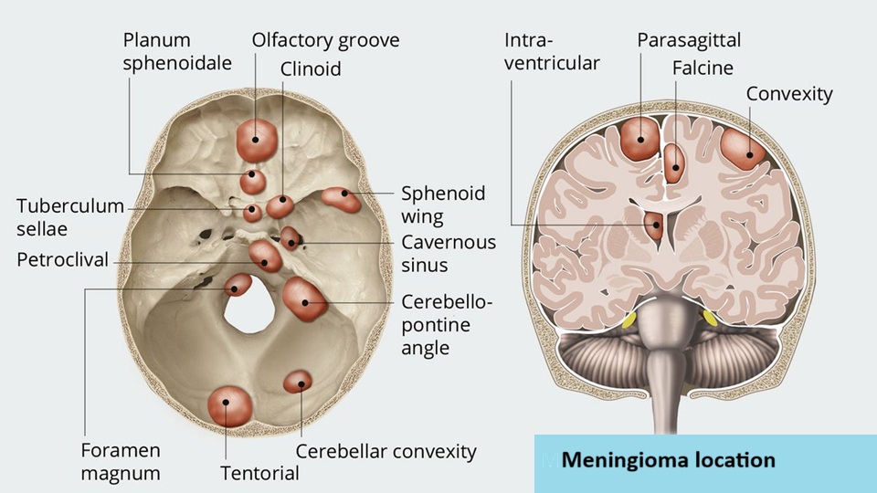
Image: Olfactory groove meningioma and meningiomas of other locations
Biological Classification of Meningiomas
The biological behavior of olfactory groove meningiomas is determined through histopathological analysis using the World Health Organization (WHO) classification.
Three main categories are recognized:
• WHO grade I — benign meningioma. This is the most common form, representing roughly 80–85% of meningiomas. These tumors typically grow slowly and often remain stable for long periods. When completely removed surgically, the chance of recurrence is relatively low.
• WHO grade II — atypical meningioma. Atypical meningiomas account for approximately 10–15% of cases in surgical series. These tumors show increased cellular activity and a higher likelihood of recurrence after surgery. After complete tumor removal, some patients may be managed with MRI follow-up rather than immediate radiation therapy.
• WHO grade III — malignant (anaplastic) meningioma. These tumors are rare, representing roughly 1–3% of cases. They grow more aggressively, recur more frequently, and usually require combined treatment with surgery and radiation therapy.
Although histological grade is important, clinical outcome also depends strongly on tumor location, the relationship to the frontal lobes, olfactory nerves, optic nerves, and anterior cerebral vessels, and the possibility of complete surgical removal.
You can read more about other brain tumors on our Brain Tumors page.
Genetic and Biological Causes
Most olfactory groove meningiomas arise from sporadic genetic mutations that occur during life rather than inherited genetic disorders. At the molecular level, tumor development usually results from two main biological mechanisms:
• inactivation of tumor suppressor genes, which normally prevent uncontrolled cell growth
• activation of oncogenic signaling pathways, which stimulate cell proliferation
The most frequently involved gene is NF2, located on chromosome 22. Loss of function of this gene disrupts normal cell growth control and allows tumor formation. Other molecular alterations identified in meningiomas include mutations involving genes such as TRAF7, AKT1, SMO, and KLF4, while additional less common alterations may involve genes such as PIK3CA or TERT.
The only clearly established environmental risk factor is exposure to ionizing radiation, particularly during childhood.
Meningiomas occur approximately two to three times more frequently in women than in men, suggesting that hormonal influences may play a role in tumor development and growth.
Although most meningiomas arise sporadically, a small proportion occur in the context of inherited genetic syndromes. The best known example is neurofibromatosis type 2 (NF2). Patients with NF2 may develop multiple meningiomas in different intracranial and spinal locations.
How Olfactory Groove Meningiomas Grow and Affect the Brain
Olfactory groove meningiomas usually grow slowly over many years. Because they arise outside the brain tissue, they affect the brain mainly through compression and do not infiltrate the brain. In the early stages, the tumor mainly compresses the olfactory bulbs and olfactory tracts. With further growth over time, it may gradually extend upward and begin to compress the inferior surfaces of the frontal lobes. Large olfactory groove meningiomas may also extend posteriorly toward the optic nerves, optic chiasm and anterior cerebral arteries.
Studies following patients with untreated meningiomas have shown that a substantial proportion of tumors remain stable for long periods. Among tumors that do grow, the increase in size is usually slow and gradual, often measured in millimeters per year rather than rapid expansion.
Another important mechanism is peritumoral brain edema, a form of swelling in the surrounding brain tissue caused by leakage of fluid from nearby blood vessels. This swelling increases intracranial pressure and often worsens symptoms.
Together with the very enlarged tumor itself, this edema can produce a mass effect, meaning that the expanding lesion compresses and displaces normal brain structures. Because the skull is a rigid, closed cavity with limited space for expansion, increasing pressure may eventually force parts of the brain to shift from their normal position. In severe cases, this may lead to brain herniation, which is a dangerous stage of mass effect.
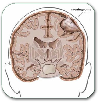
Image: A meningioma growing from the coverings of the brain and compressing surrounding parts of the brain, without infiltrating them.
Symptoms of Olfactory Groove Meningioma
The symptoms caused by olfactory groove meningiomas depend mainly on tumor size, surrounding edema, and involvement of nearby structures.
Typical symptoms may include:
•gradual loss of smell (anosmia)- often affecting both sides. This is usually the earliest symptom because the tumor initially compresses the olfactory (smell) nerves. The change typically develops slowly and often goes unnoticed by the patient. If the tumor remains small or stops growing, this may remain the only symptom. If the tumor continues to enlarge over time, additional symptoms listed below may appear years later as the tumor and surrounding edema become more significant.
• headaches, especially when the tumor becomes larger
• personality or behavioral changes, such as apathy, irritability, disinhibition, or poor judgment
• progressive decline in concentration, memory, or executive function
• visual disturbances, particularly if the tumor grows posteriorly toward the optic nerves or optic chiasm
• seizures, especially in larger tumors irritating the frontal lobes
• gait imbalance or generalized slowing, sometimes seen in advanced cases
• urinary incontinence or marked cognitive decline, occasionally in very large bilateral frontal tumors
Because frontal lobe compression may develop slowly, some patients are first evaluated for psychiatric, cognitive, or personality-related changes before the tumor is discovered on imaging.
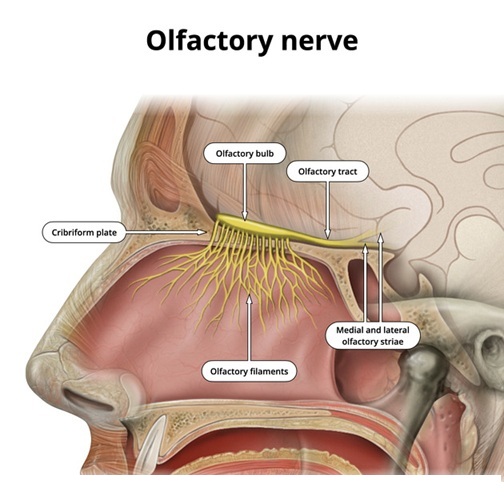
Image: The olfactory nerve (CN I) — consisting of numerous nerve fibers originating in the roof of the nasal cavity, entering the skull through the cribriform plate, and terminating via relay pathways in the evolutionary oldest part of the brain. Read more on these pages about brain anatomy and the anatomy of the cranial nerves.
Diagnosis of Olfactory Groove Meningioma
The most important diagnostic test is MRI of the brain with contrast.
During this examination, the patient lies inside the MRI scanner while magnetic fields generate highly detailed images of the brain. A contrast agent injected through a vein highlights tumor tissue.
MRI allows physicians to determine:
• tumor size
• precise location along the anterior skull base
• relationship to the frontal lobes
• relationship to the optic nerves, optic chiasm, and anterior cerebral vessels
• presence of surrounding brain edema
• possible extension toward the ethmoid region or skull base bone
Olfactory groove meningiomas typically appear as well-defined extra-axial tumors attached to the dura of the anterior cranial fossa, often with strong contrast enhancement and sometimes with a dural tail.
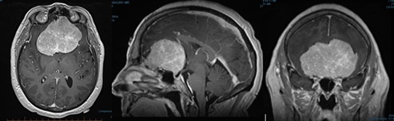
Image: A T1-weighted contrast-enhanced MRI shows a large olfactory groove meningioma in three planes as a bright (enhancing) tumor.
The final diagnosis is confirmed through histopathological analysis after surgery or biopsy.
However, several other conditions may occasionally produce similar imaging findings and should be considered in the differential diagnosis.
These include:
• other anterior skull base meningiomas, such as planum sphenoidale meningiomas
• dural metastases
• solitary fibrous tumor / hemangiopericytoma
• esthesioneuroblastoma extending intracranially in selected skull base cases
• sinonasal or skull base tumors with intracranial extension
Although MRI is the main imaging method used to diagnose olfactory groove meningiomas, CT scans may provide useful additional information. CT is particularly helpful in evaluating hyperostosis, skull base bone changes, calcification, and extension toward the paranasal sinuses. In selected cases, vascular imaging may also be required to assess the relationship of the tumor to the anterior cerebral arteries.
Monitoring Without Treatment: When an Olfactory Groove Meningioma is Only Observed
Not all olfactory groove meningiomas require immediate treatment.
Observation may be considered when:
• the tumor is small
• the patient has no clear neurological symptoms
• imaging shows little or no growth over time
• there is little or no surrounding edema
• there is no significant compression of the frontal lobes or optic pathways
• the patient is elderly or has serious medical conditions increasing surgical risk
Follow-up generally involves MRI examinations every 6–12 months initially. If a small tumor shows documented growth and remains at a safe distance from the optic nerves, treatment may in selected cases be performed with stereotactic radiosurgery (SRS) rather than surgery.
However, observation must be individualized carefully in this location. Even slowly growing bigger tumors may eventually cause subtle cognitive or behavioral deterioration, and some patients do not notice progressive loss of smell or frontal lobe dysfunction until the tumor becomes large. For this reason, the decision to observe rather than treat depends not only on tumor size, but also on growth trend, edema, cognitive symptoms, visual risk, and overall clinical context.
Symptomatic Treatment
Medications are often used to control complications caused by the tumor.
• Brain swelling (edema) is commonly treated with corticosteroids, most often dexamethasone, which reduces edema in the surrounding brain tissue.
• Seizures are treated with antiepileptic medications, such as levetiracetam or other modern antiseizure drugs.
These medications may stabilize the patient temporarily but do not eliminate the tumor itself.
Headache is common, particularly in larger tumors associated with significant edema or raised intracranial pressure. Headache in patients with small meningiomas does not necessarily mean that the tumor is the cause. Many patients have common primary headache disorders such as tension-type headache, migraine, or cervicogenic headache, which are frequent in the general population and may occur independently of the tumor. For this reason, the type of headache should be carefully evaluated and defined before attributing the symptoms to the meningioma, and the presence of headache alone is usually not a sufficient reason to recommend immediate surgical removal of a small, otherwise asymptomatic tumor.
Olfactory Groove Meningioma and Cognitive or Behavioral Change
Unlike many tumors that produce obvious focal neurological deficits early, olfactory groove meningiomas may first present with changes in personality, initiative, emotional control, or judgment.
Because the tumor compresses the frontal lobes, some patients may gradually become:
• more apathetic
• less organized
• emotionally blunted
• socially disinhibited
• forgetful or mentally slowed
Family members often notice these changes before the patient does.
This clinical pattern is important because it may delay diagnosis. In some patients, the tumor is first suspected only after progressive changes in daily functioning, work performance, or interpersonal behavior become obvious.
Surgical Treatment of Olfactory Groove Meningiomas
Surgery remains the primary treatment for symptomatic or progressively growing olfactory groove meningiomas. The procedure is usually performed through a craniotomy, which involves temporarily removing a portion of the skull to access the tumor.
In most cases the operation proceeds through several stages:
• Skin incision and craniotomy
The scalp is opened using a carefully planned incision, usually placed behind the hairline when possible for cosmetic reasons. A bone flap is created to expose the anterior cranial base and the dura overlying the tumor. Various craniotomy approaches (e.g., pterional, bicoronal, and subfrontal) can be used for olfactory groove meningioma surgery, depending on the tumor size, its dominant growth direction, and the surgeon’s preference, as shown in the image below.
• Opening of the dura
The dura mater is carefully opened to expose the tumor and surrounding frontal lobe structures.
• Internal tumor debulking
Whenever possible, the surgeon first attempts to control or detach the tumor from its dural attachment, because meningiomas receive much of their blood supply from dural vessels. The tumor is then reduced from the inside, most often using an ultrasonic aspirator (CUSA). This internal debulking decreases tumor volume and pressure, making subsequent dissection safer.
• Microsurgical dissection
The remaining peripheral parts of the tumor are then gradually separated from the surrounding brain tissue, blood vessels, and preserved olfactory structures, and especially from the optic nerves and anterior cerebral arteries if the tumor extends posteriorly. These residual portions of the tumor are removed piece by piece under microsurgical visualization.
• Treatment of the dural attachment
Because the tumor arises from the dura of the anterior skull base, the involved dura is usually removed or coagulated whenever possible to reduce recurrence risk.
• Removal of infiltrated bone when present
Some olfactory groove meningiomas produce hyperostosis or infiltration of the anterior skull base bone. In such cases, abnormal bone may need to be drilled away, and in selected situations skull base reconstruction may be required.
• Dural reconstruction and closure
After tumor removal, the dura is reconstructed if necessary. The bone flap is replaced, and the scalp is closed.
The main goals of surgery are:
• safe tumor removal
• decompression of the frontal lobes
• protection of the optic nerves and major arteries
• reduction of recurrence risk
• preservation of neurological function
In many patients, preoperative loss of smell is already permanent, and restoration of olfaction after surgery is often limited, especially when the tumor has already severely compressed or destroyed the olfactory structures.
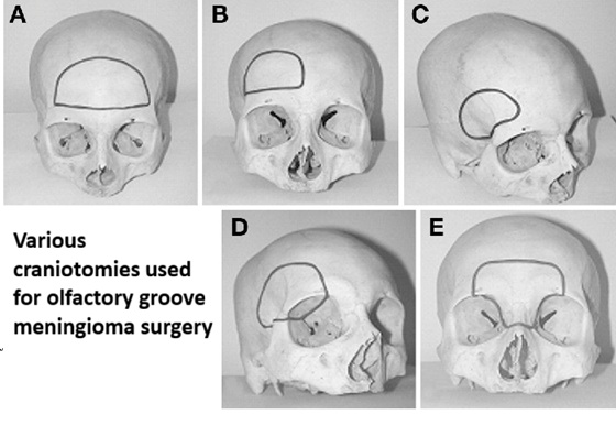
Image: Various locations and sizes of craniotomies can be used for olfactory groove meningioma surgery, depending on the tumor’s size, its dominant growth direction, or the surgeon’s preference.

Image: The image illustrates a craniotomy. Both the skin incision and the skull opening are performed within the hair-bearing area of the scalp. The dura is opened to expose the brain as part of the surgical approach to the tumor. After the procedure, the bone flap is secured and the scalp is reconstructed, ensuring no cosmetic defect remains after healing. The same principles apply to the craniotomies shown in the image above regarding olfactory groove meningioma surgery.
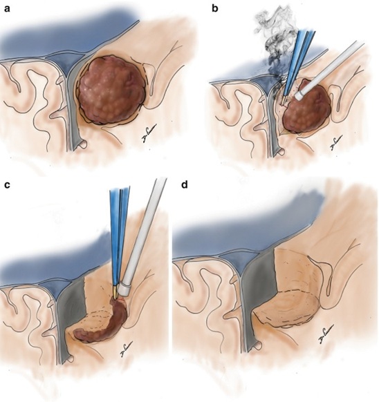
Image: Stages of surgical removal of a meningioma. After craniotomy and opening of the dura, image (a) shows the dark brown meningioma. Image (b) illustrates its separation from the brain, blood vessels, and other normal tissues, followed by its piecemeal reduction. Image (c) shows the removal of the remaining peripheral parts of the meningioma. Image (d) shows the condition after the meningioma has been completely removed. These same surgical principles are also applied in the surgery of olfactory groove meningiomas
Modern Surgical Technologies
Modern neurosurgery employs several technologies to increase precision and safety.
• Neuronavigation systems function as intraoperative GPS, allowing the surgeon to navigate within the skull with millimeter accuracy.
• Ultrasonic aspirators (CUSA) fragment and aspirate tumor tissue while preserving surrounding blood vessels and nerves.
• Intraoperative neurophysiological monitoring may be used in selected cases, depending on tumor extension and nearby critical structures.
• Microsurgical magnification allows careful dissection from arteries, optic pathways, and frontal lobe surfaces.
These technologies significantly improve precision and reduce risk during surgery.
In selected cases, preoperative embolization may be considered, but this is less common and depends on the vascular supply of the tumor.
Endoscopic Surgery for Olfactory Groove Meningiomas
In addition to classical microsurgical craniotomy, some olfactory groove meningiomas may be removed using an endoscopic endonasal approach.
This technique accesses the tumor through the nasal cavity and skull base, avoiding a standard cranial opening. It is mainly considered for selected midline anterior skull base tumors, especially when their anatomical extension is favorable for this route. Importantly, the endoscopic endonasal approach necessarily sacrifices olfactory function because it traverses the cribriform plate; patients who retain any residual smell should be informed of this before selecting this route.
The basic steps of endoscopic surgery usually include:
• endonasal access through the nasal cavity
• removal of part of the skull base bone to expose the dura
• opening of the dura covering the tumor
• internal debulking of the tumor
• careful separation of the tumor from surrounding structures
• removal of the tumor in fragments
• reconstruction of the skull base defect, usually with vascularized tissue flaps to reduce the risk of cerebrospinal fluid leakage
Potential advantages of endoscopic surgery include:
• no large scalp incision
• no standard craniotomy
• direct midline access from below
• limited brain retraction
However, this approach is suitable only for a minority of olfactory groove meningiomas. The endoscopic endonasal approach is mainly used for small to medium-sized midline tumors located at the cribriform plate or planum sphenoidale, especially when the tumor grows predominantly downward toward the skull base and does not extend far laterally over the frontal lobes or surround major arteries. Many tumors of this size may also be technically suitable for stereotactic radiosurgery (SRS) if they demonstrate growth but remain relatively small and are not producing significant brain compression. In current practice, only a minority of olfactory groove meningiomas are treated using a purely endoscopic endonasal approach, roughly around 10–15% in selected surgical series.
Important limitations include:
• difficulty removing large lateral tumor extensions
• limited control when the tumor surrounds important arteries
• challenges in managing extensive dural attachment or infiltrated bone
• increased risk of CSF leakage if skull base reconstruction is not adequate
For these reasons, microsurgical craniotomy remains the most common treatment for many olfactory groove meningiomas, especially larger tumors or tumors with more complex extension.
The choice between microsurgical and endoscopic approaches depends on tumor size, extension, vascular relationships, skull base anatomy, and the experience of the surgical team.
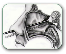
Image: Many skull base tumors, such as meningiomas, pituitary adenomas, and others, can be removed without a craniotomy using an endoscopic endonasal approach.
Extent of Tumor Removal — Simpson Classification
The completeness of tumor removal is classified using the Simpson grading system.
• Simpson Grade I — complete removal of the tumor and its dural attachment
– recurrence risk approximately 5–10% over long-term follow-up
– probability of needing another intervention ~3–5%
• Simpson Grade II — tumor removed, dural attachment coagulated
– recurrence risk approximately 10–20%
– probability of further treatment ~5–10%
• Simpson Grade III — tumor removed but dura left intact
– recurrence risk approximately 20–30%
– probability of additional treatment ~10–20%
• Simpson Grade IV — subtotal removal
– recurrence risk approximately 40–60%
– additional treatment often required in 30–50%
• Simpson Grade V — biopsy or decompression only
– tumor progression expected in most patients
– further definitive treatment usually necessary
In olfactory groove meningiomas, the extent of removal may be influenced by the relationship of the tumor to the optic apparatus, anterior cerebral arteries, frontal skull base, and bone infiltration.
Request Olfactory Groove Meningioma Second Opinion — 24-Hour Review (Priority Option Available Within Hours)
Being told that an MRI has revealed an olfactory groove meningioma often raises important questions:
Is the tumor dangerous?
Should it be monitored or surgically removed?
Is loss of smell permanent?
What is the risk of visual problems or cognitive changes?
Is radiosurgery an appropriate alternative?
An independent neurosurgical second opinion may help clarify the urgency of treatment,
expected neurological and visual outcome, likelihood of tumor growth or recurrence, and the safest balance between
observation, surgical removal, and radiation therapy based on MRI findings, tumor size, skull base extension,
relationship to the optic nerves and anterior cerebral arteries, surrounding brain edema, and the patient’s overall neurological condition.
- ✔ Send a brief message describing your current symptoms and the key findings from your MRI or CT report
- ✔ You will receive a reply within 24 hours explaining whether an online consultation is appropriate and which documentation is required
- ✔ Priority cases: rapidly worsening vision, significant brain compression, progressive neurological symptoms, proposed urgent surgery, or conflicting specialist recommendations — write PRIORITY in your first message
- ✔ MRI images (DICOM format), radiology reports, and relevant documentation can be reviewed to assess tumor size, skull base involvement, surrounding brain edema, and possible treatment strategies
- ✔ During consultation we explain whether observation, microsurgical removal, radiosurgery, or combined treatment is most appropriate — including expected neurological and visual risks and up to 10 days of follow-up clarification
Consultation fees typically range from $180–250 depending on case complexity and documentation volume.
Secure payment by credit card, PayPal invoice (USD), or bank transfer.
This corresponds to typical international specialist telehealth neurosurgical second-opinion services.
Radiation Therapy for Olfactory Groove Meningiomas
The goal of radiation therapy in olfactory groove meningioma is to stop or substantially slow further tumor growth and achieve long-term tumor control, rather than to remove the tumor immediately. Modern radiation techniques can provide long-term tumor control in approximately 85–95% of benign meningiomas, particularly when small residual, recurrent, or otherwise well-defined tumors are treated.
Radiation therapy may be used when complete surgical removal is not possible, when the tumor recurs, when a small residual tumor remains after surgery, or when a small olfactory groove meningioma demonstrates documented growth during MRI follow-up. It may also be considered when surgery carries increased risk because of the tumor’s relationship to the optic nerves, optic chiasm, anterior cerebral arteries, or other critical skull-base structures, or when the patient prefers a non-surgical treatment option after discussion of the expected benefits and risks.
Radiation therapy is generally not the preferred primary treatment for large symptomatic tumors with marked frontal lobe compression, significant surrounding edema, progressive neurological deterioration, substantial mass effect, or visual impairment caused by direct compression that requires decompression. In these situations, surgery is usually more appropriate because radiation does not immediately relieve pressure on the frontal lobes or optic pathways and may temporarily worsen surrounding edema.
The main radiation approaches are:
- Single-fraction stereotactic radiosurgery (SRS) — usually approximately 12–15 Gy in one session
- Hypofractionated stereotactic radiotherapy (HSRT/SRT) — commonly delivered in several sessions, for example approximately 25 Gy in 5 fractions or, in selected cases, 30 Gy in 5 fractions
- Conventionally fractionated radiotherapy — usually approximately 50–54 Gy, delivered in small daily fractions over several weeks
For small, well-defined olfactory groove meningiomas that are located at a safe distance from the optic nerves and optic chiasm, single-fraction stereotactic radiosurgery may be used, particularly when the tumor is smaller than approximately 2.5–3 cm, does not cause major frontal lobe compression, and has little or no significant surrounding edema. Treatment may be delivered using Gamma Knife, CyberKnife, or LINAC-based radiosurgery.
Although proximity to the optic nerves and optic chiasm may limit single-session radiosurgery, tumor size, frontal lobe compression, and surrounding edema are often the more common practical limitations in olfactory groove meningiomas, because these tumors are frequently already relatively large when diagnosed. This distinguishes them from some other anterior skull-base meningiomas, such as tuberculum sellae meningiomas, where the relationship to the optic apparatus is often the dominant factor limiting radiosurgery.
In olfactory groove meningiomas extending posteriorly toward the planum sphenoidale, the optic nerves and optic chiasm may become the main structures limiting the radiation dose. When the tumor lies too close to the optic pathways for safe single-fraction radiosurgery, hypofractionated stereotactic radiotherapy may be preferred. Dividing the total dose into several smaller fractions reduces the biological effect of each individual treatment on the optic apparatus while maintaining an effective total dose to the tumor. Hypofractionated stereotactic radiotherapy may also be preferred for tumors larger than about 3 cm or exceeding approximately 10 cm³ in volume, particularly when they have a broad interface with the brain or when single-fraction radiosurgery would expose too much normal tissue. Highly conformal fractionated treatment may be delivered using LINAC-based systems, CyberKnife, or helical tomotherapy, depending on treatment planning and available equipment.
Conventionally fractionated radiotherapy is generally considered when the treatment volume is larger, when the tumor lies too close to the optic pathways for a stereotactic hypofractionated schedule, or when stereotactic treatment is otherwise unsuitable. Smaller daily fractions are delivered over several weeks in order to reduce the risk of injury to nearby normal structures.
In some patients with a small, stable residual tumor near the optic pathways or major arteries, careful MRI surveillance may be preferred over immediate radiation, particularly when the remnant is asymptomatic and shows no evidence of growth. Radiation can then be reserved for documented progression.
Planned subtotal resection followed by radiosurgery is less common for olfactory groove meningiomas than for some other skull-base meningiomas, because complete surgical removal is often achievable. A small residual tumor is usually left only when the tumor is densely adherent to critical arteries, the optic nerves, the optic chiasm, or other structures where aggressive removal would create excessive neurological risk.
The pituitary stalk is usually not a major limiting structure in a typical olfactory groove meningioma, although it may become relevant in unusually posteriorly extending tumors approaching the tuberculum sellae or sellar region.
The final treatment decision depends on tumor size and volume, growth rate, frontal lobe compression, surrounding edema, visual function, distance from the optic nerves and optic chiasm, relationship to major arteries, previous treatment, and the radiation tolerance of surrounding tissues. In complex cases, treatment planning is usually multidisciplinary, involving a neurosurgeon, radiation oncologist, and neuroradiologist.
Complications After Radiation Therapy for Olfactory Groove Meningiomas
Complications after radiation therapy for olfactory groove meningioma depend mainly on the radiation dose, fractionation, tumor size, amount of surrounding brain exposed to radiation, and proximity to the optic pathways and frontal lobes.
One important delayed complication is vasogenic edema of the frontal lobes, which may appear weeks or months after treatment. Depending on the extent and location of the swelling, patients may develop headache, seizures, cognitive slowing, personality or behavioral change, gait disturbance, weakness, speech disturbance, or other neurological symptoms. If edema becomes extensive, it may produce significant mass effect and increased intracranial pressure, with severe headache, vomiting, somnolence, confusion, or reduced consciousness.
Because olfactory groove meningiomas may extend posteriorly toward the optic nerves and optic chiasm, radiation injury to the optic apparatus is another important potential complication. Radiation-induced optic neuropathy may cause progressive visual deterioration, visual field loss, or, in severe cases, major permanent visual impairment. This is one of the main reasons why single-fraction radiosurgery is avoided when the tumor lies too close to the optic pathways and why fractionated treatment may be preferred.
Depending on the exact radiation field, other delayed complications may include radiation necrosis, persistent injury of adjacent frontal lobe tissue, gliosis, vascular injury, and less commonly endocrine dysfunction if the radiation field extends sufficiently posteriorly toward the sellar or hypothalamic region.
New neurological or visual symptoms after treatment should not automatically be interpreted as tumor progression, since post-radiation edema, treatment-related enhancement, radiation necrosis, and true tumor growth may sometimes produce overlapping MRI findings. Careful clinical assessment and comparison with previous imaging are therefore important. Significant or progressive radiation-related changes should be recognized and managed appropriately, because prolonged edema, tissue injury, or optic pathway damage may lead to persistent neurological or visual consequences, while early recognition and treatment may reduce the risk of long-term deficits.
Recurrence of Olfactory Groove Meningiomas
The likelihood of recurrence depends on tumor grade and completeness of removal.
• After complete removal of benign meningiomas, recurrence occurs in approximately 5–10% of cases over 10–15 years.
• After subtotal removal, recurrence rates may reach 20–40%.
• Atypical meningiomas recur in approximately 30–40% of cases.
• Malignant meningiomas may recur in 50–80% of patients.
Because recurrence may occur many years after treatment, long-term MRI follow-up is essential.
When an olfactory groove meningioma recurs, treatment decisions depend on several factors:
• tumor growth rate
• tumor size
• relationship to the optic apparatus and skull base arteries
• previous treatments
• patient symptoms
• histological grade
Not all recurrences require immediate treatment. Some small stable recurrences can be monitored, while others require surgery, radiation, or both.
Observation of Small Recurrences
Small recurrent olfactory groove meningiomas that show minimal or no growth may initially be managed with MRI surveillance.
Observation is most commonly chosen when:
• the recurrent tumor is small
• the patient has no neurological or visual symptoms
• imaging shows slow or stable growth
• the recurrence is located in a higher-risk surgical area
Follow-up usually involves MRI every 6–12 months.
Repeat Surgery
Surgical reoperation is considered when:
• the recurrent tumor causes neurological, cognitive, or visual symptoms
• imaging shows significant growth
• the tumor is accessible for safe removal
• mass effect on the frontal lobes becomes clinically important
Repeat surgery aims to remove the recurrent tumor and, when possible, its dural attachment.
However, reoperation may be more technically demanding because:
• scar tissue from the previous surgery may be present
• arteries, optic structures, or frontal lobe surfaces may already be displaced
• previous skull base reconstruction may alter normal anatomy
Radiation Therapy for Recurrence
Radiation therapy is frequently used for recurrent olfactory groove meningiomas, particularly when complete reoperation is not feasible.
Radiation may be recommended when:
• residual tumor remains after surgery
• recurrence occurs after previous surgery
• the tumor lies in a surgically difficult skull base location
• the tumor shows atypical or malignant histology
Two main radiation approaches are used:
• fractionated radiotherapy, typically delivering 50–54 Gy
• stereotactic radiosurgery, usually delivering 12–16 Gy in a single focused treatment
When recurrent tumor lies close to the optic nerves or optic chiasm, fractionated treatment is often safer than single-session radiosurgery.
Treatment Strategy by Tumor Grade
Management also depends strongly on the WHO tumor grade.
• WHO grade I meningiomas. Recurrence may be managed with observation, repeat surgery, or radiosurgery, depending on tumor growth and symptoms.
• WHO grade II (atypical) meningiomas. These tumors recur more frequently. Postoperative radiotherapy is often recommended, particularly after incomplete tumor removal, although some patients may be managed with close MRI follow-up after complete resection.
• WHO grade III (malignant) meningiomas. These aggressive tumors typically require postoperative radiotherapy regardless of the completeness of surgical removal, together with close imaging follow-up.
Long-Term Monitoring
Because olfactory groove meningiomas may recur many years after treatment, long-term follow-up is essential.
Typical surveillance includes:
• annual MRI for several years after treatment
• longer imaging intervals if the tumor remains stable
Early detection of recurrence allows treatment before major neurological, cognitive, or visual deterioration develops.
Treatment Decision Summary for Olfactory Groove Meningioma
Treatment decisions for olfactory groove meningioma usually follow several general principles.
1. Before treatment
If the tumor is small, causes no clear neurological symptoms, and does not significantly compress the frontal lobes or optic pathways, MRI monitoring is often the safest first step.
If the tumor causes progressive loss of smell, personality or behavioral changes, cognitive decline, visual disturbances, significant mass effect or edema, or shows documented growth on follow-up imaging, active treatment is more often recommended.
2. Choosing the main treatment
In general, tumors smaller than about 2.5–3 cm that demonstrate documented growth and are located at a safe distance from the optic nerves may be suitable for stereotactic radiosurgery (SRS). Larger tumors, tumors causing significant frontal lobe compression or brain edema, or tumors closely related to major arteries usually require microsurgical removal through craniotomy. In selected small to medium-sized midline tumors located mainly at the cribriform plate or planum sphenoidale, an endoscopic endonasal approach may sometimes be considered.
3. After surgery
WHO Grade I + complete removal: MRI follow-up is usually sufficient.
WHO Grade I + residual tumor: follow-up or radiosurgery/radiotherapy may be considered depending on tumor growth, residual size, and proximity to the optic nerves or major arteries.
WHO Grade II: postoperative radiotherapy is usually recommended after incomplete tumor removal. After complete resection, some patients may be managed with MRI follow-up or radiotherapy depending on recurrence risk.
WHO Grade III: surgery is usually followed by radiotherapy regardless of the completeness of tumor removal, together with close imaging follow-up.
This is a simplified overview. In real clinical practice, treatment decisions also depend on tumor size, skull base extension, surrounding brain edema, relationship to the frontal lobes, optic nerves, optic chiasm, and anterior cerebral vessels, as well as patient age and overall neurological condition.
Experimental and Targeted Therapies
Several medications have been studied for recurrent or progressive meningiomas when surgery and radiotherapy are no longer sufficient. These include bevacizumab, as well as drugs acting on molecular pathways such as AKT or SMO inhibitors. Somatostatin-receptor–based treatments such as octreotide and combinations including everolimus have also been investigated.
However, current evidence remains limited, and surgery and radiation therapy remain the main treatments for most olfactory groove meningiomas.
When Expert Opinions May Differ
In many patients diagnosed with olfactory groove meningioma, different specialists may recommend different treatment strategies. This is common in neurosurgery and does not necessarily mean that one opinion is correct while another is wrong.
Treatment decisions depend on multiple clinical factors, including:
• tumor size
• tumor location and skull base extension
• growth rate observed on MRI
• presence of surrounding edema or mass effect
• patient age and overall health
• presence or absence of neurological symptoms
• degree of visual risk
• relationship of the tumor to important arteries and optic pathways
• tumor grade if histological diagnosis is already known
Because these factors interact in complex ways, reasonable specialists may arrive at different conclusions regarding the safest management strategy.
Observation vs. Early Surgery
One of the most common differences in opinion occurs when an olfactory groove meningioma is discovered incidentally on MRI and the patient has no obvious symptoms. Some specialists recommend active surveillance, particularly when the tumor is small and shows no signs of growth. Others may recommend earlier surgery, especially if the tumor is located in a position where further growth could later threaten the frontal lobes, optic pathways, or major vessels.
Differences in Surgical Strategy
Even when surgery is recommended, surgeons may disagree about the optimal operative strategy. One surgeon may aim for maximal removal including the involved dura and abnormal bone, while another may prefer a more conservative removal if the tumor is densely adherent to critical arteries or the optic apparatus.
Similarly, some tumors may be approached through microsurgical craniotomy, while others may be considered for endoscopic endonasal surgery in carefully selected cases.
Surgery vs. Radiation Therapy
Another area where opinions may differ involves the use of radiation therapy. Some specialists recommend surgery first, followed by radiation only if residual tumor remains. Others may recommend primary radiation in carefully selected small tumors or in patients with increased surgical risk.
These differences usually reflect different risk assessments, particularly regarding vision, skull base anatomy, and long-term tumor control.
How Prognosis Depends on Tumor Grade, Location, and Extent of Removal
The prognosis of olfactory groove meningioma depends primarily on the biological grade of the tumor, its anatomical relationships, and the extent to which it can be safely removed.
In most patients, olfactory groove meningiomas are WHO Grade I tumors, which usually grow slowly and have a favorable long-term prognosis when treated appropriately.
Functional recovery after treatment varies depending on the structures that were compressed before surgery.
Sense of smell (olfaction). Because the tumor usually compresses the olfactory nerves early, loss of smell is common at diagnosis. When smell has already been completely lost before surgery, recovery is uncommon. However, partial improvement of smell may occur in approximately 10–30% of patients, particularly when olfactory function was only partially impaired before treatment.
Cognitive and behavioral symptoms. Many patients develop subtle frontal lobe symptoms such as slowed thinking, reduced concentration, personality changes, or decreased motivation. After tumor removal and relief of brain compression, significant improvement in cognitive or behavioral symptoms occurs in about 60–80% of patients, although recovery may take several months.
Vision. Visual disturbances may occur when the tumor compresses the optic nerves or optic chiasm. If visual loss is mild and compression is relieved early, visual improvement is observed in roughly 50–70% of patients. When visual impairment has been severe or long-standing, recovery is less likely.
Tumor control is generally very good for benign tumors. When complete or near-complete removal is achieved, long-term tumor control is typically above 85–90%. In cases where a small residual tumor remains, additional treatment with stereotactic radiosurgery or radiotherapy can often provide effective long-term control.
Overall prognosis is most favorable in patients with benign tumors, limited brain edema, preserved vision, and safe near-complete tumor removal. Prognosis is generally less favorable in higher-grade tumors, very large tumors causing severe frontal lobe compression, or cases where only limited tumor removal is possible.
Can an Olfactory Groove Meningioma Become Life-Threatening?
Yes, an olfactory groove meningioma can become life-threatening, although this is not the usual course in most patients.
Most of these tumors grow slowly, and many remain stable for years without causing major neurological problems. However, if the tumor becomes large enough, it may produce severe pressure on both frontal lobes, marked edema, increased intracranial pressure, significant cognitive deterioration, visual compromise, or in extreme cases brain herniation.
The danger therefore does not arise only from the biological nature of the tumor itself, but from its mass effect on vital brain structures.
This risk is higher in:
• large tumors
• tumors with extensive frontal edema
• tumors extending toward the optic pathways or major arteries
• higher-grade meningiomas
For this reason, even though olfactory groove meningiomas are usually benign, they should never be considered harmless solely on the basis of histological grade. Their real clinical significance depends on tumor size, location, rate of growth, and effect on surrounding brain structures.
Frequently Asked Questions About Olfactory Groove Meningioma
Why is loss of smell often the first symptom of olfactory groove meningioma?
Loss of smell is often the first symptom of olfactory groove meningioma because the tumor arises at the anterior skull base, directly near the olfactory bulbs and olfactory tracts. In the early stages, the tumor mainly compresses these smell pathways before it becomes large enough to affect the frontal lobes, optic nerves, or other structures. The loss of smell usually develops gradually and may affect both sides, so many patients do not notice it immediately. If the tumor remains small or stops growing, anosmia may remain the only symptom. If the olfactory groove meningioma continues to enlarge, additional symptoms may appear years later as frontal lobe compression, brain edema, visual pathway involvement, or mass effect become more significant.
Can olfactory groove meningioma cause personality or behavioral changes?
Yes. Olfactory groove meningioma can cause personality or behavioral changes because it grows beneath the frontal lobes and may slowly compress them over time. Unlike tumors that immediately produce obvious weakness or speech problems, frontal lobe compression may first appear as subtle changes in initiative, judgment, emotional control, or social behavior. Patients may become more apathetic, less organized, emotionally blunted, irritable, disinhibited, forgetful, or mentally slowed. Family members often notice these changes before the patient does. This pattern can delay diagnosis because the patient may first be evaluated for psychiatric, cognitive, or personality-related problems. When the tumor and surrounding edema are removed or reduced, cognitive and behavioral symptoms may improve gradually, although recovery may take months.
How does olfactory groove meningioma affect the frontal lobes?
Olfactory groove meningioma affects the frontal lobes mainly by compression rather than direct invasion. The tumor grows outside the brain tissue, attached to the dura of the anterior skull base. As it enlarges, it extends upward and presses on the inferior surfaces of the frontal lobes. This pressure may interfere with concentration, memory, executive function, judgment, motivation, and behavior. Another important mechanism is peritumoral edema, which increases the effective mass effect of the tumor. Together, the tumor and surrounding swelling may compress and displace normal brain structures inside the closed skull. In large olfactory groove meningiomas, this can cause headaches, cognitive decline, personality change, seizures, gait slowing, or, in severe cases, dangerous intracranial pressure and brain shift.
Can olfactory groove meningioma cause memory problems or cognitive decline?
Yes. Olfactory groove meningioma can cause memory problems, slowed thinking, reduced concentration, poor organization, and broader cognitive decline when it compresses the frontal lobes. These symptoms may develop gradually and may be overlooked because they do not always appear as a sudden neurological deficit. Some patients become less efficient at work, less motivated, emotionally changed, or unable to organize daily activities as before. In very large bilateral frontal tumors, marked cognitive decline, gait imbalance, or urinary incontinence may occasionally occur. Cognitive symptoms are especially important because they may be reversible to some degree after decompression. Improvement in cognitive or behavioral symptoms after tumor removal is described in many patients, although recovery often takes several months.
Can olfactory groove meningioma affect vision or the optic nerves?
Yes. Olfactory groove meningioma can affect vision when it grows posteriorly toward the optic nerves or optic chiasm. Small tumors may mainly affect smell, but larger tumors may extend backward from the anterior skull base and compress the visual pathways. Visual disturbance is an important warning sign because the chance of recovery depends partly on how long and how severely the optic structures have been compressed. MRI with contrast is used to define the tumor’s relationship to the optic nerves, optic chiasm, anterior cerebral arteries, and surrounding edema. If visual loss is mild and decompression is performed early, visual improvement may occur in many patients. Long-standing or severe visual impairment is less likely to recover fully.
Why can olfactory groove meningioma grow for years before diagnosis?
Olfactory groove meningioma can grow for years before diagnosis because it usually grows slowly and initially compresses structures that may not produce dramatic symptoms. The earliest sign is often gradual loss of smell, but anosmia may develop slowly, affect both sides, and go unnoticed. Because the tumor arises beneath the frontal lobes, it may also cause subtle changes in personality, initiative, concentration, judgment, or emotional control before obvious neurological deficits appear. These changes may be attributed to aging, stress, psychiatric problems, or other causes. In addition, olfactory groove meningiomas may become quite large before they are discovered because the anterior skull base provides space for slow expansion. Diagnosis often occurs only when MRI or CT is performed for headaches, cognitive change, visual symptoms, seizures, or incidental findings.
Does every olfactory groove meningioma require surgery?
No. Not every olfactory groove meningioma requires surgery. Observation may be appropriate when the tumor is small, causes no clear neurological symptoms, shows little or no growth over time, has little or no surrounding edema, and does not significantly compress the frontal lobes or optic pathways. Follow-up usually involves MRI examinations every 6–12 months at first. However, observation must be individualized carefully in this location. Even slowly growing tumors may eventually cause subtle cognitive or behavioral deterioration, and patients may not notice progressive loss of smell or frontal lobe dysfunction until the tumor becomes large. Surgery becomes more likely when the tumor grows, causes symptoms, produces edema, compresses optic pathways, or creates significant frontal lobe mass effect.
When can olfactory groove meningioma be monitored without treatment?
Olfactory groove meningioma can be monitored without treatment when the tumor is small, asymptomatic, stable on imaging, and not compressing the frontal lobes, optic nerves, optic chiasm, or nearby arteries. Monitoring is also more reasonable when there is little or no surrounding brain edema and when the patient is elderly or has medical conditions that increase surgical risk. MRI follow-up is usually performed every 6–12 months initially. If the tumor remains stable, the interval may later be extended. Monitoring becomes less safe when there is documented growth, increasing edema, cognitive or behavioral change, visual risk, seizures, or significant mass effect. The decision depends not only on tumor diameter, but on growth trend, symptoms, edema, visual risk, and overall clinical context.
How is treatment decision-making performed in olfactory groove meningioma?
Treatment decision-making in olfactory groove meningioma depends on several factors considered together. The most important are tumor size, growth rate on MRI, symptoms, surrounding brain edema, frontal lobe compression, visual risk, relationship to the optic nerves and optic chiasm, relationship to anterior cerebral arteries, skull base extension, patient age, and overall neurological condition. Small tumors without symptoms or growth may be monitored. Tumors causing cognitive change, behavioral change, visual disturbance, seizures, edema, or mass effect usually require active treatment. Stereotactic radiosurgery may be considered for selected small growing tumors located safely away from the optic pathways. Larger tumors, tumors with significant frontal lobe compression, or tumors close to major arteries usually require microsurgical removal through craniotomy.
Can olfactory groove meningioma be treated with radiosurgery instead of surgery?
Olfactory groove meningioma can sometimes be treated with stereotactic radiosurgery instead of surgery, but only in selected cases. Radiosurgery is generally considered for small tumors, usually smaller than about 2.5–3 cm, that demonstrate growth and are located at a safe distance from the optic nerves and optic chiasm. It may also be used for small residual tumor after surgery or for recurrent tumor when reoperation is not ideal. Radiosurgery is usually not appropriate when the tumor is large, causes significant frontal lobe compression, produces substantial brain edema, or requires immediate decompression. In olfactory groove meningioma, many tumors are already large at diagnosis, which often makes microsurgical removal more appropriate than primary radiosurgery.
When is stereotactic radiosurgery appropriate for olfactory groove meningioma?
Stereotactic radiosurgery may be appropriate for olfactory groove meningioma when the tumor is small, shows documented growth, does not produce significant frontal lobe compression, and remains at a safe distance from the optic nerves and optic chiasm. It can also be used for small residual tumors after surgery or selected recurrences. The usual single-session dose for meningioma radiosurgery is in the range of 12–16 Gy, while fractionated radiotherapy may be used when the tumor is too close to sensitive visual structures. The key limitation is safety: even a relatively small tumor may be unsuitable for single-session radiosurgery if it lies too close to the optic apparatus. Larger tumors with edema or mass effect are usually treated surgically instead.
What types of radiation therapy are used for olfactory groove meningioma?
Radiation treatment for olfactory groove meningioma may be delivered as single-fraction stereotactic radiosurgery, hypofractionated stereotactic radiotherapy, or conventionally fractionated radiotherapy. Single-fraction stereotactic radiosurgery is usually used for small, well-defined tumors that are sufficiently separated from the optic nerves and optic chiasm. Typical single-session doses are approximately 12–15 Gy and treatment may be delivered using Gamma Knife, CyberKnife, or LINAC-based systems.
When the tumor is larger than 3 cm or 10ml in volume, or lies too close to the optic pathways for a safe single high dose, hypofractionated stereotactic radiotherapy may be used, commonly over 3–5 sessions. Conventionally fractionated radiotherapy, usually to a total dose of approximately 50–54 Gy over several weeks, may be chosen when a larger treatment volume must be covered or when stereotactic treatment is not appropriate.
What complications can occur after radiation therapy for olfactory groove meningioma?
Complications depend on tumor size, radiation dose, fractionation, and proximity to the frontal lobes and optic pathways. Delayed vasogenic edema of the frontal lobes may develop weeks or months after treatment and can cause headache, seizures, cognitive or behavioral changes, weakness, gait disturbance, or other neurological symptoms. Extensive edema may produce mass effect and increased intracranial pressure.
Radiation injury to the optic nerves or optic chiasm may cause progressive visual deterioration or visual field loss. Other delayed complications may include radiation necrosis, gliosis, vascular injury, and, less commonly, endocrine dysfunction when the radiation field extends toward the sellar or hypothalamic region. New neurological or visual symptoms after treatment should be evaluated carefully because radiation-related changes can sometimes resemble tumor progression on MRI.
How is surgery for olfactory groove meningioma performed?
Surgery for olfactory groove meningioma is usually performed through a craniotomy. The scalp incision is planned carefully, often behind the hairline when possible, and a bone flap is created to expose the anterior skull base and dura over the tumor. The dura is opened, and the surgeon usually performs internal tumor debulking first, often using an ultrasonic aspirator, to reduce tumor volume and pressure. The remaining tumor is then separated from the frontal lobes, blood vessels, optic nerves, and preserved olfactory structures under microsurgical visualization. The dural attachment is removed or coagulated when safe, and abnormal hyperostotic bone may be drilled away. The goals are safe tumor removal, frontal lobe decompression, protection of vision and arteries, recurrence reduction, and neurological preservation.
Can olfactory groove meningioma surgery preserve the sense of smell?
Olfactory groove meningioma surgery may preserve smell in selected patients, but recovery is often limited when the sense of smell has already been completely lost before treatment. The tumor usually compresses the olfactory nerves early, and in many patients this damage is already long-standing by the time the tumor is diagnosed. If olfaction is only partially impaired before surgery, partial improvement may occur in some patients, but complete recovery is not guaranteed. The surgical priority is usually safe removal of the tumor, decompression of the frontal lobes, protection of the optic nerves and anterior cerebral arteries, and reduction of recurrence risk. When olfactory structures are already severely compressed or destroyed, restoration of smell after surgery is uncommon.
Why may surgeons recommend different operations for olfactory groove meningioma?
Surgeons may recommend different operations for olfactory groove meningioma because the best approach depends on tumor size, direction of growth, skull base anatomy, relationship to the optic nerves, anterior cerebral arteries, frontal lobes, and the surgeon’s experience. Various craniotomy approaches, such as pterional, bicoronal, or subfrontal routes, may be used. Some selected small to medium midline tumors may be considered for an endoscopic endonasal approach through the nasal cavity and skull base. However, endoscopic surgery is suitable only for a minority of olfactory groove meningiomas, especially when the tumor does not extend far laterally or surround major arteries. Different recommendations may therefore reflect different risk assessments, not necessarily right versus wrong opinions.
How long is recovery after olfactory groove meningioma surgery?
Recovery after olfactory groove meningioma surgery varies depending on tumor size, frontal lobe edema, surgical approach, visual involvement, age, general health, and whether complications occur. Many patients spend several days in hospital after craniotomy, followed by several weeks of gradual recovery at home. Light daily activities may often resume within a few weeks, but full recovery can take several months, especially after large tumors with frontal lobe compression or brain edema. Cognitive, behavioral, or concentration problems may improve gradually after decompression, while smell recovery is often limited if anosmia was complete before surgery. Driving, work, exercise, and air travel should be decided individually after postoperative review and follow-up imaging.
How do doctors determine whether headaches are actually caused by an olfactory groove meningioma?
Headaches are more likely to be caused by an olfactory groove meningioma when the tumor is large, produces significant frontal lobe compression or surrounding brain edema, or contributes to increased intracranial pressure. Doctors also consider whether the headache is new, progressively worsening, more severe in the morning, associated with vomiting, visual symptoms, cognitive change, or other neurological findings.
A small olfactory groove meningioma discovered incidentally may not explain longstanding migraine, tension-type headache, or cervicogenic headache. Determining the relationship therefore requires careful review of the headache pattern, neurological examination, MRI findings, tumor size, edema, mass effect, and any accompanying loss of smell, visual deterioration, behavioral change, or cognitive slowing. Headache alone is usually not sufficient evidence that a small tumor requires surgery.
How do doctors determine whether an olfactory groove meningioma is truly causing seizures?
An olfactory groove meningioma is more likely to be responsible for seizures when it has become large enough to compress or irritate the frontal lobes, particularly when significant peritumoral edema is present. Doctors assess whether the seizure pattern is consistent with frontal lobe involvement and whether the seizures began after the tumor became clinically significant.
Evaluation usually includes a detailed seizure history, neurological examination, EEG findings, and careful review of MRI images. The relationship is more convincing when EEG abnormalities, seizure semiology, frontal lobe compression, and edema correspond to the same anatomical region. Other possible causes of epilepsy must also be considered. When seizures remain uncontrolled or the relationship is uncertain, a structured epilepsy assessment may be required. More information is available on our epilepsy surgery page.
What are the specific risks and complications of olfactory groove meningioma surgery?
The risks depend mainly on tumor size, surrounding frontal lobe edema, skull base extension, and the relationship of the tumor to the optic nerves, optic chiasm, anterior cerebral arteries, perforating vessels, and olfactory structures. Possible complications include bleeding, postoperative hematoma, brain swelling, seizures, infection, cerebrospinal fluid leakage, meningitis, stroke, frontal lobe contusion, and new neurological or cognitive deficits.
Injury to the optic nerves or optic chiasm may cause visual deterioration or, rarely, severe permanent visual loss. Injury to the anterior cerebral arteries or small perforating vessels may cause cerebral infarction, weakness, cognitive decline, or behavioral and personality changes. Loss of smell is often already present before surgery and may remain permanent; an endoscopic endonasal approach usually sacrifices remaining olfactory function. A small residual tumor may be intentionally left when it is densely adherent to critical arteries or optic structures. Residual or recurrent tumor may later require MRI monitoring, radiosurgery, radiotherapy, or further surgery. A broader explanation is available on our craniotomy and brain surgery complications page.
Can olfactory groove meningioma return after surgery or radiation treatment?
Yes. Olfactory groove meningioma can recur after surgery or radiation treatment, although recurrence risk depends strongly on tumor grade and completeness of removal. After complete removal of benign meningiomas, recurrence is relatively low, approximately 5–10% over long-term follow-up. After subtotal removal, recurrence may reach 20–40%. Atypical meningiomas recur more often, and malignant meningiomas have the highest recurrence risk. Simpson grade is important because removal or treatment of the dural attachment lowers recurrence probability. Small stable recurrences may sometimes be monitored with MRI, while growing or symptomatic recurrences may require repeat surgery, stereotactic radiosurgery, fractionated radiotherapy, or combined treatment. Long-term MRI follow-up is essential because recurrence may occur many years after treatment.
How does WHO grade influence prognosis in olfactory groove meningioma?
WHO grade is one of the most important biological factors influencing prognosis in olfactory groove meningioma. Most tumors are WHO Grade I, which usually means benign behavior, slow growth, and favorable long-term tumor control when treatment is appropriate. WHO Grade II, or atypical meningioma, has higher cellular activity and a greater tendency to recur after surgery. WHO Grade III, or malignant anaplastic meningioma, is rare but more aggressive, recurs more frequently, and usually requires combined treatment with surgery and radiation therapy. However, grade is not the only prognostic factor. Outcome also depends on tumor size, frontal lobe compression, edema, relationship to optic nerves and arteries, and whether safe complete or near-complete removal is possible.
Can olfactory groove meningioma become life-threatening despite being benign?
Yes. Olfactory groove meningioma can become life-threatening despite being benign, although this is not the usual course in most patients. The danger does not come only from the biological grade of the tumor, but from its size, location, edema, and mass effect. A large olfactory groove meningioma may compress both frontal lobes, produce marked brain edema, increase intracranial pressure, compromise vision, cause severe cognitive deterioration, or in extreme cases contribute to brain herniation. The risk is higher in large tumors, tumors with extensive frontal edema, tumors extending toward the optic pathways or major arteries, and higher-grade meningiomas. For this reason, a benign olfactory groove meningioma should not automatically be considered harmless.
What determines long-term prognosis in olfactory groove meningioma?
Long-term prognosis in olfactory groove meningioma depends mainly on tumor grade, size, anatomical relationships, edema, visual involvement, cognitive or behavioral symptoms, and the extent of safe tumor removal. Prognosis is generally best in WHO Grade I tumors with limited edema, preserved vision, and complete or near-complete removal. Functional recovery varies by structure. Loss of smell is often permanent if olfactory function was completely lost before surgery. Cognitive and behavioral symptoms may improve after frontal lobe decompression, although recovery can take months. Visual improvement is more likely when compression is relieved early and the deficit was not severe or long-standing. Tumor control is generally very good for benign tumors, especially when complete or near-complete removal is achieved.
Why may specialist opinions differ in olfactory groove meningioma treatment?
Specialist opinions may differ in olfactory groove meningioma treatment because several reasonable strategies may exist for the same tumor. Some specialists may recommend observation when the tumor is small, asymptomatic, and stable. Others may recommend earlier surgery if the tumor is growing or lies in a position where future enlargement could threaten the frontal lobes, optic pathways, or major arteries. Even when surgery is recommended, surgeons may differ in the preferred approach or in how aggressively they would remove dura, abnormal bone, or tumor adherent to critical structures. Radiation specialists may recommend radiosurgery for selected small tumors. These differences usually reflect different assessments of tumor size, edema, growth, visual risk, surgical risk, and long-term tumor control.
Can an online second opinion help clarify treatment decisions for olfactory groove meningioma?
Yes. An online second opinion can help clarify treatment decisions for olfactory groove meningioma when MRI or CT has revealed the tumor but the safest strategy is uncertain. It is especially useful when specialists recommend different options, when surgery has been proposed but the expected risks are unclear, when radiosurgery or radiotherapy is suggested, or when symptoms such as loss of smell, cognitive change, personality change, seizures, or visual disturbance raise concern. A neurosurgical review can assess tumor size, growth, skull base extension, frontal lobe compression, edema, relationship to the optic nerves and anterior cerebral arteries, and likelihood of recurrence. The goal is to clarify whether observation, microsurgical removal, radiosurgery, or combined treatment is most appropriate.