Dr Željko Kojadinović — NEUROHIRURGIJA I LEČENJE BOLA
Dr Zeljko Kojadinovic — Pain Treatment & Neurosurgery


Author:
Dr. Zeljko Kojadinovic, MD, PhD
— Consultant Neurosurgeon
Specialized Experience:
30 years of clinical expertise in neurosurgery.
Last medically reviewed:
February 28, 2026
Who This Dural Arteriovenous Fistula (DAVF) Page Is For
This page is intended for patients diagnosed with dural arteriovenous fistula (DAVF) or those in whom this condition is suspected based on imaging or symptoms such as pulsatile tinnitus, neurological deficits, or unexplained intracranial hemorrhage.
It is particularly relevant for individuals trying to understand whether a DAVF is low-risk or high-risk, whether treatment is necessary, and which option — observation, endovascular treatment, or surgery — is most appropriate.
If imaging findings are unclear, symptoms are progressing, different treatment strategies have been proposed, or there is uncertainty about the risk of hemorrhage or neurological deterioration — an individualized
neurosurgical second opinion
can help.
When patients usually seek a second opinion for dural arteriovenous fistula (DAVF)
- Pulsatile tinnitus persists, but imaging findings are unclear or no definitive diagnosis has been established
- A DAVF has been identified, but it is uncertain whether it is low-risk or high-risk
- Cortical venous reflux is suspected, but its clinical significance and urgency of treatment are unclear
- Different specialists recommend different strategies (observation vs endovascular treatment vs surgery)
- Endovascular treatment has been proposed, but there are questions about success rates or potential risks
- Symptoms such as neurological deficits, seizures, or cognitive changes are developing without clear explanation
- A hemorrhage has occurred, and there is uncertainty about whether the underlying fistula has been definitively treated
- Spinal DAVF is suspected due to progressive weakness, but diagnosis remains uncertain or delayed
- There is concern about long-term prognosis and risk of neurological deterioration
Dural Arteriovenous Fistula (DAVF) — Quick Summary (Read This First)
- DAVF is an abnormal direct connection between arteries and veins located within the dura mater. Unlike arteriovenous malformations (AVMs), it does not contain a nidus and represents a pathological shunt of blood into the venous system.
- The condition is usually acquired rather than congenital. It most often develops in adults (typically in their 40s–60s) and is rare in children. Venous sinus thrombosis and prior trauma are commonly implicated.
- DAVFs can be classified as cranial or spinal. Cranial fistulas are more common and most frequently occur in the transverse–sigmoid sinus region (occipital region, approximately 60–65% of cases) and the cavernous sinus, while spinal DAVFs arise along the dura of spinal nerve roots and primarily affect the venous drainage of the spinal cord.
- The main problem is venous hypertension, not arterial flow itself. Elevated pressure in the venous system leads to impaired drainage, venous congestion, dilation and weakening of vein walls, and potential damage to the brain or spinal cord.
- The clinical risk depends primarily on venous drainage. The presence of cortical venous reflux significantly increases the risk of intracranial hemorrhage and neurological deterioration.
- Low-risk DAVFs (without cortical venous reflux) may be managed conservatively. Regular follow-up is required, as some lesions may evolve over time.
- High-risk DAVFs require treatment. Indications include cortical venous reflux, hemorrhage, or progressive neurological symptoms (such as focal deficits — weakness, sensory loss, visual disturbance, cranial nerve deficits — as well as seizures). In selected cases, even severe, disabling pulsatile tinnitus may represent an indication for treatment due to significant impact on quality of life.
- Cranial DAVFs are primarily treated endovascularly (≈70–90% success). Microsurgical treatment is reserved for cases in which endovascular therapy is not feasible or remains incomplete, as well as in cases of hemorrhage where emergency evacuation of a hematoma is required to relieve intracranial pressure and venous congestion. During surgery, the abnormal venous drainage may be disconnected and, if possible, the fistula itself addressed; if definitive closure is not achieved, additional endovascular treatment is performed subsequently.
- Spinal DAVFs are most often treated surgically, although endovascular therapy may be used in selected cases. Surgical disconnection of the draining vein is usually definitive, while embolization can be considered if angiographic anatomy is favorable, but with lower durability compared to surgery.
- Outcome depends strongly on early recognition and correct risk assessment. Delayed diagnosis, especially in spinal DAVFs, may lead to irreversible neurological damage.
- This page is structured to guide clinical understanding and decision-making. Use the Contents section to navigate directly to symptoms, diagnosis, risk assessment, treatment options, and when a neurosurgical second opinion may be helpful.
Most readers benefit from reviewing the Quick Summary together with the sections on Symptoms, Venous Drainage and Risk, and Treatment Options. Other sections provide deeper clinical context and decision-making insight.
What Is a Dural Arteriovenous Fistula
Dural arteriovenous fistula (DAVF) is an abnormal direct connection between arteries and veins located in the dura mater (the outer covering of the brain). In many DAVFs, the abnormal connection is not a single channel but a network of small arterial feeders within the dura that converge into a venous sinus or vein. Because blood flows directly from arteries into veins (especially the venous sinus), pressure rises in the venous system. This can make it harder for blood to drain normally from the brain, leading to a “back-up” of blood, and may result in brain swelling, reduced blood flow (ischemia), or bleeding.
DAVFs can occur:
- in the brain (cranial DAVF)
- along the spine (spinal DAVF)
Cranial DAVFs are more common than spinal ones and their clinical significance depends entirely on which dural venous sinus is involved. These sinuses are the „main highways“ for blood leaving the brain, and the location of the abnormal arterial shunt determines how that blood pressure is redistributed. The most frequent site is the transverse–sigmoid sinus region, accounting for approximately 60–65% of all cases. These sinuses are located at the back and sides of the head (the occipital and region behind the ear), where the brain’s drainage system is most robust. Another common location is the cavernous sinus, situated behind the eyes (orbits), which surrounds the internal carotid artery and several cranial nerves. Less frequent but clinically critical sites include the tentorial sinus, located within the dural fold separating the cerebrum from the cerebellum, and the anterior fossa (ethmoidal region) at the very front of the skull base above the nose. Finally, fistulas can occur in the superior sagittal sinus, which runs along the top of the head from front to back. Identifying the specific location is the first step in diagnosis, as each site has a different anatomical relationship to the brain’s surface veins and determines the potential for dangerous venous reflux.
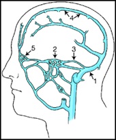
Image: Cranial DAVFs are most commonly located in the transverse–sigmoid sinus region (approximately60-65% of cases), followed by the cavernous sinus (around 20–30%), while other locations such as the tentorium, superior sagittal sinus, and anterior cranial fossa are less common (each typically around 5–10%).
The exact cause of DAVFs is not fully understood. They are considered acquired lesions, most commonly developing in adults in their 40s–60s, and are rare in children. Venous sinus thrombosis and prior trauma are frequently implicated as potential triggers, likely leading to venous hypertension and the formation of abnormal arteriovenous shunts within the dura.
This page focuses primarily on cranial DAVFs, with a separate section on spinal DAVFs below.
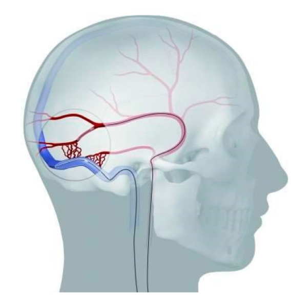
Image: Most common DAVF location between the dural artery and the dural venous sinus (transverse sinus)
Pathophysiology — Venous Hypertension and Congestion in DAVF
The key problem in dural arteriovenous fistulas is not the arterial inflow itself, but impaired venous drainage.
Arterial blood is shunted directly into the venous system and may lead to:
- increased venous pressure — direct inflow of arterial blood raises pressure in the veins
- reduced normal venous outflow — higher venous pressure makes it harder for blood to drain normally
- chronic venous congestion — persistent back-up of blood in the veins over time, leading to impaired circulation
This results in venous hypertension, which is the central mechanism of symptoms and complications. If the blood flow through the fistula is low—particularly in cases where the shunt involves a dural vein rather than a major venous sinus—these venous drainage disturbances may not develop. Such fistulas often remain stable over time without further progression or clinical worsening.
In cranial DAVFs, if venous drainage disturbances appear:
- abnormal venous drainage may reverse into cortical veins — a phenomenon known as cortical venous reflux
- elevated venous pressure leads to dilation and weakening of cortical veins, making them prone to rupture and intracranial hemorrhage (intracerebral, subdural or subarachnoid)
- Impaired venous outflow may reduce normal drainage of blood from the brain, resulting in a back-up of blood in the veins (venous congestion), which can lead to swelling (edema) and reduced oxygen supply to brain tissue (ischemic changes).
In spinal DAVFs if venous drainage disturbances appear:
- Venous congestion affects the spinal cord — impaired venous drainage leads to a build-up of blood in the spinal veins, increasing pressure and making it harder for the spinal cord to receive normal blood flow.
- Leads to chronic ischemia and progressive neurological decline — over time, reduced oxygen supply damages the spinal cord, resulting in gradually worsening symptoms such as leg weakness, difficulty walking, sensory loss, and bladder or bowel dysfunction.
Symptoms of DAVF
If there is no disturbance in the brain’s venous drainage, many dural AV fistulas remain asymptomatic and may be discovered incidentally on imaging. In such cases, they can often simply be monitored over time.
Common symptoms include:
- pulsatile tinnitus — a rhythmic “whooshing” sound caused by turbulent blood flow, most often in transverse or sigmoid sinus fistulas (the occipital and region behind the ear), transmitted through adjacent bone structures
- headache — related to venous hypertension and impaired cerebral venous drainage
- visual disturbances — particularly in cavernous sinus fistulas, due to orbital venous congestion
- seizures — may occur as a result of cortical irritation caused by venous congestion or ischemia
- cognitive changes — including slowed thinking or confusion, often due to chronic venous congestion
- neurological deficits — such as weakness or speech problems, typically caused by venous ischemia or intracranial hemorrhage
- intracranial hemorrhage — may present suddenly with severe symptoms, especially in high-risk fistulas with cortical venous reflux
Pulsatile Tinnitus — Hearing Blood Flow in AV Fistula
Pulsatile tinnitus is one of the most common and characteristic symptoms.
Patients typically describe:
- a rhythmic “whooshing” sound
- synchronized with the heartbeat
- often louder at night or in quiet environments
This symptom is most commonly associated with fistulas located in the transverse sinus (occipital region) and the sigmoid sinus (mastoid region, behind the ear).
It occurs because turbulent blood flow is transmitted to nearby structures and becomes audible.
Headache or Seizures and DAVF — When Is It the Cause?
Headache or seizures without a clear cause on MRI should raise suspicion for a dural arteriovenous fistula (DAVF), especially when symptoms are atypical, progressive, or associated with pulsatile tinnitus or fluctuating neurological findings. Small or complex DAVFs may not be visible on standard imaging and may require more detailed studies such as MRA, CTA, or DSA.
On the other hand, if a DAVF is incidentally detected in a patient with headache or seizures, it must be confirmed that it is truly the cause of symptoms. In headache, this is often not straightforward, as headache is very common and frequently related to primary disorders such as migraine or tension-type headache. A DAVF is more likely to be relevant if there are associated features such as pulsatile tinnitus, atypical or progressive symptoms, or signs of venous hypertension on imaging.
In patients with seizures, it is important to determine whether the DAVF is actually responsible for cortical irritation, venous congestion, or ischemic changes that could trigger epilepsy. This requires correlation between imaging findings (especially cortical venous reflux, edema, ischemic changes, or hemorrhagic lesions), the anatomical location of the DAVF, the clinical seizure pattern, and EEG findings. A DAVF is more likely to be epileptogenic when the vascular lesion and its secondary brain changes involve the same cortical region from which seizures appear to arise. Not all detected DAVFs are epileptogenic, and some may represent incidental findings.
When Is a Dural AV Fistula Dangerous
The risk of complications depends primarily on the pattern of venous drainage.
Low-risk DAVFs:
- drain into dural venous sinuses
- do not involve cortical veins
High-risk DAVFs:
- show cortical venous reflux, where arterial blood flows retrogradely into cortical veins instead of normal venous drainage
- this leads to arterialization of cortical veins, with increased flow and pressure within the venous system
- progressive venous hypertension causes dilation and weakening of vein walls, making them prone to rupture
- as a result, there is a significantly increased risk of intracranial hemorrhage and neurological deterioration
This classification is commonly described using systems such as Borden or Cognard, but the key clinical concept is simple: The presence of abnormal cortical venous drainage determines the risk. In higher-grade DAVFs with cortical venous reflux, the annual risk of hemorrhage is about 8%, and the annual risk of new neurological deficits is about 7%.
Symptoms of the Most Common Cranial DAVF Locations
Transverse and Sigmoid Sinus DAVF
These are the most common locations, with the fistula typically situated at the junction of the transverse and sigmoid sinuses.
Typical features:
- pulsatile tinnitus — the most common presentation, often unilateral and synchronous with the heartbeat
- audible bruit in the occipital region, sometimes detectable on examination
- symptoms may initially be limited to tinnitus, especially in low-risk fistulas without cortical venous reflux
However, in more advanced or high-risk cases:
- neurological deficits may occur due to impaired venous drainage (venous congestion — build-up of blood in the veins), leading to reduced blood flow (ischemia) and resulting in focal deficits such as visual disturbances and cognitive changes
- seizures can develop as a result of cortical venous hypertension
- intracranial hemorrhage may occur, particularly when cortical venous reflux is present
Cavernous Sinus (Carotid–Cavernous Fistula)
These fistulas have a distinct clinical presentation due to their effect on the orbit and cranial nerves. They are most commonly caused by trauma.
Typical features:
- red eye due to venous congestion
- protrusion of the eye (proptosis)
- double vision (diplopia) caused by cranial nerve dysfunction
- pulsatile noise, sometimes perceived by the patient
These symptoms result from venous congestion in the cavernous sinus and orbital veins.
Important considerations:
Cavernous sinus fistulas may be dural (indirect) or direct high-flow fistulas
- indirect (dural) fistulas are often less aggressive and may present mainly with ocular symptoms
- direct fistulas, typically involving the internal carotid artery within the cavernous sinus, represent a high-flow lesion and may lead to rapid symptom progression
The severity depends primarily on venous drainage patterns and flow dynamics, rather than location alone
Diagnosis — MRI, MRA, CTA and Angiography
MRI and MRA (MR angiography) as well as CTA (CT angiography) may suggest the diagnosis.
Findings may include:
- dilated veins — enlarged veins due to increased pressure and abnormal blood flow
- signs of venous congestion — impaired venous drainage with build-up of blood in the veins
- brain edema — swelling of brain tissue caused by venous hypertension and reduced circulation
In spinal DAVF:
- spinal cord edema — swelling of the spinal cord due to chronic venous congestion
- serpiginous flow voids — tortuous, abnormal vessels around the spinal cord seen as dark, snake-like structures on MRI
However, the gold standard is:
Digital Subtraction Angiography (DSA)
This is the gold standard. DSA is a minimally invasive imaging technique performed under local anesthesia, often with light conscious sedation to ensure the patient is comfortable and relaxed. A neuro-radiologist inserts a thin catheter, typically through a puncture in the femoral artery (in the groin) or the radial artery (in the wrist). The catheter is then carefully navigated through the major blood vessels up to the arteries supplying the brain. Once positioned, a contrast agent is injected. This substance allows high-resolution, real-time X-ray images to visualize the blood flow, which clearly outlines the arteries and identifies DAVFs, and other vascular pathologies such as AVMs and aneurysms.
This allows:
- precise identification of the fistula
- evaluation of venous drainage
- assessment of hemorrhage risk
Treatment Options for DAVF
Treatment depends on symptoms and venous drainage pattern.
Options include:
- observation (in low-risk cases)- Approximately 20–40% of DAVFs, typically those without cortical venous reflux, may be managed conservatively with observation and regular follow-up.
- endovascular treatment
- microsurgical disconnection
- radiosurgery
In more complex cases, a combination of therapeutic methods is used.
Treatment is generally recommended:
- if the fistula has caused hemorrhage
- if cortical venous reflux is present
- if symptoms are progressive
In contrast:
- asymptomatic DAVFs without cortical venous reflux may be managed conservatively, with regular clinical and imaging follow-up
- these lesions are generally considered low-risk, although a small proportion may change over time and therefore require reassessment
Endovascular treatment is the most commonly used approach and is successful in approximately 70–90% of cases, depending on location and anatomy. This procedure involves navigating microcatheters through the blood vessels to reach the site of the fistula and closing the abnormal connection.
The goal is not only to eliminate the abnormal arteriovenous shunt, but to disconnect the pathological venous drainage.
This can be done:
- transarterial approach — by injecting liquid embolic agents (Onyx or glue) to penetrate and seal the fistulous connection, ideally reaching the venous side
- transvenous approach — treatment is performed from the venous side by targeting the venous end of the fistula, including the fistulous point, draining vein, or the involved venous sinus. This may involve selective occlusion of the fistula or complete occlusion of the affected sinus segment. In advanced DAVFs (especially transverse–sigmoid) the sinus is often already functionally excluded from normal drainage, so sinus occlusion is commonly performed, in a substantial proportion of high-grade cases (approximately 50–70%).
- combined approach — where both arterial and venous routes are used in the same procedure or in staged treatments, particularly in complex fistulas with multiple feeders or difficult anatomy
Whenever possible, normal venous drainage is preserved, but in many high-grade DAVFs the involved sinus can be safely sacrificed because it no longer serves normal cerebral venous outflow.
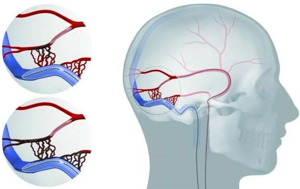
Image: Schematic illustration of an occipital dAVF and its management via combined transarterial and transvenous endovascular occlusion.
In cases where a fistula has caused intracranial hemorrhage, emergency surgery may be required to remove the hematoma and relieve pressure on the brain. During the same procedure, an attempt may be made to identify and disconnect the fistula, particularly if it is surgically accessible. However, this is not always possible. If the fistula cannot be definitively treated during surgery, it is subsequently managed using endovascular techniques.
Definitive treatment requires closure of the arteriovenous shunt itself. This is most commonly achieved through endovascular embolization, but in some cases microsurgical treatment is used as a primary approach, particularly when endovascular access is not feasible or is unlikely to be effective, such as in anterior cranial fossa DAVFs.
The choice between endovascular and surgical treatment is made by a multidisciplinary team, typically including neurosurgeons and interventional neuroradiologists, based on the location of the fistula, venous drainage pattern, and overall risk profile, in order to prevent further bleeding or neurological deterioration.
Spinal Dural Arteriovenous Fistula — A Frequently Missed Cause of Progressive Leg Weakness
Spinal DAVF is a separate but related condition.
It is caused by abnormal shunting in the dura surrounding spinal nerve roots, leading to venous congestion of the spinal cord.
Typical features:
- progressive weakness
- thoracic lesions — weakness of the legs (paraparesis), often with gradual deterioration of walking ability
- cervical lesions — weakness may involve both arms and legs (quadriparesis), sometimes with hand clumsiness
- difficulty walking — typically due to progressive myelopathy and spasticity
- sensory disturbances
- thoracic lesions — sensory level on the trunk and lower limbs
- cervical lesions — involvement of upper limbs, often with numbness or loss of fine sensation in the hands
- worsening with physical activity — symptoms may fluctuate or worsen due to increased venous congestion
This condition is often misdiagnosed as:
- spinal stenosis
- peripheral neuropathy
Delayed diagnosis can lead to permanent deficits. Spinal DAVFs are treated by either endovascular embolization or, more definitively, by surgical disconnection of the fistulous draining vein to eliminate the abnormal shunt and relieve venous hypertension.
Prognosis After Treatment
Outcome depends on:
- early diagnosis — recognizing the condition before significant damage occurs
- prompt treatment after diagnosis — starting treatment without delay to prevent progression
- type of venous drainage — especially the presence of cortical venous reflux, which increases risk
Cranial DAVF:
- good outcome when treated early
- higher risk if cortical venous reflux is present
- pulsatile tinnitus often improves or resolves after treatment
- neurological deficits or seizures may improve if caused by venous congestion and edema
- recovery is less complete if prolonged venous hypertension has led to ischemic brain damage
Spinal DAVF:
- improvement possible if treated early
- delayed diagnosis may lead to irreversible ischemic damages
Why Specialist Opinions May Differ in DAVF
Interpretation of Venous Drainage and Cortical Venous Reflux
The presence or absence of cortical venous reflux often determines whether observation is safe or treatment is required. Specialist recommendations for Dural Arteriovenous Fistula may differ even when based on the same imaging findings. This does not necessarily indicate error, but rather reflects the complexity of the condition. One of the most important sources of disagreement is the interpretation of venous drainage patterns, especially regarding the presence and clinical significance of cortical venous reflux, which directly determines the risk of hemorrhage and neurological deterioration.
Variation in Perceived Risk and Timing of Treatment
Variation in perceived risk is another key factor. Some specialists may recommend earlier intervention, while others may favor observation in borderline cases, particularly when imaging findings are not clearly high-risk. These differences arise from how each specialist weighs the risk of hemorrhage or neurological deterioration against the risks associated with treatment.
Differences in Treatment Experience and Strategy
Recommendations may also differ based on treatment experience. Centers with strong endovascular expertise may prefer embolization, while others may recommend surgery in selected cases. These differences often reflect familiarity with specific techniques and expected outcomes in different anatomical situations.
Anatomical Complexity of DAVF
DAVFs often show anatomical complexity, including multiple arterial feeders, partial thrombosis, or unusual venous pathways, which can influence decision-making and the feasibility of different treatment approaches.
Patient-Specific Factors
Patient-specific factors such as age, symptoms, comorbidities, and tolerance of procedural risk also play an important role in determining the most appropriate management strategy.
Why Different Recommendations Are Often Reasonable
In practice, these differences often represent reasonable clinical judgment rather than conflicting standards.
For this reason, obtaining a second opinion can be particularly valuable when the diagnosis is uncertain, when treatment options differ, or when the risks and benefits are not clearly understood.
Request Dural Arteriovenous Fistula (DAVF) Neurosurgery Consultation — 24-Hour Review or Priority Option (Usually Within 3 Hours)
When a dural arteriovenous fistula (DAVF) is diagnosed or suspected, patients often face critical questions:
whether the fistula is low-risk or high-risk, whether treatment is necessary, and which approach — observation, endovascular therapy, or surgery — is most appropriate.
An independent neurosurgical second opinion can help.
- ✔ Send a short message describing your condition, symptoms (pulsatile tinnitus, neurological deficits, visual changes, seizures, or progressive weakness), and what your MRI or angiography report states
- ✔ You’ll receive a reply within 24 hours explaining if and how we can help in your specific DAVF situation
- ✔ Time-sensitive cases: if hemorrhage has occurred, symptoms are worsening, cortical venous reflux is suspected, or urgent treatment has been proposed — write PRIORITY in your first message
- ✔ If available, MRI, MRA, or angiography images (DICOM format) and specialist reports can be reviewed after initial contact
- ✔ During the consultation, we clarify risk level, need for treatment, timing, expected outcomes, and the most appropriate next steps — with up to 10 days of follow-up for brief questions
Consultation fees typically range from $180–250, depending on case complexity and imaging findings.
Secure payment by credit card, PayPal invoice (USD), or bank transfer.
This is within the usual range for international specialist telehealth second opinions in neurosurgery.
Frequently Asked Questions About Dural Arteriovenous Fistula (DAVF)
Why is cortical venous reflux dangerous in dural arteriovenous fistula (DAVF)?
Cortical venous reflux is dangerous in dural arteriovenous fistula (DAVF) because arterial blood is redirected backward into cortical veins instead of draining normally through the venous system. This increases pressure inside veins that are not designed to tolerate arterial flow. Over time, these veins may become dilated, weakened, and prone to rupture. The main clinical danger is therefore not only the abnormal connection itself, but the way it changes venous drainage from the brain. When cortical venous reflux is present, the risk of intracranial hemorrhage and neurological deterioration rises significantly. This is why DAVFs with cortical venous reflux are usually considered high-risk and commonly require active treatment rather than simple observation.
Can a dural arteriovenous fistula (DAVF) cause intracranial hemorrhage even without warning symptoms?
Yes. A dural arteriovenous fistula (DAVF) can cause intracranial hemorrhage, especially when cortical venous reflux is present. Some patients may first have symptoms such as pulsatile tinnitus, headache, seizures, or neurological deficits, but hemorrhage can sometimes be the first dramatic presentation. The mechanism is venous hypertension: arterial blood enters the venous system directly, raises venous pressure, and may cause cortical veins to dilate and weaken. If these veins rupture, bleeding may occur inside or around the brain, including intracerebral, subdural, or subarachnoid hemorrhage. For this reason, the clinical risk of DAVF is determined less by symptoms alone and more by the venous drainage pattern seen on angiographic evaluation.
How does dural arteriovenous fistula (DAVF) produce venous hypertension and brain congestion?
Dural arteriovenous fistula (DAVF) produces venous hypertension because arterial blood is shunted directly into veins or venous sinuses within the dura. Veins normally carry low-pressure blood away from the brain, but in DAVF they receive blood under higher arterial pressure. This makes normal venous drainage more difficult and can create a “back-up” of blood in the venous system. The result is venous congestion, impaired blood outflow, brain edema, reduced oxygen supply to brain tissue, and sometimes neurological deficits or seizures. In high-risk DAVFs, abnormal drainage may reverse into cortical veins, further increasing hemorrhage risk. This is why the key problem in DAVF is venous drainage failure, not simply the presence of abnormal arterial flow.
Can pulsatile tinnitus be the only symptom of dural arteriovenous fistula (DAVF)?
Yes. Pulsatile tinnitus can be the only symptom of dural arteriovenous fistula (DAVF), especially in low-risk fistulas located in the transverse or sigmoid sinus region. Patients often describe a rhythmic “whooshing” sound synchronized with the heartbeat, sometimes louder at night or in quiet surroundings. This happens because turbulent blood flow from the fistula is transmitted through nearby bone and venous structures around the ear and occipital region. If there is no cortical venous reflux and no disturbance of brain venous drainage, symptoms may remain limited to tinnitus. However, persistent pulsatile tinnitus should not be dismissed automatically, because some DAVFs with tinnitus may still require vascular imaging to clarify venous drainage and risk level.
Why may pulsatile tinnitus from dural arteriovenous fistula (DAVF) be misdiagnosed for months or years?
Pulsatile tinnitus from dural arteriovenous fistula (DAVF) may be misdiagnosed because many patients first report only a rhythmic sound in the ear, without obvious neurological deficits. This may be interpreted as an ear problem, benign tinnitus, vascular noise, or another non-neurosurgical condition. Standard imaging may also miss small or complex DAVFs if the venous drainage pattern is not clearly visible. The suspicion becomes stronger when the sound is synchronized with the heartbeat, is unilateral, persists over time, or is associated with headache, visual symptoms, seizures, cognitive change, or fluctuating neurological findings. In such cases, MRI, MRA, CTA, or digital subtraction angiography may be needed to determine whether a DAVF is present.
When is dural arteriovenous fistula (DAVF) considered low-risk and suitable for observation?
Dural arteriovenous fistula (DAVF) is usually considered low-risk when it drains into a dural venous sinus without cortical venous reflux and without evidence of dangerous venous congestion. In such cases, the fistula may produce symptoms such as pulsatile tinnitus but may not significantly disturb cortical venous drainage. Asymptomatic DAVFs without cortical venous reflux may sometimes be managed conservatively with clinical and imaging follow-up. Observation is not the same as ignoring the lesion. Regular reassessment is needed because a small proportion of DAVFs may change over time. The decision to observe depends on symptoms, location, venous drainage, imaging findings, and whether there is any sign of progression, hemorrhage risk, or neurological deterioration.
When does dural arteriovenous fistula (DAVF) require treatment rather than monitoring?
Dural arteriovenous fistula (DAVF) usually requires treatment when it has caused hemorrhage, when cortical venous reflux is present, or when neurological symptoms are progressive. Treatment is also considered when venous congestion produces seizures, focal deficits, cognitive changes, visual disturbance, or other signs of brain or spinal cord dysfunction. In selected cases, severe disabling pulsatile tinnitus may also justify treatment because of its impact on quality of life. The decision is based mainly on venous drainage pattern and risk, not only on symptom intensity. A DAVF with cortical venous reflux is generally high-risk because elevated pressure in cortical veins increases the chance of bleeding and neurological deterioration. Low-risk DAVFs without cortical venous reflux may sometimes be observed.
How is treatment decision-making performed in dural arteriovenous fistula (DAVF)?
Treatment decision-making in dural arteriovenous fistula (DAVF) depends on symptoms, fistula location, venous drainage pattern, and overall risk profile. The most important factor is whether cortical venous reflux is present, because this determines the risk of hemorrhage and neurological deterioration. Doctors also consider whether the DAVF has caused bleeding, seizures, visual symptoms, cognitive change, focal deficits, or progressive symptoms. Imaging must define the arterial feeders, the venous sinus or vein involved, and whether normal venous drainage can be preserved. Endovascular embolization is usually the first treatment for cranial DAVFs, while surgery is used when embolization is not feasible, incomplete, or when hematoma evacuation is required. Complex cases often need a multidisciplinary decision.
Can a dural arteriovenous fistula (DAVF) become dangerous even when symptoms seem mild?
Yes. A dural arteriovenous fistula (DAVF) can be dangerous even when symptoms seem mild, because the true risk depends mainly on venous drainage rather than how dramatic the symptoms appear. A patient may have only pulsatile tinnitus, headache, or vague symptoms, yet imaging may show cortical venous reflux or venous congestion. When arterial blood flows backward into cortical veins, pressure rises in veins that are vulnerable to rupture. This increases the risk of intracranial hemorrhage and neurological deterioration. Conversely, some DAVFs may cause annoying symptoms but remain low-risk if cortical venous reflux is absent. This is why risk assessment requires careful imaging analysis, often including DSA, rather than relying only on symptom severity.
How does venous drainage determine prognosis in dural arteriovenous fistula (DAVF)?
Venous drainage determines prognosis in dural arteriovenous fistula (DAVF) because it shows whether the abnormal shunt is disturbing normal outflow from the brain or spinal cord. Low-risk DAVFs drain into dural venous sinuses without involving cortical veins. These lesions may sometimes remain stable and be monitored. High-risk DAVFs show cortical venous reflux, meaning blood flows abnormally into cortical veins. This causes venous hypertension, dilation and weakening of veins, and a higher risk of hemorrhage or neurological decline. In spinal DAVF, impaired venous drainage produces congestion of the spinal cord, chronic ischemia, and progressive weakness. Early recognition and correct classification of venous drainage are therefore central to prognosis and treatment planning.
Can headache or seizures be caused by dural arteriovenous fistula (DAVF)?
Headache or seizures can be caused by dural arteriovenous fistula (DAVF), but the relationship must be evaluated carefully. Headache is common in the general population and may be unrelated to an incidentally discovered DAVF. A DAVF is more likely to be relevant when headache is atypical, progressive, associated with pulsatile tinnitus, or accompanied by imaging signs of venous hypertension, brain edema, or impaired venous drainage. Seizures may occur when venous congestion, cortical irritation, ischemic changes, or hemorrhagic lesions affect the same brain region from which seizures appear to arise. This requires correlation between clinical symptoms, imaging findings, DAVF location, cortical venous reflux, and sometimes EEG. Not every detected DAVF is the cause of headache or epilepsy.
Why can dural arteriovenous fistula (DAVF) remain invisible or unclear on standard MRI?
Dural arteriovenous fistula (DAVF) can remain unclear on standard MRI because the abnormal connection may be small, complex, or located within dural venous structures that are not easily recognized on routine images. MRI may show indirect signs such as dilated veins, venous congestion, brain edema, or ischemic changes, but it may not always define the exact fistula or venous drainage pattern. MRA or CTA may suggest the diagnosis, but the gold standard is digital subtraction angiography (DSA). DSA shows real-time blood flow, identifies arterial feeders, demonstrates the fistulous connection, and defines venous drainage. This is especially important when cortical venous reflux is suspected, because risk assessment and treatment decisions depend on precise vascular anatomy.
How is digital subtraction angiography (DSA) performed for dural arteriovenous fistula (DAVF)?
Digital subtraction angiography (DSA) is the gold standard test for dural arteriovenous fistula (DAVF). It is a minimally invasive imaging procedure usually performed under local anesthesia, often with light conscious sedation. A neuroradiologist inserts a thin catheter through an artery, commonly in the groin or wrist, and carefully navigates it through the blood vessels toward the arteries supplying the brain or spine. Contrast material is then injected, allowing high-resolution real-time X-ray imaging of blood flow. DSA can precisely identify the fistula, show arterial feeders, define the venous drainage pattern, detect cortical venous reflux, and assess hemorrhage risk. This information is crucial for deciding between observation, embolization, surgery, or combined treatment.
What is the difference between endovascular and surgical treatment for dural arteriovenous fistula (DAVF)?
Endovascular treatment for dural arteriovenous fistula (DAVF) is performed through blood vessels using microcatheters. The goal is to close the abnormal arteriovenous shunt and disconnect pathological venous drainage, often using liquid embolic agents such as Onyx or glue, or by treating the venous side of the fistula. This is the most common treatment for cranial DAVFs and is successful in many cases, depending on location and anatomy. Surgical treatment is different: the neurosurgeon directly disconnects the abnormal venous drainage, or removes a hematoma when bleeding has occurred. Surgery is usually reserved for DAVFs that cannot be safely or completely treated endovascularly, for selected anterior cranial fossa lesions, or for emergencies requiring hematoma evacuation.
When is surgery necessary for dural arteriovenous fistula (DAVF)?
Surgery for dural arteriovenous fistula (DAVF) is necessary in selected situations. It may be used when endovascular treatment is not feasible, when embolization remains incomplete, or when the anatomy makes microsurgical disconnection more reliable. Some DAVFs, such as selected anterior cranial fossa fistulas, may be better suited for surgical treatment. Surgery may also be urgently required if the fistula has caused intracranial hemorrhage and the hematoma must be removed to relieve pressure on the brain. During such surgery, the abnormal venous drainage may be disconnected if accessible. If the fistula cannot be definitively closed during the operation, additional endovascular treatment may be performed afterward. The choice depends on location, venous drainage, and risk.
Can endovascular embolization permanently cure dural arteriovenous fistula (DAVF)?
Endovascular embolization can permanently cure many cranial dural arteriovenous fistulas (DAVFs), but success depends on the fistula location, arterial feeders, venous drainage, and whether the pathological venous connection can be completely closed. The goal is not simply to reduce blood flow, but to eliminate the arteriovenous shunt and disconnect abnormal venous drainage. This may be performed through an arterial route, a venous route, or a combined approach. In some high-grade transverse–sigmoid DAVFs, the involved venous sinus may already be functionally excluded from normal drainage and can sometimes be occluded safely. If embolization does not achieve definitive closure, surgery or additional staged endovascular treatment may be needed to prevent hemorrhage or neurological deterioration.
How do doctors know whether my dural arteriovenous fistula (DAVF) has been completely closed after embolization?
Complete closure cannot be confirmed only because pulsatile tinnitus or other symptoms improve. The key question is whether the abnormal arteriovenous shunt has disappeared and whether dangerous venous drainage, especially cortical venous reflux, is no longer present. This requires follow-up imaging after treatment.
Digital subtraction angiography (DSA) is usually the most reliable test for confirming complete closure because it shows the arterial feeders, fistulous point, draining veins, and venous flow in real time. MRI, MRA, or CTA may be useful during follow-up, but small residual shunts can be missed. If a residual dural arteriovenous fistula remains, the patient may require further embolization, surgery, or another individualized treatment strategy.
What happens if endovascular treatment does not completely close my dural arteriovenous fistula (DAVF)?
Incomplete closure does not always mean that treatment has failed, but the remaining fistula must be reassessed carefully. The clinical importance depends on whether abnormal cortical venous reflux persists, whether symptoms remain, and whether the residual shunt still creates venous hypertension or a risk of hemorrhage.
Further treatment may include another transarterial or transvenous embolization, a combined endovascular approach, or microsurgical disconnection when the anatomy is unsuitable for complete endovascular closure. In selected low-risk residual fistulas without cortical venous reflux, observation may occasionally be considered. The decision should be based on follow-up angiography and the remaining venous drainage pattern, not only on whether tinnitus or other symptoms have improved.
What are the specific risks of endovascular treatment for dural arteriovenous fistula (DAVF)?
The risks of endovascular DAVF treatment depend on the fistula location, arterial feeders, venous drainage, treatment route, and whether normal arteries or venous sinuses are closely involved. Possible complications include bleeding, ischemic stroke, vessel perforation, unintended blockage of a normal artery or vein, cranial nerve injury, venous thrombosis, contrast-related complications, and incomplete fistula closure.
Transarterial treatment carries particular concern when dangerous connections exist between dural arteries and vessels supplying cranial nerves or the brain. Transvenous treatment may require occlusion of an involved venous sinus, which is safe only when that sinus no longer provides essential normal drainage. Careful angiographic planning and experienced neurointerventional treatment reduce these risks but cannot eliminate them completely.
Will pulsatile tinnitus, seizures, or neurological symptoms improve after dural arteriovenous fistula (DAVF) treatment?
Many symptoms improve after successful DAVF closure, but the degree and speed of recovery depend on how long venous hypertension and congestion were present before treatment. Pulsatile tinnitus often improves rapidly or disappears once the abnormal flow is eliminated.
Seizures, cognitive slowing, weakness, visual symptoms, or other neurological deficits may improve more gradually as venous congestion and brain edema resolve. Recovery may be incomplete if prolonged venous hypertension has already caused ischemic injury, hemorrhage, gliosis, or permanent damage to the brain or spinal cord. Early diagnosis and complete treatment therefore offer the best chance of neurological improvement, while delayed treatment may stop further deterioration without fully reversing existing deficits.
Why is spinal dural arteriovenous fistula (DAVF) often diagnosed late?
Spinal dural arteriovenous fistula (DAVF) is often diagnosed late because its symptoms usually develop gradually and resemble more common disorders. Patients may develop progressive leg weakness, difficulty walking, sensory disturbance, stiffness, or bladder and bowel problems. These symptoms may be mistaken for spinal stenosis, peripheral neuropathy, degenerative spine disease, or other neurological conditions. The underlying mechanism is venous congestion of the spinal cord: abnormal shunting raises venous pressure, impairs drainage, and reduces oxygen supply to spinal cord tissue. MRI may show spinal cord edema and abnormal serpiginous vessels, but the diagnosis can still be missed if DAVF is not suspected. Delayed diagnosis matters because prolonged venous hypertension can cause irreversible spinal cord damage.
Can spinal dural arteriovenous fistula (DAVF) mimic spinal stenosis or peripheral neuropathy?
Yes. Spinal dural arteriovenous fistula (DAVF) can mimic spinal stenosis or peripheral neuropathy because it often causes slowly progressive walking difficulty, leg weakness, sensory symptoms, and sometimes bladder or bowel dysfunction. In thoracic spinal DAVF, patients may develop progressive weakness of the legs and worsening gait. In cervical lesions, both arms and legs may be affected. Symptoms can fluctuate or worsen with physical activity because venous congestion increases. This pattern can look similar to degenerative spine disease or nerve damage, especially early in the course. The difference is that spinal DAVF causes impaired venous drainage of the spinal cord itself. If diagnosis is delayed, neurological deficits may become permanent even after treatment.
What happens if spinal dural arteriovenous fistula (DAVF) is not treated early?
If spinal dural arteriovenous fistula (DAVF) is not treated early, chronic venous congestion may continue to damage the spinal cord. The abnormal shunt raises pressure in the spinal veins, making it harder for blood to drain normally and reducing oxygen supply to spinal cord tissue. Over time, this can cause progressive myelopathy: worsening leg weakness, difficulty walking, sensory loss, spasticity, and bladder or bowel dysfunction. Symptoms may slowly advance and become less reversible as ischemic damage accumulates. Early treatment can improve or stabilize neurological function, but delayed diagnosis may leave permanent deficits. Treatment usually involves surgical disconnection of the draining vein, which is often definitive, or endovascular embolization in selected favorable anatomy.
Why may specialists recommend different treatments for dural arteriovenous fistula (DAVF)?
Specialists may recommend different treatments for dural arteriovenous fistula (DAVF) because management depends on complex anatomy and risk interpretation. The most important issue is venous drainage, especially whether cortical venous reflux is present. Some specialists may recommend observation in borderline low-risk cases, while others may favor earlier treatment if they believe hemorrhage or neurological deterioration risk is significant. Recommendations may also differ because of local experience: centers with strong endovascular expertise may prefer embolization, while others may recommend surgery in selected anatomical situations. Multiple arterial feeders, partial thrombosis, unusual venous pathways, patient age, symptoms, and procedural risk all influence the decision. Different recommendations may therefore reflect reasonable judgment rather than medical error.