Dr Željko Kojadinović — NEUROHIRURGIJA I LEČENJE BOLA
Dr Zeljko Kojadinovic — Pain Treatment & Neurosurgery


Author:
Dr. Zeljko Kojadinovic, MD, PhD
— Neurosurgeon
Specialized Experience:
30 years of clinical expertise in neurosurgery.
Last medically reviewed:
June 08, 2026
Who This Spontaneous CSF Leak Page Is For
This page is intended for patients with suspected or confirmed spontaneous cerebrospinal fluid (CSF) leak, including nasal or sinus-related cranial CSF leak, otogenic (ear) or temporal bone-related cranial CSF leak, and spinal CSF leak with spontaneous intracranial hypotension.
It is especially relevant if you have clear nasal drainage, fluid running into the throat, persistent middle ear or mastoid fluid, hearing symptoms, recurrent meningitis, orthostatic headache, brain sagging on MRI, or symptoms that worsen when upright and improve when lying down.
If the diagnosis remains unclear, tests are negative despite suspicious symptoms, imaging reports are contradictory, the leak cannot be localized, treatment has failed, or surgery has been proposed, an individualized neurosurgical second opinion can help clarify the most reasonable next step.
When patients seek a second opinion for a suspected spontaneous CSF leak
• Clear nasal drainage, throat drainage, or ear fluid is present, but it is unclear whether the fluid is truly cerebrospinal fluid
• Beta-2 transferrin or beta-trace protein testing is negative, but the symptoms and clinical history remain suspicious
• Persistent middle ear fluid (effusion), mastoid fluid, hearing loss, or recurrent ear symptoms raise concern for an ear (otogenic) or temporal bone CSF leak
• Orthostatic headache, brain sagging, or MRI signs of intracranial hypotension suggest a possible spinal CSF leak
• MRI, CT myelography, CT cisternography, or digital subtraction myelography reports are difficult to interpret or do not clearly locate the leak
• Treatment has failed, symptoms have returned, or a new headache pattern appears after blood patch, skull base repair, or spinal leak treatment
• Surgery has been proposed, but the patient is unsure whether the diagnosis, leak location, or surgical strategy is clear enough
CSF leak diagnosis can be difficult because nasal leaks, ear-related CSF leaks, and spinal leaks may present in very different ways.
If your situation involves uncertainty about fluid testing, imaging, leak localization, treatment failure, or surgical planning, you may request an individualized neurosurgical review here:
Request Second Opinion
Nasal or Sinus-Related Cranial Spontaneous CSF Leak — Quick Summary (Read This First)
- A nasal or sinus-related cranial CSF leak occurs when cerebrospinal fluid escapes through a defect in the dura and skull base toward the nose, sinuses, or throat. The defect may involve the anterior skull base, ethmoid region, sphenoid sinus, frontal sinus, or other paranasal sinus-related areas.
- The most typical presentation is clear, watery nasal drainage. The fluid may appear from one nostril or both nostrils, may be intermittent, and may become more obvious when bending forward, straining, coughing, lifting, or changing head position.
- Nasal CSF leak is often mistaken for chronic rhinitis, sinus drainage, allergy, or a persistent clear “runny nose.” This is especially likely when the drainage is intermittent, when the patient has no fever, or when symptoms do not respond to usual sinus or allergy treatment.
- Some patients do not see obvious nasal fluid but feel fluid running into the throat. This can happen when cerebrospinal fluid drains backward into the nasopharynx, especially in certain head positions or during periods of active leakage.
- Clear nasal drainage should not automatically be assumed to be CSF. Ordinary rhinitis, allergy, sinus disease, tears, mucus, and postoperative secretions can look similar, so the fluid should be tested when the history is suspicious.
- Glucose testing was used more often in the past but is not specific enough to reliably confirm or exclude CSF leak. Beta-2 transferrin is the most specific commonly used laboratory test, while beta-trace protein is another useful marker in some centers.
- A negative fluid test does not always exclude CSF leak. The result may be falsely negative if the leak is intermittent, if only a very small amount of fluid is collected, or if the sample is diluted with nasal secretion, mucus, blood, tears, or other fluids.
- High-resolution skull base imaging is usually needed to identify the defect. Depending on the suspected site, evaluation may include CT of the skull base, MRI, and sometimes CT cisternography or other specialized imaging.
- The main risk of an untreated cranial CSF leak is meningitis. Persistent clear drainage, recurrent meningitis, suspicious skull base imaging, or drainage that repeatedly returns after treatment should not be ignored.
- Treatment depends on the exact location of the skull base defect. Many nasal or sinus-related CSF leaks can be repaired endoscopically through the nose, while more complex, recurrent, lateral, large, or difficult-to-access defects may require a different surgical approach.
Readers with clear nasal drainage, fluid running into the throat, persistent “runny nose,” recurrent meningitis, or suspicious skull base imaging should focus first in the text on the nasal or sinus-related cranial CSF leak sections. The remaining sections provide deeper clinical detail about fluid testing, imaging, treatment, surgery, recurrence, and second opinion.
Ear-Related (Otogenic) or Temporal Bone-Related Cranial Spontaneous CSF Leak — Quick Summary (Read This First)
- An otogenic or temporal bone-related cranial CSF leak occurs when cerebrospinal fluid escapes through a defect in the dura and bone near the ear, middle ear, mastoid region, or temporal bone. This is still a cranial CSF leak, but it may present very differently from a typical nasal or sinus-related leak.
- The leak may drain from the ear, but it may also remain hidden behind the eardrum. In many patients, there is no obvious clear fluid coming out of the ear. Instead, CSF may collect in the middle ear or mastoid air cells.
- Otogenic CSF leak can mimic chronic ear disease. It may look like persistent middle ear effusion, recurrent serous otitis, chronic otitis media, mastoiditis, unexplained mastoid fluid on imaging, ear fullness, reduced hearing, or recurrent ear infections.
- A temporal bone CSF leak may cause a salty or metallic taste when fluid drains through the Eustachian tube toward the throat. In certain head positions, the same pathway can allow fluid to reach the nasopharynx and then appear as clear nasal drainage, even though the actual defect is ear-related or temporal bone-related.
- Hearing symptoms are common clues. Patients may describe ear fullness, hearing loss, fluctuating hearing, pressure in the ear, pulsatile sensations, or persistent fluid behind the eardrum despite treatment.
- Testing the fluid may require an ENT procedure if there is no obvious drainage. If fluid is trapped behind the eardrum, a sample may sometimes be obtained during placement of an ear ventilation tube, also called a tympanostomy tube, or by tympanocentesis, in which a thin needle and syringe are used to aspirate fluid through the tympanic membrane.
- Beta-2 transferrin and beta-trace protein can help confirm whether the fluid is CSF. Glucose testing was used more often in the past but is not specific enough. A negative test does not always exclude CSF leak if the sample is very small, diluted, intermittent, or collected at the wrong time.
- Imaging should focus on the temporal bone, middle ear, mastoid region, and skull base. High-resolution CT of the temporal bone, MRI, and sometimes CT cisternography may be needed to identify the defect and distinguish CSF leak from ordinary ear disease.
- The main risks are persistent leakage, recurrent ear problems, and meningitis. Recurrent meningitis, persistent middle ear fluid, or clear ear/nasal drainage with suspicious imaging should raise concern for cranial CSF leak.
- Treatment depends on the exact location and accessibility of the defect. Some temporal bone leaks can be repaired through a transmastoid approach — an operation performed by ENT through the mastoid bone behind the ear — while others may require a middle cranial fossa craniotomy to identify and close tegmen or temporal bone defects.
Readers with persistent middle ear fluid, ear fullness, reduced hearing, clear ear drainage, recurrent ear infections should focus in the text first on the otogenic or temporal bone-related CSF leak sections. The remaining sections provide deeper clinical detail about fluid testing, temporal bone imaging, surgical repair, recurrence, and second opinion.
Spinal Spontaneous CSF Leak — Quick Summary
- A spontaneous spinal CSF leak occurs when cerebrospinal fluid escapes through a defect in the spinal dura. Unlike cranial CSF leaks, the fluid usually remains internal rather than leaking from the nose or ear.
- The main consequence is loss of CSF volume, which may cause spontaneous intracranial hypotension. The most characteristic symptom is orthostatic headache that worsens when upright and improves when lying down.
- Brain or spine MRI may be normal or inconclusive. Difficult leaks, including CSF-venous fistulas, may require specialized imaging such as CT myelography, dynamic CT myelography, or digital subtraction myelography.
- Treatment depends on the leak mechanism and location. Options may include epidural blood patching, targeted treatment, embolization for selected CSF-venous fistulas, or surgical repair.
For detailed information about spinal CSF leak symptoms, normal or negative MRI, CSF-venous fistulas, myelography, blood patching, rebound intracranial hypertension, and surgical treatment, read the dedicated guide: Spontaneous Spinal CSF Leak and Intracranial Hypotension .
Contents
- Who this is for
- Nasal leak summary
- Ear leak summary
- Spinal leak summary
- Leak types
- Cranial CSF leak
- Spinal CSF leak
- Typical symptoms
- Orthostatic headache
- Fluid testing
- Cranial diagnosis
- Brain MRI signs
- Chiari mimic
- Post-LP headache
- Spinal imaging
- Difficult leak localization
- Conservative treatment
- Targeted surgical repair
- Persistent symptoms after treatment
- Hidden hydrocephalus and CSF leak
- Recovery prognosis after treatment
- Second opinion
- FAQ
A spontaneous CSF leak means that cerebrospinal fluid (CSF) escapes from its normal closed space around the brain or spinal cord without a recent operation, trauma, lumbar puncture, or another obvious trigger. The leak may occur through the skull base toward the nose, sinuses, throat, middle ear, or mastoid region, or it may occur along the spinal dura and cause spontaneous intracranial hypotension.
This condition can be difficult to recognize because symptoms may look like migraine, cervical spine disease, sinus disease, ear disease, chronic fatigue, or nonspecific neurological complaints. In some patients, the most important clue is clear watery drainage from the nose, throat, or ear. In others, the key clue is an orthostatic headache — symptoms that worsen when upright and improve when lying down.
The key clinical question is not only whether cerebrospinal fluid is leaking, but whether the leak is cranial or spinal, where it is located, and whether it truly explains the patient’s symptoms.
Read more about traumatic cerebrospinal fluid (CSF) leaks on this page.
Types of Spontaneous CSF Leak: Cranial and Spinal
Spontaneous CSF leaks can be divided into two main groups: cranial CSF leaks and spinal CSF leaks.
A cranial CSF leak usually manifests as cerebrospinal fluid drainage through the nose or into the ear, depending on the leak location. For this to happen, there must usually be both a dural defect — an opening in the membrane that contains the CSF — and a bone defect at the skull base, creating a pathway between the intracranial CSF space and the nasal cavity, paranasal sinuses, middle ear, or mastoid air cells. Patients may describe clear, watery fluid, sometimes with a salty or metallic taste.
A spinal CSF leak occurs along the spinal dura. The patient does not see any fluid leaving the body. Instead, the leak reduces the normal CSF volume around the brain and spinal cord. This can lead to spontaneous intracranial hypotension, where the brain is no longer supported in the usual way by the cerebrospinal fluid. The result may be orthostatic headache, neck pain, nausea, dizziness, hearing symptoms, visual symptoms, fatigue, or cognitive fog.
Cranial CSF leak is often recognized because of visible nasal or ear drainage. Spinal CSF leak is often suspected because of intracranial hypotension and symptoms that worsen when upright.
Some spontaneous CSF leaks are associated with underlying weakness of connective tissue. This may be seen in patients with hypermobility spectrum disorders, Ehlers-Danlos syndrome, Marfan syndrome, dural ectasia, or other conditions that make the dura more fragile. In these patients, leaks may be multiple, recurrent, or harder to localize, and the clinical history may include joint hypermobility, unusual flexibility, easy bruising, poor wound healing, scoliosis, tall slender body habitus, or family history of vascular or connective tissue disease.
Connective tissue disorders do not prove that a CSF leak is present, but they can increase suspicion when symptoms, imaging, and recurrence pattern fit the diagnosis.
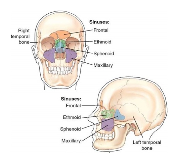
Image: Paranasal sinuses are cavities within the bones that communicate with the nose. In cases of fractures or defects involving the frontal, ethmoid, or sphenoid sinuses, a connection can form between the CSF space and the nose, leading to CSF rhinorrhea (dripping of clear CSF from the nose, especially in provocative positions, such as leaning the head forward).
Spontaneous Cranial CSF Leak: When Fluid Leaks Through the Nose or Into the Ear
A cranial CSF leak occurs when cerebrospinal fluid escapes through a defect in the skull base both the dura and the bone. The fluid may drain through the nose or into the ear, depending on whether the defect is related to the areas around the paranasal sinuses or the middle ear (temporal bone, mastoid region).
In nasal or sinus-related cranial CSF leak, patients may notice clear, watery fluid from the nose, often more pronounced when bending forward, straining, coughing, or changing head position. This type of leakage can be mistaken for chronic rhinitis, sinus drainage, allergy, or a persistent “runny nose.” The main risk of an untreated nasal or sinus-related cranial CSF leak is meningitis, because the defect creates a direct communication between the nasal/sinus spaces and the intracranial compartment; in some patients it may also reflect or contribute to abnormal intracranial pressure dynamics.
In ear-related or temporal bone cranial CSF leak, the presentation can be more confusing. CSF may first collect behind the eardrum or within the mastoid air cells, causing persistent middle ear or mastoid fluid that can be mistaken for chronic ear inflammation. If there is a tympanic membrane defect, ear tube, or previous ear surgery, the fluid may drain outward through the ear. However, when the eardrum is intact, CSF may pass through the Eustachian tube toward the nasopharynx and throat, and sometimes appear as clear nasal drainage. For this reason, a temporal bone CSF leak can present with ear fullness, persistent mastoid or middle ear fluid, postnasal drainage, salty fluid in the throat, or clear nasal drainage, even though the actual defect is located near the ear. If there is no obvious external fluid leakage the main clues may be persistent middle ear effusion, mastoid fluid on imaging, aural fullness, reduced hearing, recurrent ear infections, or symptoms that resemble chronic otitis media or mastoid disease. An untreated temporal bone CSF leak can lead to recurrent meningitis, persistent middle ear or mastoid fluid, hearing problems, tinnitus or ear fullness, and in some cases may be associated with abnormal intracranial pressure or progressive skull base/temporal bone defects.
This is why cranial CSF leak should be considered not only in patients with clear nasal drainage, but also in selected patients with unexplained persistent ear fluid, mastoid opacification on images, hearing loss, or recurrent meningitis.
The key diagnostic question is whether the fluid is truly cerebrospinal fluid. Glucose testing was used more often in the past, but it is not specific enough to reliably confirm or exclude a CSF leak. Today, beta-2 transferrin is the most specific commonly used test, while beta-trace protein is another modern marker used in some centers. However, if the leak is intermittent, the collected sample is very small, or the fluid is diluted with nasal secretion, ear fluid, blood, mucus, or tears, these tests may be negative even when a CSF leak still exists.
Clear fluid from the nose or ear should not automatically be assumed to be CSF, but if the history is suspicious, it should be properly tested because an untreated cranial CSF leak can increase the risk of meningitis.
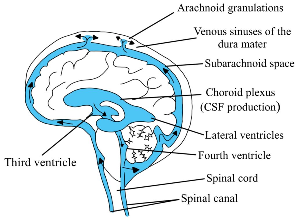
Image: Cerebrospinal fluid (CSF) flow: from production within the ventricles, circulation through the ventricular system, and exit into the subarachnoid space, where it flows before being resorbed into the venous system (specifically the dural venous sinuses). CSF can leak into the nose if there is a defect in the bone and dura at the front base of the skull.
Spinal CSF Leak and Spontaneous Intracranial Hypotension
A spinal CSF leak occurs when cerebrospinal fluid escapes through a defect in the spinal dura, most often in the thoracic or cervicothoracic spine. Unlike cranial CSF leaks, the fluid usually remains internal rather than leaking from the nose or ear.
The resulting loss of CSF volume can cause spontaneous intracranial hypotension, often producing orthostatic headache that worsens when sitting or standing and improves when lying down. Other symptoms may include neck pain, dizziness, nausea, hearing symptoms, visual changes, fatigue, and cognitive difficulty.
Brain or spine MRI may be normal or inconclusive. Some spinal leaks, including CSF-venous fistulas, require specialized myelographic imaging for localization.
For detailed information about spinal leak mechanisms, MRI-negative cases, CSF-venous fistulas, myelography, blood patching, embolization, surgery, and rebound intracranial hypertension, see the dedicated page on Spontaneous Spinal CSF Leak and Intracranial Hypotension.
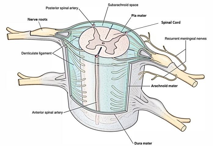
Image: The subarachnoid space is shown between the spinal cord, the spinal nerves, and the arachnoid membrane. Cerebrospinal fluid (CSF) flows through this space, and spinal nerves pass through it before they receive their individual nerve coverings.
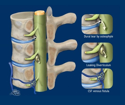
Image: Types of spinal CSF leak
Typical Symptoms of Spontaneous CSF Leak
Symptoms of spontaneous CSF leak vary depending on whether the leak is spinal or cranial.
Cranial CSF leak may cause clear nasal drainage, salty or metallic taste, ear fullness, recurrent meningitis, headaches, or symptoms that seem similar to sinus or ear disease.
Spinal CSF leak often causes headache, neck pain, nausea, dizziness, imbalance, tinnitus, hearing changes, visual blurring, fatigue, cognitive fog, and sensitivity to upright posture. Some patients feel significantly better when lying flat. Some patients do not present in a textbook way. Headache may become less clearly positional over time. Symptoms may fluctuate. Imaging may be incomplete or interpreted as nonspecific.
The most important point is to connect the symptoms with the CSF leak. When clear fluid is actively draining from the nose or ear, fluid testing can help confirm that it is CSF, and imaging can then be used to search for the skull base defect. When there is no obvious external drainage, especially in temporal bone/ear-related CSF leaks or spinal CSF leaks, suspicion must be based on the clinical pattern, otoscopic findings when relevant, persistent middle ear or mastoid fluid, hearing symptoms, positional headaches, and targeted CT/MRI findings. In spinal CSF leak, the fluid usually does not leave the body; instead, it leaks into the spinal extradural space and may cause intracranial hypotension with orthostatic headache, neck pain, nausea, dizziness, tinnitus, or other neurological symptoms. In these cases, diagnosis is often delayed unless the doctor actively looks for the leak pattern and uses the available tests to confirm it. Otherwise, patients may be treated for years under nonspecific diagnoses such as chronic ear inflammation, sinus disease, migraine, tension headache, hearing disorder, anxiety, or depression, while the underlying CSF leak remains unrecognized.
Orthostatic Headache: Why Body Position Matters
Orthostatic headache is one of the most important clues in suspected spinal CSF leak.
This means that the headache becomes worse when the patient is upright and improves after lying down. The improvement may happen quickly or gradually. Some patients describe pressure, pulling, heaviness, or pain in the head, neck, or upper spine.
The reason body position matters is mechanical. When CSF volume is reduced, the brain and its supporting structures are affected more when the patient is upright. Lying down may reduce traction and temporarily improve symptoms.
However, not every patient has a perfect orthostatic headache. In chronic cases, symptoms may become less clearly positional. Some patients may have more neck pain, dizziness, ear symptoms, or cognitive fatigue than classic headache.
A positional pattern is very important, but the absence of a perfect positional headache does not always exclude a CSF leak.
Clear Nasal or Ear Fluid Leak: When Beta-2 Transferrin Testing Is Needed
In suspected cranial CSF leak, the first question is whether the fluid is truly cerebrospinal fluid. However, the fluid is not always easy to collect. A cranial CSF leak may appear as obvious clear nasal drainage, clear ear drainage, fluid running into the throat, or persistent fluid trapped behind the eardrum or in the mastoid air cells.
If the suspected leak drains toward the nose or throat, the patient may sometimes be placed in a position that makes the drainage more likely to appear, such as leaning forward or turning the head in a way that reproduces the leakage. The sample should be collected during active drainage whenever possible, because testing a dry period or a very small diluted sample may be misleading.
If the suspected leak is temporal bone or ear-related, there may be no obvious fluid coming out of the ear. CSF may collect behind the tympanic membrane and look like persistent middle ear effusion, recurrent serous otitis, mastoid fluid on imaging, ear fullness, or reduced hearing. In these cases, the sample may sometimes be obtained during placement of an ear ventilation tube, also called a tympanostomy tube, if the patient is being treated for suspected persistent middle ear effusion. In selected cases, an ENT specialist may obtain fluid by tympanocentesis, a procedure in which a thin needle and syringe are used to aspirate fluid through the tympanic membrane. If fluid reaccumulates behind the eardrum and suspicion remains high, repeat sampling may be considered by an ENT specialist, especially if the result would change management. The decision depends on symptoms, otoscopic findings, imaging, infection risk, and whether a safer or more definitive diagnostic step is available.
Glucose testing of nasal or ear fluid was used more often in the past, but it is not specific enough to reliably confirm or exclude CSF leak. Today, beta-2 transferrin is usually the most specific commonly used laboratory test. Beta-trace protein is another useful marker used in some centers, depending on local laboratory availability. These tests help answer the same first question: is this fluid really CSF?
A negative result does not always exclude CSF leak. If the leak is intermittent, if only a very small amount of fluid is collected, if the sample is diluted with nasal secretion, middle ear fluid, mucus, blood, or tears, or if the sample is not taken during active leakage, beta-2 transferrin or beta-trace protein testing may be falsely negative.
Testing the fluid is often the first step when there is suspicious clear drainage from the nose or ear. Imaging is then used to find the defect.
How Spontaneous Cranial CSF Leak Is Diagnosed
Diagnosis begins with the clinical history. The doctor must understand how symptoms started, whether they change with position, whether there is visible fluid drainage, whether there was previous surgery or trauma, and whether there are neurological, ear, sinus, or spine symptoms.
After that, imaging is selected according to the suspected type of leak.
If a cranial CSF leak is suspected (fluid leaking from the nose or ear), several tests may be used to confirm it and find the leak. These include a beta-2 transferrin test (a simple lab test of the fluid), a high-resolution CT scan to look for tiny bone defects at the base of the skull, an MRI, and sometimes a CT cisternography (a specialized scan that uses a safe contrast dye to track the exact path of the leak).
A CT report may mention tegmen dehiscence, tegmen tympani or tegmen mastoideum defect, cribriform plate defect, ethmoid roof defect, sphenoid sinus defect, frontal sinus posterior table defect, bone thinning, meningocele, or encephalocele. These findings mean that the skull base bone is thin, open, or abnormal in a place where CSF leakage could occur. However, they do not automatically prove an active CSF leak. Some bony defects are incidental, old, small, or not currently leaking. The diagnosis becomes stronger when the CT defect matches the patient’s drainage pattern, ear or nasal findings, beta-2 transferrin or beta-trace protein result, MRI findings, and clinical history.
ICHD-3 diagnostic criteria are also useful when spontaneous intracranial hypotension is suspected. According to ICHD-3, the diagnosis of headache attributed to spontaneous intracranial hypotension requires headache related to low CSF pressure or CSF leakage, absence of a procedure or trauma that could explain the leak, temporal relationship between the headache and the CSF leak or low CSF pressure, and exclusion of another better headache diagnosis. Evidence may include low CSF pressure or imaging signs of CSF leakage.
In spontaneous nasal CSF leaks, doctors should also consider idiopathic intracranial hypertension, especially in patients with obesity, pulsatile tinnitus, visual symptoms, papilledema, empty sella, transverse sinus stenosis, or recurrent skull base defects. An active leak may temporarily mask raised intracranial pressure, which can become more obvious only after the defect is repaired. When raised intracranial pressure or IIH is suspected, evaluation should include attention to papilledema and visual function, because untreated intracranial hypertension can threaten vision and increase the risk of recurrent leak after repair.
Brain MRI Signs of Intracranial Hypotension
Brain MRI can provide important indirect signs of spinal CSF leak and intracranial hypotension.
Typical findings may include diffuse pachymeningeal enhancement, downward displacement or sagging of the brain, enlargement of venous structures, subdural fluid collections, and changes around the pituitary gland. These signs suggest that the intracranial compartment is reacting to reduced CSF volume.
However, brain MRI does not always show all signs clearly. Some patients with real CSF leaks may have subtle or incomplete findings. Others may have imaging changes that need careful interpretation in the context of symptoms.
Brain MRI can support the diagnosis of intracranial hypotension, but it does not always locate the spinal leak itself.
Important Differential Diagnosis in Spinal CSF Leak
Spontaneous intracranial hypotension can cause downward displacement of the cerebellar tonsils and mimic Chiari I malformation. The distinction is important because, in SIH, the tonsillar descent is secondary to loss of CSF volume and brain sagging rather than a primary posterior fossa abnormality. Low-lying tonsils should therefore be interpreted together with the clinical history and the wider MRI pattern. More about Chiari differential diagnosis.
Post-lumbar puncture headache may also resemble spontaneous intracranial hypotension because both can worsen when upright and improve when lying down. The key difference is that post-lumbar puncture headache follows a known dural puncture, while spontaneous intracranial hypotension occurs without a recent procedure or trauma and may result from an occult spinal CSF leak or CSF-venous fistula.
When a spinal CSF leak is suspected, spinal imaging may be needed to identify the leak site and mechanism.
Spine MRI may show extradural fluid collections or other indirect signs. If MRI is inconclusive, CT myelography, dynamic CT myelography, or digital subtraction myelography may be required to detect difficult leaks or CSF-venous fistulas.
A negative or unclear first imaging study does not always exclude a spinal CSF leak, particularly when the clinical pattern remains strongly suggestive.
The choice of imaging depends on the suspected leak type, previous studies, and available expertise.
For detailed information about imaging strategies and difficult-to-localize leaks, see the dedicated page on Spontaneous Spinal CSF Leak and Intracranial Hypotension.
When the CSF Leak Is Not Easily Found
Some spinal CSF leaks are difficult to localize because they may be intermittent, slow, or require a specific imaging technique. CSF-venous fistulas can be particularly difficult to detect because they may not produce a large extradural fluid collection.
Spinal meningeal diverticula and perineural cysts, including Tarlov cysts, may be associated with a leak but do not by themselves prove that active CSF leakage is present.
If the first imaging study is negative or unclear, the diagnosis should be reconsidered carefully and the imaging strategy matched to the suspected leak mechanism.
Conservative Treatment and Epidural Blood Patch for Spontaneous CSF Leak
Treatment depends on the type of leak, severity of symptoms, imaging findings, and clinical course.
Some mild and recent-onset nasal or ear-related cranial CSF leaks may initially be managed conservatively, especially if symptoms are improving. Conservative measures may include bed rest, hydration, caffeine in selected patients, avoiding straining, and close follow-up. The goal is to reduce pressure fluctuations and give the dural and arachnoid layers time to seal if the leak is small and the clinical course is favorable.
If conservative measures do not help, more active CSF diversion may be considered in specialised institutions before surgery. In selected cases, lumbar puncture with controlled CSF drainage may be performed once or, rarely, repeated a few times, depending on the clinical situation. However, repeated lumbar punctures are not usually used as a planned long-term treatment for CSF leak, because each puncture creates a new dural opening and may theoretically provoke or maintain another dural defect.
If controlled diversion of CSF is needed, a lumbar drain may be placed instead. This allows CSF to be drained continuously or intermittently under supervision for several days (often about 3 to 5 days). The purpose is to temporarily reduce CSF pressure across the leak site and give the dura and arachnoid time to adhere and seal. Before removal, the drain is often closed for a trial period to see whether leakage, headache, or other symptoms return. If the patient remains stable and there is no recurrent leak, the drain may be removed.
Lumbar drainage should not be continued casually for a long time, because prolonged drainage increases the risk of infection, overdrainage, pneumocephalus, subdural collections, headache, and neurological deterioration.
In confirmed persistent cranial CSF leak, many centers also consider vaccination against common meningitis pathogens according to local protocols, because the defect creates a pathway for ascending infection.
However, prolonged severe symptoms, progressive findings, recurrent leakage, or clear intracranial hypotension usually require more active evaluation and targeted treatment rather than prolonged observation alone.
In suspected spinal CSF leak with spontaneous intracranial hypotension, conservative management should not continue indefinitely if significant symptoms persist. Epidural blood patching is an important treatment option and may be non-targeted or targeted, depending on whether the leak site has been identified.
Some patients improve after one procedure, while others require repeated or more targeted treatment. The result depends in part on whether the leak mechanism and location have been correctly identified.
When Surgery Is Considered in Spontaneous CSF Leak
More direct treatment may be considered when conservative measures and blood patching fail, when the leak is clearly localized, or when the underlying defect requires repair.
For cranial CSF leaks, surgery may be needed to close a skull base defect, especially when the leak is persistent, recurrent, clearly localized, or associated with meningitis risk. The surgical approach depends on the location of the defect.
When the leak comes from the anterior skull base, paranasal sinuses, sphenoid sinus, ethmoid region, or another nasal/sinus-related area, surgical repair is often performed endoscopically through the nose. If the exact site of leakage is difficult to identify, intrathecal fluorescein may sometimes be used in selected centers to help localize the leak during endoscopic surgery. (Intrathecal use of fluorescein is off-label in some countries and regulatory systems, requires specific informed consent, and should only be performed according to experienced institutional protocols). In more complex, recurrent, large, or endoscopically inaccessible defects, an open cranial approach with craniotomy may be required.
When the leak is temporal bone or ear-related, the repair may be performed through a transmastoid approach (an operation performed through the mastoid bone behind the ear, allowing access to the middle ear and mastoid air cells), especially when the defect can be reached through the mastoid and middle ear region. In other cases, a middle cranial fossa craniotomy may be needed. This approach allows the surgeon to elevate the brain temporal lobe, inspect the floor of the middle cranial fossa, identify tegmen or temporal bone defects, and close the site of leakage.
The goal of surgery is not only to stop visible fluid leakage, but also to close the communication between the intracranial CSF space and the nose, sinuses, middle ear, or mastoid region, thereby reducing the risk of recurrent leakage and meningitis.
For spinal leaks, targeted treatment may include a targeted epidural patch, fibrin sealant in selected cases, endovascular treatment for some CSF-venous fistulas, or surgery to repair a dural defect.
Why Symptoms May Persist or Return After Treatment of CSF Leak
Symptoms may persist or return after treatment for several reasons:
• The leak may not have been fully sealed.
• The treated site may not have been the true source of the leak.
• There may be more than one leak.
• A CSF-venous fistula may have been missed on initial imaging.
• Another condition may coexist with the CSF leak and confuse the clinical picture.
There are also situations where symptoms are not caused only by CSF leak. Migraine, cervical spine disease, vestibular disorders, sinus disease, neuralgia, or central sensitization may coexist and confuse the picture.
Another reason for a new or different headache after treatment is rebound intracranial hypertension, especially after successful treatment of a spinal CSF leak. The original low-pressure headache may improve, but a new headache pattern related to increased CSF pressure can appear, sometimes becoming worse when lying down. Recognizing this change is important because it may require a different diagnostic and treatment approach.
Persistent symptoms after treatment do not always mean that nothing was done correctly. They mean the diagnosis and mechanism must be rechecked carefully.
Hidden Hydrocephalus or Raised Intracranial Pressure Before Cranial CSF Leak Repair
In cranial CSF leak, an active leak may sometimes mask coexisting hydrocephalus or raised intracranial pressure. In that situation, the leak acts like a pressure-release pathway: cerebrospinal fluid escapes through the skull base defect, and intracranial pressure may appear lower than it would be if the defect were closed.
When the leak is repaired, this pressure-release pathway is closed. Intracranial pressure may then rise, and previously hidden hydrocephalus or intracranial hypertension may become clinically apparent. This can lead to a new headache pattern, visual symptoms, nausea, papilledema, or recurrence of the leak if the underlying pressure problem is not recognized.
For this reason, brain MRI should not be used only to look for the leak. It should also be reviewed for ventricular size and signs of raised intracranial pressure.
Recovery and Prognosis After Treatment of Spontaneous CSF Leak
The prognosis depends on the type of CSF leak, how long symptoms have been present, whether the leak is cranial or spinal, whether the site can be localized, and whether there are complications such as meningitis, subdural collections, severe brain sagging, or recurrent leakage.
Some mild leaks may improve with conservative measures, especially when symptoms are recent and gradually improving. Persistent spinal CSF leaks often require an epidural blood patch, targeted patching, embolization in selected CSF-venous fistulas, or surgical repair. Cranial CSF leaks may require skull base repair when the leak is persistent or when meningitis risk is significant.
Recovery is not always immediate. Headache may improve before fatigue, dizziness, hearing symptoms, cognitive fog, or neck pain fully settle. In long-standing cases, symptoms may fluctuate for some time even after the leak is treated.
The most useful prognostic question is not only whether a CSF leak exists, but whether the leak type, location, treatment response, and remaining symptoms all fit together.
Request CSF Leak Neurosurgical Second Opinion — 24-Hour Review (Priority Option Available)
Suspected CSF leak can be difficult to interpret because nasal leaks, ear-related or temporal bone leaks, and spinal leaks may present in very different ways.
Patients may have clear nasal drainage, fluid running into the throat, persistent middle ear or mastoid fluid, hearing loss, recurrent meningitis, orthostatic headache, brain sagging on MRI, or symptoms that worsen when upright.
An independent neurosurgical second opinion can help clarify whether the findings truly suggest a CSF leak, whether the suspected leak is cranial or spinal, which diagnostic steps may still be missing, and whether conservative treatment, blood patch, targeted repair, or surgery is the most reasonable next step.
- ✔ Send a brief message describing your main symptoms, including nasal drainage, throat drainage, ear fluid, hearing symptoms, recurrent meningitis, positional headache, or MRI findings suggesting intracranial hypotension.
- ✔ You’ll receive a reply within 24 hours explaining whether we can help with an online neurosurgical consultation.
- ✔ Priority cases: if there is recurrent meningitis, active clear fluid leakage, severe orthostatic headache, brain sagging, subdural collections, suspected hydrocephalus, or urgent surgical recommendation, consultations can often be arranged within a few hours — write PRIORITY in your first message.
- ✔ Radiologist reports and/or MRI images, CT skull base or temporal bone scans, CT cisternography, CT myelography, digital subtraction myelography, fluid test results, and previous specialist reports can be reviewed once initial contact is established.
- ✔ During the consultation, we explain whether the suspected leak pattern fits the symptoms, whether additional testing may be needed, and whether blood patch, targeted repair, skull base surgery, or continued observation is most appropriate — with up to 10 days of follow-up for brief clarification questions.
Consultation fees typically range from $180–250, depending on case complexity and imaging findings.
Secure payment by credit card, PayPal invoice (USD), or bank transfer.
This is within the usual range for international specialist telehealth neurosurgical second opinions.
Frequently Asked Questions About Spontaneous CSF Leak and Intracranial Hypotension
What are the main types of spontaneous CSF leak?
Spontaneous CSF leak can be divided into three clinically important patterns: nasal or sinus-related CSF leak, ear-related or temporal bone CSF leak, and spinal CSF leak with spontaneous intracranial hypotension. Cranial leaks may produce visible or hidden drainage toward the nose, throat, middle ear, or mastoid region, while spinal leaks usually remain internal and reduce CSF volume. Each type requires a different diagnostic pathway and treatment strategy.
How can I tell if clear fluid from the nose is a CSF leak?
Clear nasal fluid may be CSF, but it should not automatically be assumed to be cerebrospinal fluid. A nasal CSF leak may cause clear watery drainage, sometimes worse with bending forward, straining, coughing, or changing head position. Allergy, rhinitis, sinus disease, tears, and mucus can look similar. When active drainage is available, beta-2 transferrin or beta-trace protein testing can help determine whether the fluid is CSF, followed by imaging to locate the defect.
Can a CSF leak drain into the throat instead of coming out of the nose?
Yes. A nasal or sinus-related CSF leak may drain backward into the nasopharynx and be felt as clear or salty fluid in the throat. An ear-related or temporal bone CSF leak may also reach the throat through the Eustachian tube. The location of the perceived drainage therefore does not always identify the actual site of the defect.
How is a nasal or sinus-related CSF leak diagnosed?
Diagnosis begins with the drainage pattern, clinical history, and fluid testing when a sample is available. Beta-2 transferrin or beta-trace protein can help confirm that the fluid is CSF. High-resolution CT of the skull base and MRI are then used to identify the defect, while CT cisternography or other specialized localization methods may be considered in selected cases.
How is a nasal or sinus-related CSF leak treated?
Treatment depends on the location, duration, and behavior of the leak. Some recent mild leaks may initially be observed when symptoms are improving, while persistent, recurrent, clearly localized, or meningitis-associated leaks often require repair. Many skull base defects can be treated endoscopically through the nose, although more complex defects may require another surgical approach.
Can an untreated nasal CSF leak cause meningitis?
Yes. A cranial CSF leak creates an abnormal communication between the intracranial CSF space and the nose or paranasal sinuses, allowing bacteria to potentially reach the meninges. Persistent clear drainage, recurrent meningitis, or repeatedly recurrent leakage therefore requires proper confirmation and localization of the defect.
Can middle ear or mastoid fluid be caused by a CSF leak?
Yes. In an ear-related or temporal bone CSF leak, cerebrospinal fluid may collect behind the eardrum or within the mastoid air cells rather than draining visibly from the ear. This may resemble persistent middle ear effusion, serous otitis, mastoid disease, ear fullness, or unexplained hearing loss.
How can an ear-related CSF leak look like chronic otitis or mastoiditis?
CSF may remain trapped in the middle ear or mastoid and appear similar to persistent sterile effusion or chronic ear disease. Clues include recurrent or persistent middle ear fluid, mastoid opacification, ear fullness, reduced hearing, salty drainage toward the throat, or recurrent meningitis that does not fit ordinary ear infection.
Can an ear-related CSF leak cause clear nasal drainage?
Yes. CSF from a temporal bone or ear-related leak may pass from the middle ear through the Eustachian tube into the nasopharynx and throat, and may sometimes appear as clear nasal drainage. For this reason, the apparent drainage site does not always correspond to the location of the skull base defect.
How is an ear-related or temporal bone CSF leak diagnosed?
Diagnosis is based on ear findings, drainage pattern, fluid testing when possible, and temporal bone imaging. Beta-2 transferrin or beta-trace protein may confirm that collected fluid is CSF. High-resolution temporal bone CT and MRI can then help identify defects involving the tegmen, mastoid, middle ear, or adjacent skull base.
How is an ear-related or temporal bone CSF leak treated?
Treatment depends on the location, size, and accessibility of the defect. Persistent or clearly localized temporal bone leaks often require surgical repair. Depending on the anatomy, this may involve a transmastoid approach, a middle cranial fossa approach, or occasionally a combined procedure.
Why can beta-2 transferrin or beta-trace protein be negative if a CSF leak is still suspected?
A negative result does not always exclude CSF leak. False-negative or misleading results may occur when leakage is intermittent, the sample is very small, or the fluid is diluted with nasal secretion, middle ear fluid, mucus, blood, or tears. The result should therefore be interpreted together with the clinical history and imaging findings.
What is a spinal CSF leak and why is there usually no visible fluid?
A spinal CSF leak occurs when cerebrospinal fluid escapes through a defect in the spinal dura. Unlike nasal or ear-related leaks, the fluid usually remains inside the body rather than draining externally. Loss of CSF volume may cause spontaneous intracranial hypotension, commonly associated with orthostatic headache and other neurological symptoms.
Can spinal CSF leak be present if brain or spine MRI is normal?
Yes. A normal or inconclusive brain or spine MRI does not always exclude a spinal CSF leak. Some leaks are small, intermittent, difficult to visualize, or caused by a CSF-venous fistula that does not produce a large extradural fluid collection. Further targeted imaging may be considered when clinical suspicion remains strong.
How is spinal CSF leak with intracranial hypotension diagnosed and treated?
Diagnosis is based on the clinical pattern, brain and spine MRI, and, when necessary, specialized myelographic imaging to localize the leak. Treatment depends on the leak mechanism and may include epidural blood patching, targeted procedures, endovascular treatment for selected CSF-venous fistulas, or surgical repair. For detailed diagnostic and treatment pathways, see the dedicated Spontaneous Spinal CSF Leak and Intracranial Hypotension page.
When should a patient seek a second opinion for suspected CSF leak?
A second opinion may be useful when the diagnosis remains uncertain, tests are negative despite suspicious symptoms, imaging reports are contradictory, the leak cannot be localized, treatment has failed, symptoms have returned, or surgery has been proposed. The goal is to determine whether the clinical pattern fits a cranial or spinal CSF leak, whether additional testing is justified, and which treatment strategy is most appropriate. Request a CSF leak second opinion.