Dr Željko Kojadinović — NEUROHIRURGIJA I LEČENJE BOLA
Dr Zeljko Kojadinovic — Pain Treatment & Neurosurgery

Author:
Dr. Zeljko Kojadinovic, MD, PhD
— Neurosurgeon and Pain Management Specialist
Specialized Experience:
30 years of clinical expertise in neurosurgery
Last medically reviewed:
January 8, 2026
What Are Nerves?
Nerves are the body’s communication cables.
They transmit signals between the brain, spinal cord, and the rest of the body, allowing us to feel, move, see, hear, speak, swallow, and regulate internal organs.
There are two main groups:
- Spinal nerves – exit the spinal cord
- Cranial nerves – exit directly from the brain
What Are Cranial Nerves?
Cranial nerves are nerves that arise directly from the brain or brainstem.
There are 12 pairs (I–XII), and they mainly supply the head and neck, although some (especially the vagus nerve) extend to the chest and abdomen.
They can be:
- Sensory (sensation)
- Motor (movement)
- Mixed (both sensory and motor)
Where Do Cranial Nerves Originate?
Most cranial nerves originate from the brainstem, which includes:
- the midbrain
- the pons
- the medulla oblongata
After emerging from the brainstem, they pass through specific openings in the base of the skull (foramina) to reach their target structures.
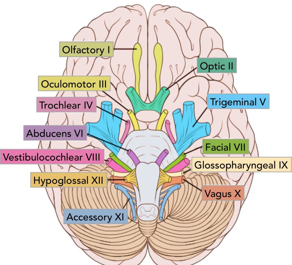
Image: Origin of cranial nerves

Image: Skull base foramina — openings through which cranial nerves pass to reach their target structures.
The Twelve Cranial Nerves (I–XII)
I – Olfactory Nerve
- Function: sense of smell
- Anatomy: travels from the nasal cavity through the cribriform plate
- Type: sensory only
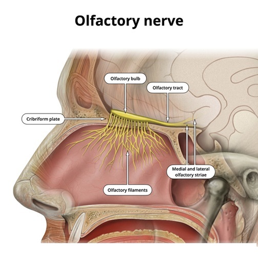
Image: The olfactory nerve (CN I) — consisting of numerous nerve fibers originating in the roof of the nasal cavity, entering the skull through the cribriform plate, and terminating via relay pathways in the evolutionary oldest part of the brain.
II – Optic Nerve
- Function: vision
- Anatomy: carries visual information from the retina to the brain
- Clinical note: damage may cause visual loss

Image: The visual pathway — originating from the eye where the optic nerve emerges, entering the brain to form the optic tract fibers, and terminating in the occipital lobe for visual processing.
III – Oculomotor Nerve
- Function: eye movements, eyelid elevation, pupil constriction
- Anatomy: originates in the midbrain
- Clinical note: damage may cause drooping eyelid and dilated pupil
IV – Trochlear Nerve
- Function: controls a muscle that moves the eye downward and inward
- Anatomy: the thinnest cranial nerve
- Clinical note: difficulty looking down (e.g., on stairs)
V – Trigeminal Nerve
- Function:
- facial sensation
- chewing muscles
- Anatomy: three branches (ophthalmic, maxillary, mandibular)
- Clinical note: the primary cause of Trigeminal Neuralgia, a condition characterized by intense facial pain.

Image: The trigeminal nerve (CN V) originating from the brainstem and dividing into its three major branches. Each branch exits the skull through a separate foramen to provide innervation to different regions of the face: the Ophthalmic (V1), Maxillary (V2), and Mandibular (V3) nerves.
VI – Abducens Nerve
- Function: moves the eye outward
- Anatomy: arises from the lower pons
- Clinical note: vulnerable to increased intracranial pressure
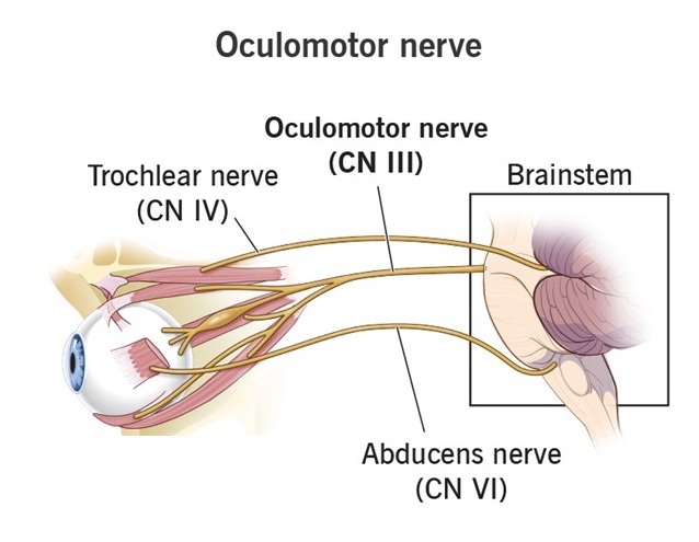
Image: The extraocular (bulbomotor) nerves responsible for eye movement: Oculomotor (III), Trochlear (IV), and Abducens (VI).
VII – Facial Nerve
- Function:
- facial expression
- tear and saliva production
- taste from the front two-thirds of the tongue
- Anatomy: passes through the temporal bone
- Clinical note: damage causes facial weakness or paralysis
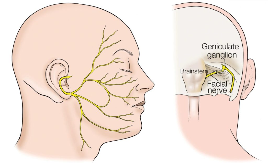
Figure: The image on the right shows the facial nerve (CN VII) originating from the brainstem and entering the facial canal within the petrous part of the temporal bone. The image on the left displays the facial nerve after exiting the canal, branching out to provide innervation to the muscles of facial expression.
VIII – Vestibulocochlear Nerve
- Function: hearing and balance
- Anatomy: connects the inner ear to the brain
- Clinical note: dizziness, hearing loss, tinnitus. Example is vestibular schwannoma (acoustic neuroma)
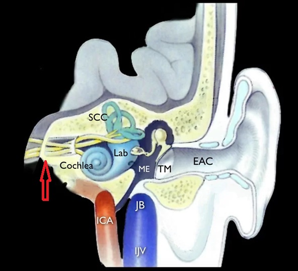
Image: The red arrow indicates the vestibulocochlear nerves as they enter the internal auditory meatus (inner ear).
IX – Glossopharyngeal Nerve
- Function:
- swallowing
- taste from the back of the tongue
- throat reflexes
- Anatomy: arises from the medulla oblongata
X – Vagus Nerve (Wandering nerve)
- Function:
- voice and swallowing
- regulation of heart, lungs, and digestive system
- Anatomy: the longest cranial nerve
- Clinical note: key nerve of the autonomic nervous system
XI – Accessory Nerve
- Function: head and shoulder movement
- Anatomy: supplies the sternocleidomastoid and trapezius muscles
- Clinical note: weakness turning the head or lifting the shoulder
XII – Hypoglossal Nerve
- Function: tongue movements
- Anatomy: arises from the medulla
- Clinical note: tongue deviation when damaged
Image: Anatomy of the last four cranial nerves after exiting the endocranium: Glossopharyngeal (IX), Vagus (X), Accessory (XI), and Hypoglossal (XII). Because of their close anatomical relationship, these nerves are often illustrated together.
Why Are Cranial Nerves Clinically Important?
Cranial nerve abnormalities may indicate:
- brainstem lesions
- tumors
- inflammation or infection
- vascular disorders
- neuralgias and neuropathic pain
For this reason, cranial nerve examination is a core part of every neurological assessment.