Dr Željko Kojadinović — NEUROHIRURGIJA I LEČENJE BOLA
Dr Zeljko Kojadinovic — Pain Treatment & Neurosurgery


Author:
Dr. Zeljko Kojadinovic, MD, PhD
— Neurosurgeon
Specialized Experience:
30 years of clinical expertise in neurosurgery.
Last medically reviewed:
July 16, 2026
WHO THIS PAGE IS FOR
This page is for patients who continue to have neck, shoulder, arm, interscapular, or hand pain after cervical discectomy, ACDF, cervical disc replacement, or another cervical spine operation. It also explains possible causes of persistent numbness, tingling, weakness, or myelopathic symptoms after surgery.
You will learn why symptoms may never improve, return after an initial recovery, or appear in a new form, and how doctors distinguish residual compression, incomplete nerve recovery, fusion or implant problems, surgical complications, and pain generators outside the cervical spine.
Need help identifying the most likely pain generator and planning the next diagnostic or treatment step? Book a brief telehealth pain consultation.
If revision cervical spine surgery has been suggested, we can review your symptoms, MRI, CT, dynamic X-rays, operative report, and other available findings to clarify whether another operation is likely to help through an online neurosurgical second opinion.
The specialist who can identify the true pain generator does not necessarily need to be located in your country.
When patients usually seek a second opinion for persistent pain after cervical discectomy or ACDF
- Neck, shoulder, arm, interscapular, or hand pain never improved after surgery
- Pain improved initially but later returned or developed in a different location
- MRI or CT findings are unclear, incomplete, or do not match the symptoms
- Numbness, tingling, weakness, hand clumsiness, or gait problems persist or worsen after surgery
- Pseudarthrosis, cage subsidence, implant problems, adjacent segment disease, or recurrent compression has been suggested
- Revision surgery, injections, or long-term medication has been recommended without a clear explanation of the pain generator
- The pain has remained unresolved despite examinations and treatment in your country, and the appropriate specialist is not available locally
If this reflects your situation, a focused telehealth review can help identify the most likely cause of the remaining symptoms, such as residual or recurrent compression, incomplete nerve recovery, a fusion or implant-related problem, facet or myofascial pain, shoulder or peripheral nerve disease, or neuropathic sensitization. It can also clarify which diagnostic and treatment options are reasonable and whether another cervical spine operation is likely to help: Request Consultation
Persistent Pain After Cervical Discectomy or ACDF (FNSS) — Quick Summary (Read This First)
- Persistent pain after cervical discectomy or ACDF is not one diagnosis — the true cause must be identified. Failed Neck Surgery Syndrome (FNSS) is a broad term for pain or neurological symptoms that persist, return, or newly develop after cervical spine surgery. It does not explain whether the problem comes from residual compression, incomplete nerve recovery, failed fusion, an implant-related problem, another pain generator, or several overlapping causes.
- The most useful starting point is the pattern of symptoms after surgery. Patients usually describe one of four patterns: (1) the original pain never improved, (2) pain improved and later returned, (3) a new type or location of pain appeared, or (4) numbness, tingling, weakness, hand clumsiness, gait problems, or other neurological symptoms persisted or developed. Each pattern points toward a different group of possible causes.
- If the original arm pain never improved, residual compression or an unrecognized pain generator must be considered. Possible causes include incomplete nerve-root decompression, residual foraminal stenosis (persistent narrowing where the nerve exits), an untreated osteophyte, surgery at the wrong symptomatic level, a second active level, persistent radiculitis, or pre-existing nerve damage that has not recovered. Shoulder disease, peripheral nerve entrapment, brachial plexus disorders, facet pain, and myofascial pain may also have been present before surgery.
- If pain improved and later returned, the cause may be recurrent or newly developed pathology. Important possibilities include progressive foraminal stenosis, adjacent segment disease, pseudarthrosis (failure of the operated level to fuse), cage subsidence, implant loosening or migration, a new disc–osteophyte complex, or degeneration at another cervical level. New facet, muscular, shoulder, or peripheral nerve pain may also become clinically important after an initially successful operation.
- A new pain pattern after surgery does not automatically mean recurrent disc compression. New axial neck pain, interscapular pain, shoulder pain, scar sensitivity, or burning neuropathic pain may result from altered biomechanics (changed movement and load distribution in the cervical spine), muscle injury related to the surgical approach, facet-joint overload, scar-related nerve irritation, central sensitization, complex regional pain syndrome, or another local complication.
- Persistent numbness or weakness may remain even after technically adequate decompression. A chronically compressed nerve root or spinal cord cannot always recover immediately or completely. Arm pain often improves before numbness, weakness, muscle wasting, hand dysfunction, gait imbalance, or spasticity. Myelomalacia or long-standing neural damage may limit recovery even when postoperative imaging shows adequate decompression.
- MRI, CT, dynamic X-rays, and EMG answer different questions. MRI is most useful for the spinal cord, nerve roots, discs, soft tissues, infection, and residual or recurrent compression. CT better shows fusion, osteophytes, foraminal bone narrowing, cage position, hardware, and heterotopic ossification. Dynamic flexion–extension X-rays assess motion, pseudarthrosis, instability, alignment, and artificial-disc movement. EMG and nerve-conduction studies help distinguish active or chronic radiculopathy from plexus or peripheral nerve disorders.
- A postoperative abnormality on imaging is not automatically the cause of pain. Pseudarthrosis, adjacent-level degeneration, scar tissue, loss of lordosis, minor cage subsidence, or facet degeneration may be asymptomatic. A finding becomes clinically important only when it matches the timing, location, neurological pattern, examination, and other diagnostic evidence.
- Pain with a normal-looking MRI can still have a real and treatable cause. Bony foraminal narrowing may be better seen on CT, while EMG may reveal nerve-root or peripheral nerve injury. Facet-joint pain, myofascial pain, shoulder disease, occipital neuralgia, scar neuroma, peripheral nerve entrapment, and central sensitization are usually not visible on routine cervical imaging, including MRI.
- Treatment must match the confirmed pain generator. Revision surgery may help when a clear residual or recurrent compressive lesion, symptomatic pseudarthrosis, implant failure, instability, or adjacent segment disease matches the symptoms. Medication and time may be more appropriate for radiculitis or incomplete nerve recovery. Facet, muscular, shoulder, peripheral nerve, scar-related, and neuropathic pain require targeted non-surgical treatment.
- Another cervical operation is unlikely to help when no surgically correctable lesion has been identified. Surgery is less likely to improve diffuse axial pain, permanent nerve or spinal-cord damage, central sensitization, myofascial pain, shoulder disease, or peripheral nerve entrapment. Repeating surgery without a clear anatomical target may add scar tissue, biomechanical changes, and further risk without treating the actual cause.
- Contributing factors can maintain pain even when they are not the primary cause. Poor posture, prolonged screen use, deconditioning, weak cervical stabilizers, disturbed sleep, obesity, diabetes, thyroid disease, vitamin deficiencies, prolonged opioid use, fear of movement, psychological stress, and central sensitization may increase pain and slow recovery. Successful treatment often requires addressing both the main pain generator and the factors that continue to aggravate it.
- How to use this page: Start with the section that matches your postoperative pattern: “pain never improved,” “pain improved and returned,” “new pain appeared,” or “neurological symptoms persisted or developed.” Then use the Contents box to review the most likely causes, the appropriate diagnostic tests, and the treatment options. If revision surgery has been suggested, focus particularly on Residual Compression, Fusion and Implant Problems, Diagnostic Imaging, When Revision Surgery Is Appropriate, and When Another Operation Is Unlikely to Help.
Contents
- Who This Page Is For
- Quick Summary: Read First
- Persistent Pain After Cervical Surgery
- Pain Never Improved
- Pain Improved, Then Returned
- New Pain After Surgery
- Persistent Neurological Symptoms
- Residual or Recurrent Compression
- Incomplete Nerve-Root Decompression
- Wrong or Missed Level
- Symptoms Without Residual Compression
- Persistent Cervical Radiculitis
- Preoperative Nerve-Root Damage
- Persistent Cervical Myelopathy
- Surgical Neural Injury
- Fusion and Implant Problems
- Pseudarthrosis or Failed Fusion
- Cage Subsidence
- Cage, Plate, or Screw Problems
- Instability or Alignment Loss
- Adjacent Segment Disease
- Cervical Disc Prosthesis Problems
- Heterotopic Ossification
- Prosthesis Malposition or Failure
- Facet Overload After Replacement
- Scar and Neuropathic Pain
- Epidural or Perineural Fibrosis
- Nerve Tethering or Neuroma
- Central Sensitization
- Complex Regional Pain Syndrome
- Other Pain Generators
- Cervical Facet-Joint Pain
- Myofascial Neck and Shoulder Pain
- Shoulder Disease
- Peripheral Nerve Entrapment
- Brachial Plexus and Thoracic Outlet
- Occipital Neuralgia
- Pain From Surgical Approach
- Anterior Scar and Tissue Pain
- Posterior Muscle Injury
- Complications That Must Be Excluded
- Factors Maintaining Postoperative Pain
- Diagnostic Evaluation
- Cervical MRI
- Dynamic Cervical X-Rays
- Targeted Diagnostic Blocks
- When MRI Looks Normal
- Treatment of Persistent Pain
- Residual Compression Treatment
- Persistent Radiculitis Treatment
- Pseudarthrosis Treatment
- Cage and Hardware Treatment
- Scar-Related Pain Treatment
- When Revision Surgery Helps
- When Another Operation Will Not Help
- Prognosis and Recovery
- Revision Surgery Prognosis
- Less Favorable Outcome Factors
- Why Pain Treatment May Fail
- Why Specialists May Disagree
- Start Telehealth Consultation
- Frequently Asked Questions
Persistent Pain After Cervical Discectomy, ACDF, or Other Cervical Spine Surgery
Cervical spine surgery is usually performed to decompress a cervical nerve root and/or the spinal cord. Depending on the underlying condition, the operation may include anterior cervical discectomy and fusion (ACDF), cervical disc replacement, posterior cervical foraminotomy, laminectomy, laminoplasty, or corpectomy.
When the indication is correct and the compressed neurological structure is adequately decompressed, radiating arm pain often improves rapidly. However, neck, shoulder, arm, interscapular, or hand symptoms may sometimes persist, return after an initial improvement, or appear in a new form after surgery.
Persistent symptoms after cervical spine surgery do not represent one diagnosis. The pain may be caused by:
- a residual or recurrent structural problem;
- incomplete recovery of a previously compressed nerve root or spinal cord;
- a fusion or implant-related problem;
- a postoperative complication;
- another pain generator that was present before surgery but was not recognized;
- a new condition unrelated to the operation;
- a combination of several mechanisms.
Persistent or recurrent pain after cervical spine surgery is sometimes described as Failed Neck Surgery Syndrome (FNSS), the cervical counterpart of Failed Back Surgery Syndrome (FBSS). However, FNSS is only an umbrella term, not a specific diagnosis. It does not explain whether symptoms arise from residual compression, nerve injury, pseudarthrosis, an implant-related problem, another pain generator, or several overlapping causes.
The most useful first step is to determine how the symptoms changed after surgery. Persistent symptoms after cervical spine surgery generally follow four main clinical patterns, each associated with a characteristic group of possible causes:
- the original pain never improved after surgery;
- the pain improved initially but later returned;
- a new type or location of pain appeared after surgery;
- neurologic deficits like numbness, tingling, weakness, or myelopathic symptoms persisted or developed after surgery.
These patterns may overlap, but each directs the evaluation toward a different group of possible causes.
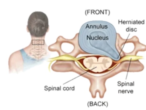
Image: A cervical vertebra showing an intervertebral disc with a herniation. The nucleus pulposus (the soft inner part) has penetrated the annulus fibrosus (the tough outer ring) and is compressing the nerve root. Following surgical treatment of this disc, symptoms may persist because the nerve has not recovered, inflammation has developed at the surgical site, the disc was not completely removed, there is additional bony stenosis that was not addressed, or there is an additional cause of pain outside of the findings shown in this image.
Pain That Never Improved After Cervical Spine Surgery — Causes
When the original pain remains almost unchanged immediately after surgery, possible explanations include:
- incomplete decompression of the nerve root or spinal cord;
- residual foraminal stenosis (persistent narrowing of the nerve-exit opening caused by bony overgrowth from the vertebrae) or an unremoved osteophyte (bone spur);
- an additional symptomatic level that was not treated;
- surgery at a level that did not correspond to the true symptomatic lesion;
- irreversible or slowly recovering nerve damage;
- an incorrect original diagnosis, such as shoulder disease, peripheral nerve entrapment, brachial plexus pathology, facet pain, or myofascial pain.
Persistent symptoms do not automatically prove that the procedure was technically unsuccessful. A chronically compressed nerve may remain painful or dysfunctional even after adequate decompression, while numbness and weakness usually recover more slowly than arm pain.
Pain That Improved and Later Returned — Causes
A pain-free or substantially improved interval followed by recurrent symptoms suggests a different group of causes. These include:
- progressive or recurrent foraminal stenosis;
- adjacent segment disease;
- pseudarthrosis after fusion;
- cage subsidence with loss of foraminal height;
- progression of degeneration at another cervical level;
- a new disc herniation or osteophyte complex;
- new facet-joint, muscular, shoulder, or peripheral nerve pain;
- delayed implant-related or infectious complications.
The longer the symptom-free interval, the more important it becomes to investigate new or progressive pathology, rather than automatically attributing the symptoms to the original operated level.
A New Type of Pain After Cervical Spine Surgery — Causes
Some patients develop a different pain pattern after surgery. For example, preoperative arm pain may improve, but new axial neck pain, interscapular pain, shoulder pain, scar sensitivity, or burning neuropathic pain may appear.
A new pattern may reflect:
- altered cervical biomechanics (changed movement and load distribution in the cervical spine);
- muscular and fascial injury related to the operative approach;
- facet-joint overload;
- shoulder pathology that becomes more apparent after radicular pain improves;
- irritation of small sensory nerves in the surgical scar;
- C5 palsy or another new neurological deficit;
- complex regional pain syndrome;
- central sensitization;
- a local surgical complication.
The distinction between persistent original pain, recurrent pain after improvement, and genuinely new pain is central to the diagnostic process.
Persistent Numbness, Tingling, Weakness, or Myelopathic Symptoms — Causes
Pain is only one possible postoperative symptom. Some patients continue to experience:
- numbness or tingling in the arm, hand, or fingers;
- burning or electric sensations;
- weakness of the shoulder, arm, wrist, or hand;
- loss of hand dexterity;
- gait imbalance;
- leg stiffness or spasticity;
- urinary urgency or other symptoms of cervical myelopathy.
These symptoms may result from residual compression, a surgical complication, or direct injury to the nerve root or spinal cord during surgery. They may also reflect pre-existing neural damage that cannot recover immediately or completely after decompression. The duration and severity of compression before surgery strongly influence neurological recovery.
The possible causes of persistent, recurrent, or newly developed pain and neurological symptoms after cervical spine surgery have been briefly outlined in the clinical patterns above. The following sections examine these causes in greater detail, including how they typically present and why symptoms may remain despite surgery.
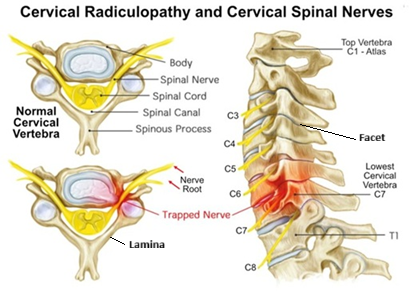
Image: Anatomy of the cervical spine showing seven vertebrae (C1–C7), eight nerve roots, intervertebral discs that act as flexible shock absorbers, and posterior articulations between vertebrae called facets. Also presented is a disc herniation at the C6–C7 level compressing the C7 cervical nerve root. For more details on spinal anatomy, please visit this page.
Residual or Recurrent Compression After Cervical Spine Surgery
Residual Disc Material or Incomplete Nerve-Root Decompression
Persistent radicular pain after cervical discectomy may result from incomplete decompression of the affected nerve root. The disc material may have been removed, but a lateral osteophyte, uncovertebral hypertrophy, foraminal narrowing, or another compressive component may remain. Foraminal stenosis is particularly important after ACDF. Although the disc space may have been opened and the main disc–osteophyte complex removed, a root can remain compressed by lateral bony narrowing.
Typical features include:
- the same arm-pain distribution as before surgery;
- persistent numbness or weakness in the same dermatome or myotome;
- symptoms that never fully improved;
- pain aggravated by cervical extension, rotation, or foraminal compression;
- imaging evidence of persistent narrowing at the clinically corresponding level.
In some cases, standard postoperative MRI does not show the lateral bony anatomy clearly enough. CT may better demonstrate uncovertebral osteophytes and residual foraminal stenosis, while MRI is more useful for assessing the spinal cord, nerve roots, discs, and soft tissues.
Wrong-Level or Missed-Level Cervical Spine Surgery
Operating at a level that does not correspond to the symptomatic lesion can result in unchanged postoperative symptoms. In cervical spondylosis, several levels often appear abnormal on MRI, but only one or two may be clinically active.
A related problem is one-level bias: the most obvious radiological lesion is treated while another symptomatic level is overlooked.
Clinical suspicion is greater when:
- symptoms remain identical after surgery;
- the operated level does not match the pain distribution or neurological deficit;
- multilevel disease was present before surgery;
- another level better corresponds to the affected dermatome, muscle group, or reflex;
- postoperative imaging shows adequate treatment of the operated level without explaining the persistent symptoms.
The diagnosis should not be based only on the postoperative MRI report. The original clinical findings, preoperative images, operative report, and current neurological pattern must be reviewed together.
Progressive or Recurrent Foraminal and Spinal Canal Stenosis After Cervical Spine Surgery
Symptoms can return after an initially successful operation because degenerative disease continues to progress.
Possible mechanisms include:
- new osteophyte formation, the development of new bony overgrowths that may narrow the space around a nerve root or the spinal cord;
- recurrent or progressive foraminal stenosis, narrowing that returns or gradually worsens in the opening through which a cervical nerve exits;
- ligamentous hypertrophy, thickening of spinal ligaments that may contribute to renewed spinal-canal narrowing;
- subsidence of an interbody cage, sinking of the implant into the adjacent vertebral bone, with possible loss of disc or foraminal height;
- loss of segmental alignment, loss of the normal position or curvature of the operated spinal segment;
- adjacent-level degeneration, progressive deterioration of the disc or joints above or below the operated level;
- heterotopic ossification after disc arthroplasty, abnormal bone formation around an artificial disc that may restrict movement or contribute to renewed narrowing.
Recurrent radiculopathy may again cause arm pain, tingling, numbness, weakness, or reflex changes. Recurrent central canal narrowing may produce hand clumsiness, gait disturbance, hyperreflexia, leg stiffness, or other signs of cervical myelopathy.
Persistent Pain and Neurological Symptoms After Cervical Disc or Spondylosis Surgery Without Residual Compression
Persistent Cervical Radiculitis After Adequate Surgical Decompression
A nerve root may remain painful after being adequately decompressed because of postoperative inflammation, edema, chemical irritation, or pre-existing sensitization.
This commonly produces:
- burning or electric arm pain;
- hypersensitivity or dysesthesia;
- pain without a clear new compressive lesion;
- gradual improvement over weeks or months;
- symptoms that are more pronounced than objective weakness or structural findings.
Persistent radiculitis must be separated from residual mechanical compression because treatment differs. Inflammatory or sensitized root pain may improve with medication, selective injections, and time, whereas a clear compressive lesion may require surgical reassessment.
Persistent Cervical Radiculopathy From Preoperative Nerve-Root Damage
When a cervical nerve root has been compressed for a prolonged period, decompression removes the ongoing mechanical pressure but cannot immediately reverse structural nerve injury.
Persistent findings may include:
- numbness;
- altered sensation;
- weakness;
- muscle wasting;
- neuropathic pain;
- reduced reflexes.
Pain often improves before sensory loss and weakness. Neurological recovery may continue for 6–12 months or longer, but severe or long-standing deficits may remain incomplete. Electromyography and nerve-conduction studies can help distinguish chronic radiculopathy from active denervation, brachial plexopathy, or peripheral nerve entrapment.
Persistent Cervical Myelopathy After Adequate Spinal-Cord Surgical Decompression
Cervical myelopathy results from spinal-cord compression. Surgery is often performed to prevent further neurological deterioration, and complete recovery cannot always be expected.
Persistent symptoms may include:
- hand clumsiness;
- difficulty fastening buttons or handling small objects;
- impaired handwriting;
- gait imbalance;
- leg stiffness or weakness;
- hyperreflexia;
- sensory disturbance in the hands or legs;
- urinary urgency or other sphincter symptoms.
The prognosis depends on the duration and severity of preoperative compression, the patient’s age, and the extent of established spinal-cord damage. Myelomalacia—an MRI sign of structural spinal-cord injury caused by chronic or severe compression—is associated with a greater likelihood of persistent spasticity, hand dysfunction, gait disturbance, weakness, and sensory deficits.
A technically adequate decompression may successfully prevent further deterioration while producing only partial improvement in previously established spinal-cord deficits. Persistent symptoms in this situation do not necessarily mean that the operation failed; the compression may have been removed, while the pre-existing spinal-cord injury remains only partly reversible.
Nerve-Root or Spinal-Cord Injury During Cervical Spine Surgery
New neurological symptoms may occasionally result from injury to a cervical nerve root or the spinal cord during surgery. The clinical presentation depends on the affected neural structure and may include new dermatomal pain or sensory loss, weakness in the muscles supplied by a specific cervical root, reduced reflexes, or new signs of cervical myelopathy.
C5 palsy is the most frequently recognized root-specific postoperative deficit and is reported more often after posterior cervical decompression, although it can also occur after anterior surgery. It usually causes new deltoid and sometimes biceps weakness. However, other cervical roots may also be affected, producing the corresponding patterns of pain, sensory loss, weakness, and reflex changes described in the section on symptoms of cervical disc herniation, radiculopathy, and myelopathy.
Any new or worsening neurological deficit after surgery requires prompt clinical assessment and imaging to exclude residual compression, postoperative hematoma, implant displacement, or another surgically correctable complication.
Fusion and Implant-Related Causes After ACDF
Pseudarthrosis or Failure of Fusion
During ACDF, the damaged cervical disc is removed to decompress the nerve root or spinal cord. A cage or interbody spacer is then placed into the empty disc space to restore disc height and maintain the opening through which the nerve exits. In many procedures, a plate and screws are also placed on the front of the cervical vertebrae to provide additional stability while bone fusion develops.
The goal is for new bone to grow through and around the cage, creating a solid fusion between the operated vertebrae. Pseudarthrosis means that this bone fusion has not become mechanically solid, so movement may persist at the operated level.
Possible symptoms include:
- persistent or recurrent axial neck pain;
- pain during neck movement;
- a sensation of mechanical instability;
- recurrent arm pain;
- symptoms appearing months after surgery.
Pseudarthrosis is assessed using dynamic flexion–extension X-rays and CT. However, not every radiological nonunion causes pain. Revision surgery should be considered only when the failure of fusion is clinically significant and corresponds to the patient’s symptoms.
Cage Subsidence and Loss of Foraminal Height
Cage subsidence occurs when an interbody cage sinks into the adjacent vertebral endplates.
This may lead to:
- loss of disc-space and foraminal height;
- recurrent nerve-root compression;
- segmental kyphosis;
- local mechanical pain;
- altered load on the adjacent segments;
- recurrence of symptoms after an initially successful operation.
Minor subsidence may remain asymptomatic. Its clinical importance depends on the degree of collapse, alignment, foraminal narrowing, neurological findings, and symptom progression.
Cage Migration, Plate Prominence, or Screw Loosening
Implant-related abnormalities may include:
- cage migration;
- plate displacement or prominence;
- screw loosening;
- screw breakage;
- loss of fixation;
- irritation of the esophagus or surrounding soft tissues;
- recurrent neural compression.
Clinical manifestations depend on the location and direction of the change. Patients may experience neck pain, recurrent radiculopathy, dysphagia, a foreign-body sensation, or progressive neurological symptoms. Imaging must show not only that an implant abnormality exists, but also that it plausibly explains the clinical problem.
Segmental Instability, Loss of Lordosis, or Postoperative Kyphosis
Postoperative changes in cervical alignment may alter the mechanical load across the cervical muscles, discs, and facet joints.
Potential symptoms include:
- axial neck pain;
- interscapular pain;
- muscular fatigue;
- pain aggravated by sustained posture;
- progressive stiffness;
- recurrent neural compression in more severe cases.
A reversal or loss of cervical lordosis on imaging is common and is not automatically a pain generator. Its relevance must be judged in combination with dynamic movement, segmental instability, deformity progression, and clinical findings.
Adjacent Segment Degeneration and Adjacent Segment Disease in FNSS
Adjacent segment degeneration refers to radiological changes above or below a previously operated level. Adjacent segment disease means that these changes have become clinically symptomatic.
Possible manifestations include:
- new neck or arm pain after a prolonged period of improvement;
- a new dermatomal pain pattern;
- progressive hand numbness or weakness;
- renewed signs of cervical myelopathy;
- stenosis or instability at a neighboring level.
Radiological degeneration alone does not prove that the adjacent segment is responsible for pain. The findings must match the new neurological and pain pattern before another operation is considered.
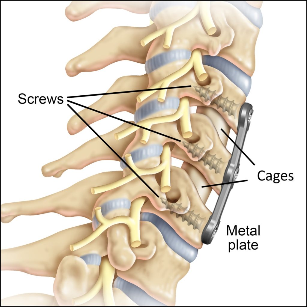
Image: ACDF complications explained – Instead of the removed discs, cages were inserted, and subsequently, a metal plate was screwed in from the front with screws to increase the stability of both segments. Sometimes, backward displacement of these cages can occur, which may lead to compression of the spinal cord or nerve roots. Instability can also occur despite the plate.
Prosthesis-Related Causes of Pain After Cervical Disc Replacement
Unlike a cage used in ACDF, which supports fusion and eliminates movement at the operated level, an artificial cervical disc is designed to preserve motion between the vertebrae.
Heterotopic Ossification and Loss of Prosthesis Motion
After cervical disc arthroplasty, bone may form around the artificial disc. This is called heterotopic ossification.
It can result in:
- reduced prosthesis movement;
- functional fusion of the operated level;
- recurrent foraminal or central narrowing;
- stiffness;
- increased load on the facet joints or adjacent levels.
Heterotopic ossification is often visible on X-ray or CT and may remain asymptomatic. Treatment is considered only when it produces a corresponding structural and clinical problem.
Prosthesis Malposition, Migration, or Mechanical Failure
An artificial cervical disc may become:
- incorrectly positioned;
- displaced;
- tilted;
- mechanically worn;
- unstable;
- associated with progressive endplate change.
Symptoms may include local neck pain, recurrent radiculopathy, restricted motion, or neurological deterioration. CT, dynamic X-rays, and MRI are used together to determine whether the device is stable and whether neural structures remain adequately decompressed.

Image: Cervical spine X-ray showing that the disc prosthesis allows some physiological movement between the vertebrae, which is the main purpose of cervical disc replacement.
Facet-Joint Overload After Cervical Disc Replacement
After cervical discectomy, altered movement at the operated level may increase stress on the posterior facet joints.
Facet-mediated pain is usually:
- axial rather than dermatomal;
- worse with extension and rotation;
- referred to the occipital area, shoulder, or scapula;
- associated with local tenderness;
- not necessarily accompanied by numbness or weakness.
Diagnostic medial-branch blocks may help determine whether the facet joints are the dominant pain generator. For more details on spinal anatomy, please visit this page.
Postoperative Scar, Neuropathic, and Pain-Modulation Disorders
Epidural or Perineural Fibrosis
Postoperative scar tissue may form around the dura or nerve root. It is usually of limited clinical importance after an anterior cervical procedure such as ACDF because the surgical corridor causes relatively little direct disruption of the posterior epidural and perineural tissues. Scar-related adhesions may be more relevant after posterior cervical decompression, repeated surgery, or procedures involving direct exposure of the dura or nerve root. For this reason, contrast-enhanced cervical MRI is not routinely required after uncomplicated anterior cervical surgery solely to assess postoperative scar tissue.
Scar tissue is common after surgery and is often an incidental finding. It may contribute to symptoms when it:
- adheres to or tethers a nerve root;
- combines with limited residual foraminal space;
- surrounds a recurrent or residual compressive lesion;
- develops after repeated or posterior cervical operations.
Scar removal alone has uncertain benefit and may expose the nerve root and dura to additional injury. Therefore, scar-related pain should not be diagnosed solely because postoperative fibrosis appears on imaging.
Nerve-Root Tethering or Traumatic Neuroma
Rarely, a nerve may be injured directly during surgery or become incorporated into postoperative scar tissue.
This can cause:
- focal burning pain;
- electric-shock sensations;
- allodynia;
- pain triggered by movement, pressure, or stretching;
- persistent neuropathic symptoms without major compression on MRI.
A traumatic neuroma or focal scar-related nerve injury is generally considered only after more common structural and extraspinal causes have been excluded.
Central Sensitization and Altered Pain Modulation
After prolonged pain, the central nervous system can become more responsive to sensory input. This may amplify pain even after the original structural problem has improved.
Typical features include:
- pain disproportionate to current structural findings;
- sensitivity to light touch or pressure;
- pain spreading beyond the original distribution;
- multiple failed procedures or treatments;
- sleep disturbance;
- reduced activity and increasing pain-related disability.
Central sensitization is a real biological pain mechanism, but it must not be used as a substitute for a proper search for residual compression, instability, joint pain, nerve injury, shoulder disease, or other anatomical generators.
Complex Regional Pain Syndrome
Complex regional pain syndrome is rare after cervical spine surgery but should be considered when severe limb pain is accompanied by autonomic and trophic changes.
Possible findings include:
- disproportionate burning pain;
- marked allodynia;
- swelling;
- skin-color or temperature changes;
- altered sweating;
- stiffness and reduced limb use.
Early recognition is important because delayed treatment can lead to progressive functional limitation.
Pain Generators Not Corrected by Cervical Spine Surgery
For more information on the causes of neck pain, please visit this page.
Cervical Facet-Joint Pain in FNSS
Facet joints are common sources of axial neck pain. Their pain may have existed before surgery but been overshadowed by severe radiculopathy, or it may become more prominent after fusion or altered cervical biomechanics.
Typical features include:
- pain in the neck rather than a clear dermatomal arm distribution;
- pain with extension and rotation;
- tenderness over the facet region;
- referred pain toward the occiput, shoulder, scapula, or upper arm;
- absence of a corresponding motor or sensory deficit.
Imaging may show facet arthropathy, but radiological changes alone do not establish the diagnosis. Targeted medial-branch or facet-joint blocks may be required.
Myofascial Neck, Shoulder, and Interscapular Pain in FNSS
The cervical and shoulder region contains numerous muscles and fascial structures that may produce persistent pain, including:
- trapezius;
- levator scapulae;
- rhomboid muscles;
- scalene muscles;
- suboccipital muscles;
- deep cervical extensors;
- cervical and thoracic paraspinal muscles.
Myofascial pain is commonly:
- localized or referred rather than dermatomal;
- reproduced by pressure over a specific point;
- aggravated by prolonged posture, computer work, or static neck loading;
- associated with muscle fatigue, tightness, or spasm;
- poorly explained by MRI.
After a posterior operation, muscle detachment and postoperative fibrosis may be important. After an anterior procedure, deconditioning, protective spasm, altered posture, and persistent pre-existing muscular pain may dominate.
Shoulder Disease Mistaken for Cervical Radiculopathy in FNSS
Shoulder pathology can closely mimic cervical nerve-root pain. Conditions include:
- rotator-cuff tendinopathy or tear;
- shoulder impingement;
- adhesive capsulitis;
- acromioclavicular joint disease;
- biceps-tendon pathology.
Shoulder pain is more likely when symptoms are provoked by shoulder movement, abduction, rotation, or lying on the affected side. Cervical radiculopathy is more likely when symptoms follow a dermatomal distribution and are accompanied by corresponding sensory loss, weakness, or reflex change.
Both conditions can coexist. In some patients, cervical surgery successfully relieves the radicular component, while the previously unrecognized shoulder pain remains.
Peripheral Nerve Entrapment and Double-Crush Syndrome in FNSS
Persistent hand numbness or arm symptoms after cervical surgery may arise from:
- carpal tunnel syndrome;
- ulnar neuropathy at the elbow or wrist;
- radial nerve entrapment;
- suprascapular neuropathy;
- other peripheral nerve lesions.
A peripheral nerve disorder does not follow the same anatomical pattern as a single cervical root. Local provocative tests and nerve-conduction studies can help distinguish these conditions.
In double-crush syndrome, a cervical root lesion and a peripheral nerve entrapment coexist. Treating the cervical lesion may improve only part of the symptoms because the distal nerve compression remains active.
Brachial Plexus and Thoracic Outlet Disorders
Brachial plexopathy, neuralgic amyotrophy, and thoracic outlet syndrome can cause neck, shoulder, arm, or hand symptoms that resemble radiculopathy.
Features suggesting an extraspinal source include:
- symptoms involving several nerve territories rather than one cervical root;
- pain or paresthesia provoked by arm position;
- marked shoulder-girdle weakness;
- a mismatch between cervical imaging and clinical findings;
- EMG abnormalities localizing outside the cervical root.
Brachial neuritis may cause severe shoulder pain followed by weakness and muscle wasting. It should not be mistaken for persistent compression requiring revision cervical surgery.
Occipital Neuralgia and Upper Cervical Pain
Pain after cervical surgery may arise from upper cervical joints, muscles, or occipital nerves.
Typical features include:
- pain beginning in the upper neck and radiating toward the occiput;
- stabbing, shooting, or burning pain;
- tenderness along the greater or lesser occipital nerve;
- headache provoked by neck position or pressure;
- coexistence with upper cervical facet or muscular pain.
This is particularly relevant after posterior cervical procedures, although it may also occur independently of surgery.
Pain Related to the Cervical Spine Surgical Approach and Local Tissues
Anterior Neck Scar and Soft-Tissue Pain after Cervical Spine Surgery
After an anterior approach, patients may experience localized pain or tightness related to the incision and deeper tissues.
Possible mechanisms include:
- scar sensitivity;
- irritation of small cutaneous nerves;
- platysma or fascial adhesions;
- persistent soft-tissue inflammation;
- tension during swallowing or neck movement.
This pain is usually localized and reproducible by pressure or specific movement. It should be distinguished from dysphagia, implant prominence, infection, or esophageal injury.
Posterior Cervical Muscle Injury and Chronic Muscle Spasm after Cervical Spine Surgery
Posterior cervical operations require varying degrees of muscle dissection and detachment.
Persistent symptoms may include:
- axial neck pain;
- interscapular pain;
- fatigue while holding the head upright;
- painful muscle spasm;
- reduced cervical range of motion;
- localized tenderness over the operative region.
These symptoms may be maintained by deconditioning, altered alignment, protective muscle guarding, and postoperative fibrosis.
Local Surgical Complications After Cervical Spine Surgery That Must Be Excluded
Some complications primarily cause local neck, swallowing, voice, or wound symptoms rather than chronic radicular pain, but they remain important because delayed recognition may lead to neurological injury, infection, airway compromise, or implant failure.
Important complications include:
- Postoperative hematoma or airway compression — neck swelling, respiratory difficulty, rapidly progressive dysphagia, voice change, or neurological deterioration.
- Superficial or deep infection — wound redness, drainage, fever, increasing pain, elevated CRP or ESR, or delayed implant infection.
- Discitis or osteomyelitis — persistent deep pain, fever or inflammatory markers, and characteristic MRI changes.
- Cerebrospinal-fluid leak or pseudomeningocele — wound fluid collection, positional headache, clear drainage, or neurological symptoms.
- New nerve-root or spinal-cord injury — new weakness, sensory loss, gait deterioration, or bladder dysfunction.
- Persistent dysphagia — commonly transient after anterior surgery, but progressive or long-lasting swallowing difficulty requires evaluation.
- Hoarseness or recurrent laryngeal-nerve injury — persistent voice change, weak voice, coughing during swallowing, or aspiration symptoms.
- Esophageal irritation, perforation, or delayed erosion — severe or progressive swallowing pain, infection, neck swelling, or symptoms related to implant prominence.
- Vascular injury — acute neurological symptoms, severe neck swelling, bleeding, or stroke-related signs.
- Horner syndrome — drooping eyelid, smaller pupil, and reduced facial sweating on one side.
- Implant displacement or failure — progressive local symptoms, dysphagia, deformity, or new neural compression.
These conditions require direct postoperative or urgent in-person evaluation rather than treatment based only on telehealth assessment.
Contributing Factors That Can Maintain Pain after Cervical Spine Surgery
Even after the primary pain generator is identified, other factors may maintain irritation, slow recovery, or amplify symptoms.
These include:
- prolonged computer or smartphone use;
- forward-head posture;
- poor cervical support during sleep;
- weakness of deep cervical stabilizers;
- overactivity of the trapezius and levator scapulae;
- reduced activity and deconditioning;
- obesity and mechanical overload;
- insulin resistance and low-grade systemic inflammation;
- vitamin D, B12, iron, or magnesium deficiency;
- vitamin B6 excess or deficiency;
- diabetes, thyroid disease, or autoimmune disorders;
- poor sleep;
- Prolonged use of certain medications;
- prolonged opioid use and opioid-induced hyperalgesia;
- anxiety or stress-related muscle activation;
- central sensitization.
The risk of pseudarthrosis, implant-related problems, and other complications generally increases with the number of fused levels, while older age, smoking, diabetes, osteoporosis, and impaired general health may further reduce healing and neurological recovery.
These factors rarely explain the entire pain syndrome by themselves. Their role is usually to aggravate a specific structural, neural, joint, muscular, or scar-related generator, and treatment is most effective when both levels are addressed.
Diagnostic Evaluation of Persistent Pain After Cervical Spine Surgery
Clinical History, Timing, and Change in Symptom Pattern
The diagnostic process begins with a detailed comparison of symptoms before and after surgery.
Important questions include:
- Did the original arm or neck pain improve at all?
- Was there a pain-free interval?
- Is the current pain identical to the preoperative pain or different?
- Is pain axial, radicular, interscapular, muscular, or shoulder-related?
- Has numbness, weakness, gait imbalance, or hand clumsiness improved or worsened?
- Which movements, positions, or pressure points reproduce the symptoms?
- Was the operation ACDF, disc arthroplasty, posterior foraminotomy, laminectomy, laminoplasty, or corpectomy?
- Were one or several levels treated?
- Was fusion achieved and were any postoperative complications documented?
The operative report, preoperative examination, preoperative imaging, and current symptoms should be evaluated together. A postoperative MRI alone cannot reconstruct whether the original diagnosis and surgical target were correct.
Neurological Examination of the Nerve Roots and Spinal Cord
The examination should assess both radiculopathy and myelopathy.
Nerve-root evaluation includes:
- dermatomal sensory testing;
- motor strength;
- muscle atrophy;
- biceps, brachioradialis, and triceps reflexes;
- comparison of symptoms with C5, C6, C7, or C8 distribution.
Spinal-cord evaluation includes:
- gait and balance;
- leg tone and spasticity;
- hand dexterity;
- pathological reflexes;
- urinary or bowel symptoms;
- progression of bilateral or multilevel deficits.
The pattern of deficit is often more informative than the degree of degeneration described in the radiology report.
Cervical MRI in FNSS
MRI of the cervical spine without contrast is usually the initial MRI examination for persistent or recurrent symptoms after cervical spine surgery.
MRI evaluates:
- residual or recurrent disc and soft-tissue compression;
- central canal and foraminal narrowing;
- spinal-cord compression;
- myelomalacia;
- adjacent-level pathology;
- postoperative infection;
- epidural or soft-tissue collections;
- scar-related changes;
- new lesions unrelated to the operation.
Contrast is added selectively when infection, tumor, cerebrospinal-fluid leakage, inflammatory change, or clinically relevant postoperative scar tissue—particularly after posterior or repeated surgery—is suspected. It is not routinely required after every cervical operation.
Metallic implants may reduce image quality. In these cases, CT or CT myelography may provide additional information.
Postoperative abnormalities on MRI do not automatically identify the cause of pain. Residual narrowing, scar tissue, adjacent-level degeneration, and spinal-cord signal changes must be interpreted together with the symptom pattern, neurological examination, operative history, and other imaging findings.
CT for Fusion, Osteophytes, Cage Position, and Hardware in FNSS
CT is particularly useful for assessing:
- pseudarthrosis;
- bony fusion;
- uncovertebral osteophytes;
- residual foraminal stenosis;
- cage position;
- cage subsidence;
- plate and screw integrity;
- heterotopic ossification after disc replacement;
- postoperative bone anatomy.
A radiological abnormality must still be correlated with the clinical presentation. Small degrees of subsidence, incomplete fusion, or hardware prominence may be asymptomatic.
CT myelography may be considered when MRI is contraindicated or when extensive implant artifact prevents adequate evaluation of the spinal canal and neural foramina.
Dynamic Flexion–Extension X-Rays in FNSS
Standard cervical X-rays provide a weight-bearing overview of the operated spine and may show implant position, hardware loosening or migration, loss of disc or vertebral height, cervical alignment, and obvious degenerative changes. Dynamic flexion–extension views add information by showing how the operated and adjacent segments behave during movement.
Dynamic cervical X-rays can help assess:
- motion at an intended fusion level;
- possible pseudarthrosis or failure of fusion;
- segmental or instrument-related instability;
- abnormal translation or angulation;
- segmental kyphosis or loss of cervical alignment;
- movement above and below a fusion;
- motion or loss of motion of an artificial cervical disc.
These studies are most useful when symptoms suggest mechanical pain, instability, nonunion, implant dysfunction, or abnormal motion that may not be visible on static MRI or CT images. They may also help show whether an apparently fused segment moves during flexion and extension.
However, flexion–extension X-rays must be interpreted cautiously. Limited neck movement because of pain, fear, or muscle spasm may make the examination less reliable, while small differences in patient positioning or measurement technique can affect the apparent degree of motion. A suspected pseudarthrosis or implant problem should therefore be assessed together with the clinical findings, standard X-rays, and CT when needed.
Electromyography and Nerve-Conduction Studies in FNSS
EMG and nerve-conduction studies may help distinguish:
- active cervical radiculopathy;
- chronic nerve-root injury;
- peripheral nerve entrapment;
- carpal tunnel syndrome;
- ulnar neuropathy;
- brachial plexopathy;
- neuralgic amyotrophy;
- generalized neuropathy.
These tests are particularly useful when symptoms and cervical imaging do not clearly match.
Timing is important when interpreting EMG. Denervation changes may not yet be visible when the examination is performed very early after a new nerve-root injury or postoperative neurological deficit. Changes in distal arm muscles often require approximately two to three weeks or longer to develop. Therefore, a normal early EMG does not reliably exclude a recent cervical radiculopathy, and repeat testing may sometimes be appropriate.
Shoulder, Brachial Plexus, and Peripheral Nerve Evaluation
Persistent arm pain should not automatically be attributed to the operated cervical level.
Evaluation may include:
- shoulder range of motion;
- rotator-cuff and impingement tests;
- local tenderness;
- provocative tests for carpal tunnel and ulnar neuropathy;
- thoracic outlet maneuvers;
- detailed sensory and motor mapping;
- ultrasound or shoulder imaging;
- EMG when indicated.
This is essential before another cervical procedure is considered.
Laboratory Testing
Laboratory tests are indicated when infection, inflammatory disease, metabolic dysfunction, or impaired healing is suspected.
Depending on the clinical context, evaluation may include:
- complete blood count;
- CRP and ESR;
- glucose and HbA1c;
- renal and liver function;
- vitamin D and B12;
- thyroid function;
- calcium, phosphate, magnesium, and parathyroid hormone;
- autoimmune testing when clinically indicated.
Laboratory findings do not identify the pain generator by themselves, but they may reveal factors that impair fusion, nerve recovery, muscle function, or treatment response.
Targeted Diagnostic Blocks in FNSS
When imaging does not clearly identify the dominant source, targeted blocks may confirm a specific clinical hypothesis.
Possible procedures include:
- selective cervical nerve-root block;
- cervical facet medial-branch block;
- occipital nerve block;
- trigger-point or fascial injection;
- peripheral nerve block;
- diagnostic shoulder injection.
In patients with previous cervical surgery, normal anatomical landmarks may be altered by decompression, fusion, implants, or postoperative scar tissue. Current imaging and the previous operative approach should therefore be reviewed before any cervical epidural or nerve-root procedure. Image guidance and contrast confirmation are particularly important when planning the safest level and route.
A block should not be selected randomly. It is useful only after the history, examination, and imaging narrow the differential diagnosis to one or two likely structures.
Diagnostic Strategy When MRI Looks Normal in FNSS
Real pain may persist even when postoperative MRI shows no major compression.
In this situation, evaluation should include:
- direct review of the MRI images, not only the report;
- CT for bony foraminal narrowing, fusion, and hardware;
- dynamic X-rays;
- EMG and nerve-conduction studies;
- examination of the shoulder and peripheral nerves;
- assessment of facet joints and myofascial generators;
- evaluation for neuropathic pain and nerve injury;
- targeted diagnostic blocks;
- investigation of central sensitization and contributing systemic factors.
A normal-looking MRI does not prove that symptoms are non-organic. It means that the diagnostic process must extend beyond major compressive lesions.
Treatment of Persistent Pain After Cervical Discectomy or ACDF
Treatment should be directed at the confirmed or most probable dominant cause, not merely at the fact that the patient has undergone previous surgery.
Treatment of Residual Compression or Incomplete Decompression in FNSS
Revision decompression may be appropriate when:
- a residual disc, osteophyte, or foraminal stenosis is clearly demonstrated;
- the structural finding matches the pain distribution and neurological deficit;
- symptoms remain severe or progressive;
- conservative treatment is unlikely to correct the mechanical problem.
The choice of anterior or posterior revision depends on the location of compression, previous approach, fusion status, alignment, number of levels, and implant position.
Revision surgery should not be recommended solely because postoperative imaging shows degeneration or narrowing. The lesion must explain the current symptoms.
Treatment of Persistent Radiculitis in FNSS
When the nerve root remains irritated without major residual compression, treatment may include:
- a short course of anti-inflammatory medication;
- neuropathic pain medication;
- temporary activity modification;
- gradual rehabilitation;
- selective nerve-root or epidural injection in carefully selected cases;
- monitoring of sensory and motor recovery
- detection and treatment of pain and inflammatory contributing factors.
The aim is to reduce inflammation and neural hypersensitivity while preserving function and avoiding unnecessary revision surgery.
Treatment of Persistent Nerve Damage and Neuropathic Pain in FNSS
Persistent neuropathic symptoms may require:
- gabapentin or pregabalin;
- duloxetine, amitriptyline, or another appropriate neuropathic agent;
- carefully selected medication combinations;
- desensitization;
- functional rehabilitation;
- treatment of sleep disturbance;
- detection and treatment of pain and inflammatory contributing factors;
- management of coexisting peripheral neuropathy or entrapment.
Medication should be adjusted according to efficacy and tolerability rather than abandoned after a single inadequate trial.
Cervical spinal cord stimulation may be considered in carefully selected patients with persistent neuropathic arm or neck pain after cervical spine surgery when no surgically correctable lesion remains and medication, rehabilitation, and targeted interventional treatments have failed. It requires specialist assessment and a successful temporary stimulation trial before permanent implantation. It is not an alternative to decompression when residual nerve-root or spinal-cord compression is present.
Treatment of Persistent Cervical Myelopathy in FNSS
When myelopathic symptoms persist, the first step is to determine whether the spinal cord has been adequately decompressed.
If residual or adjacent compression exists, further surgery may be necessary. If decompression is adequate and symptoms reflect previous cord injury, treatment focuses on:
- neurological rehabilitation;
- gait and balance training;
- hand-function therapy;
- spasticity management;
- prevention of falls;
- treatment of bladder dysfunction;
- realistic functional goals.
The purpose of surgery in advanced myelopathy may have been primarily to prevent further deterioration rather than guarantee full neurological recovery.
Treatment of Pseudarthrosis after ACDF
Asymptomatic or mildly symptomatic nonunion may be observed.
Treatment may include:
- smoking cessation;
- optimization of bone metabolism;
- correction of vitamin D or calcium-related abnormalities;
- osteoporosis management when appropriate;
- activity modification;
- targeted pain treatment.
Revision fusion may be indicated when pseudarthrosis is clearly associated with mechanical pain, instability, implant failure, deformity, or recurrent neural compression.
Treatment of Cage Subsidence and Hardware Problems in ACDF
Minor stable changes may require observation rather than intervention.
Revision may be considered when there is:
- progressive migration;
- significant loss of alignment;
- recurrent foraminal or spinal-cord compression;
- instability;
- severe dysphagia related to implant prominence;
- implant breakage or loosening;
- progressive neurological symptoms.
Treatment decisions must be based on clinical consequences, not merely the radiological appearance of the implant.
Treatment of Adjacent Segment Disease after Cervical Discectomy
Initial treatment may include:
- anti-inflammatory or neuropathic medication;
- targeted rehabilitation;
- ergonomic correction;
- selective nerve-root injections;
- facet diagnostic blocks or radiofrequency treatment where appropriate.
Surgery is reserved for clear symptomatic compression, instability, progressive neurological deficit, or disabling pain that correlates with the adjacent-level pathology.
Treatment of Facet-Joint Pain in FNSS
When facet pain is suspected, management may include:
- activity and posture modification;
- anti-inflammatory treatment;
- targeted rehabilitation;
- medial-branch blocks;
- radiofrequency ablation after a convincing diagnostic response.
Fusion or revision surgery should not be used to treat isolated or presumed facet-joint pain unless clinically significant instability, deformity, or another surgically correctable structural lesion is demonstrated.
Treatment of Myofascial and Interscapular Pain in FNSS
Treatment should target the specific muscle, attachment, or fascial structure involved.
Options include:
- temporary anti-inflammatory treatment;
- short-term muscle relaxation when protective spasm is prominent;
- trigger-point or enthesis injections;
- ultrasound-guided treatment;
- correction of scapular mechanics;
- strengthening of deep cervical stabilizers;
- ergonomic modification;
- correction of sleep position;
- graded return to activity.
Generic neck exercises may fail or worsen symptoms when they do not match the actual pain generator.
Treatment of Shoulder and Peripheral Nerve Disorders in FNSS
When the remaining symptoms originate from the shoulder, brachial plexus, or peripheral nerve, treatment should be directed at that diagnosis rather than followed by another cervical operation.
This may involve:
- shoulder rehabilitation or injection;
- treatment of rotator-cuff or adhesive-capsulitis pathology;
- carpal tunnel or ulnar nerve treatment;
- treatment of thoracic outlet syndrome;
- peripheral nerve decompression in selected cases;
- treatment of brachial neuritis or plexopathy.
Treatment of Scar-Related Pain after Cervical Discectomy
Scar-related pain may be treated with:
- neuropathic medication;
- desensitization;
- targeted local injection;
- ultrasound-guided treatment of a superficial scar neuroma;
- selective nerve blocks;
- rehabilitation.
Surgical scar removal or adhesiolysis is rarely indicated as an isolated procedure. It should be considered only when scar-related tethering clearly matches the symptoms and no more plausible pain generator exists, because reoperation may create additional fibrosis and carries an increased risk of nerve-root or dural injury.
Treatment of Central Sensitization and CRPS in FNSS
Treatment generally requires a multimodal approach:
- reduction of any ongoing peripheral pain generator;
- neuropathic medication;
- graded rehabilitation;
- desensitization;
- sleep treatment;
- restoration of normal limb use;
- psychological support where appropriate;
- sympathetic or peripheral nerve blocks in selected cases.
CRPS should be treated early. Central sensitization should be addressed only after active structural and neurological causes have been properly investigated.
Treatment of Local Surgical Complications after Cervical Discectomy
Treatment depends on the specific complication:
- compressive hematoma may require urgent evacuation;
- infection requires microbiological evaluation, antibiotics, and sometimes surgical debridement;
- CSF leak may require observation, drainage, blood patch, or surgical repair;
- progressive dysphagia or esophageal injury requires direct surgical and otolaryngological evaluation;
- implant displacement may require revision;
- new neurological deterioration requires urgent imaging and neurosurgical assessment.
These conditions should not be managed as ordinary chronic pain.
When Revision Cervical Spine Surgery Is Appropriate
Revision surgery has the best chance of helping when there is:
- one clearly identified structural lesion;
- a neurological or pain pattern that matches that lesion;
- residual or recurrent neural compression;
- symptomatic pseudarthrosis;
- implant migration or failure;
- progressive deformity or instability;
- adjacent segment disease with corresponding deficits;
- a reasonable expectation that correcting the lesion will improve symptoms.
When Another Cervical Operation Is Unlikely to Help
Another operation is less likely to help when:
- imaging does not show a surgically correctable lesion;
- pain does not match the level or side of the radiological finding;
- symptoms are primarily facet, muscular, shoulder, or peripheral-nerve related;
- the main problem is permanent nerve or spinal-cord injury;
- neuropathic pain persists without compression;
- central sensitization dominates;
- surgery is proposed only because degenerative changes are visible on imaging.
In these cases, repeating surgery may add scar tissue, biomechanical changes, and new risks without treating the actual pain generator.
Treatment of Contributing Factors That Maintain Pain After Cervical Spine Surgery
Contributing factors may not be the primary cause of pain, but they can maintain symptoms, increase pain sensitivity, and slow functional recovery. Treatment should therefore address both the main pain generator and the factors that continue to aggravate it.
Poor posture, prolonged screen use, inadequate neck support during sleep, weakness of the deep cervical stabilizers, and persistent overactivity of the trapezius or levator scapulae muscles may be addressed through ergonomic changes, graded exercise, and targeted rehabilitation. General deconditioning and fear of movement should be treated with gradual return to activity rather than prolonged avoidance.
Sleep disturbance, obesity, diabetes, thyroid disease, vitamin deficiencies, inflammatory disorders, and other systemic problems should be identified and treated when relevant. Long-term opioid use may also increase pain sensitivity in some patients and may require supervised reduction or modification.
When stress, pain catastrophizing, central sensitization, or prolonged disability significantly maintain symptoms, multidisciplinary pain rehabilitation may be more useful than repeatedly targeting the cervical spine alone.
Prognosis and Recovery in Pain after Cervical Discectomy
Recovery of Arm Pain in FNSS
When arm pain is caused by persistent mechanical nerve-root compression and the compression is successfully corrected, improvement may occur rapidly.
When symptoms are caused by radiculitis, chronic nerve injury, or neuropathic sensitization, improvement is usually more gradual.
Arm pain generally recovers earlier and more completely than:
- numbness;
- weakness;
- muscle wasting;
- long-standing dysesthesia.
Recovery of Numbness and Weakness in FNSS
Sensory and motor recovery depends on:
- duration of preoperative compression;
- severity of nerve injury;
- age and general health;
- presence of diabetes or neuropathy;
- degree of muscle atrophy;
- whether the nerve remains compressed;
- rehabilitation.
Recovery may continue for many months. Long-standing severe weakness or muscle atrophy may remain partly permanent despite technically successful decompression.
Recovery After Cervical Myelopathy Surgery in FNSS
Neurological recovery after spinal-cord decompression is less predictable than recovery from isolated radiculopathy.
Recovery is generally less favorable when myelopathic symptoms were severe or present for a long time before surgery, when the patient is older, or when there is marked gait impairment, spasticity, hand dysfunction, weakness, or bladder symptoms. MRI findings such as extensive spinal-cord signal change, myelomalacia, or T1 hypointensity also suggest more advanced structural cord injury and a lower likelihood of complete neurological recovery.
Prognosis is also worse when residual or recurrent spinal-cord compression remains, several cervical levels are involved, or diabetes and other systemic disorders impair neural recovery. Even after technically adequate decompression, improvement may be partial because surgery can stop further injury but cannot always reverse established spinal-cord damage.
Possible outcomes include:
- meaningful improvement in hand function;
- improved balance and walking;
- stabilization without further deterioration;
- partial persistence of spasticity or sensory deficits;
- limited recovery when myelomalacia or long-standing cord injury is present.
The most important benefit of surgery in advanced cases may be preventing progressive disability.
Prognosis of Pseudarthrosis and Implant-Related Pain in FNSS
Prognosis is favorable when:
- the implant-related problem is clearly demonstrated;
- symptoms correspond anatomically;
- revision restores stability and decompression;
- bone-healing factors are optimized.
The prognosis is less predictable when a radiological abnormality exists but the pain is diffuse, predominantly muscular, centrally sensitized, or not mechanically reproduced.
Prognosis After Revision Cervical Spine Surgery
Revision outcomes are generally better when:
- there is a single clear lesion;
- symptoms and imaging correlate;
- neurological deterioration is recent;
- significant permanent nerve or cord injury has not developed;
- the pain is radicular rather than nonspecific axial neck pain;
- the revision directly corrects the identified mechanism.
Results are less predictable after multiple previous operations, extensive scarring, long-standing neurological deficits, widespread pain, or revision performed without a clear anatomical target.
Factors Associated With a Less Favorable Outcome in FNSS
The outcome after primary or revision cervical spine surgery depends not only on postoperative imaging but also on the condition of the patient before surgery.
Factors associated with less favorable pain or functional outcomes include:
- long-standing pain or neurological symptoms before decompression;
- severe preoperative nerve-root or spinal-cord damage;
- predominant axial neck pain without a clearly defined surgical target;
- prolonged opioid use before surgery;
- depression, anxiety, pain catastrophizing, or unrealistic expectations;
- smoking, diabetes, osteoporosis, or impaired bone healing;
- an incorrect or incomplete preoperative diagnosis;
- several previous cervical spine operations.
These factors do not mean that pain is psychological or that improvement is impossible. They may reduce neurological recovery, impair fusion, increase pain sensitivity, or make it more difficult to identify one surgically correctable pain generator.
Each additional cervical operation also increases anatomical complexity because of scar tissue, altered biomechanics, previous implants, and fewer available surgical options. Revision surgery therefore has the best chance of helping when one clearly correctable structural cause has been identified.
Prognosis When No Surgically Correctable Lesion Exists in FNSS
The absence of a surgical lesion does not mean that pain cannot improve.
Meaningful recovery is still possible when the actual generator is identified, such as:
- facet-joint inflammation;
- a myofascial or ligamentous source;
- shoulder pathology;
- peripheral nerve entrapment;
- postoperative radiculitis;
- scar neuroma;
- neuropathic sensitization;
- central sensitization with an ongoing peripheral trigger.
The prognosis then depends on the precision of the diagnosis, duration of symptoms, extent of permanent nerve damage, treatment of contributing factors, and whether therapy is directed at the true pain generator rather than repeatedly at the operated cervical level.
The central principle is:
Persistent pain after cervical discectomy or ACDF does not automatically mean that the operation failed or that another operation is required. The correct treatment depends on identifying whether the remaining symptoms come from persistent compression, incomplete neurological recovery, a fusion or implant problem, a local complication, or another pain generator that cervical surgery was never designed to treat.
Why Pain Treatment May Fail in Failed Neck Surgery Syndrome
Treatment of pain after cervical spine surgery may fail when the true cause of the symptoms has not been identified, when the identified cause has not been treated appropriately, or when contributing factors that continue to maintain pain have been overlooked. These problems may occur separately or together.
Persistent symptoms are sometimes attributed automatically to postoperative scar tissue, nerve damage, or failed surgery without confirming whether residual compression, pseudarthrosis, implant-related problems, adjacent-level disease, facet pain, shoulder pathology, or peripheral nerve entrapment is actually present. Treatment directed at the wrong presumed cause is unlikely to provide lasting improvement.
Even when the main diagnosis is correct, treatment may be incomplete or poorly matched to the underlying mechanism. Medication alone cannot correct significant mechanical compression or instability, while revision surgery is unlikely to help pain caused predominantly by chronic nerve damage, central sensitization, myofascial pain, or an extraspinal condition.
Pain may also continue when contributing factors such as deconditioning, poor posture, sleep disturbance, prolonged opioid use, metabolic disease, fear of movement, psychological distress, or central sensitization are not addressed. Successful treatment therefore depends on identifying the principal pain generator, selecting treatment that matches that cause, and managing the factors that continue to amplify or maintain symptoms.
Why Specialists May Have Different Opinions About Pain After Cervical Spine Surgery
Specialists may give different opinions because persistent symptoms after cervical spine surgery rarely have one obvious explanation. Postoperative MRI and CT findings may show several abnormalities, but not every abnormality is responsible for the patient’s pain or neurological symptoms.
A spine surgeon may focus primarily on residual compression, fusion, alignment, and implants, while a neurologist may give greater importance to chronic nerve damage, neuropathy, brachial plexus disease, or another neurological disorder. A pain specialist may identify facet-joint, myofascial, neuropathic, or sensitization-related pain, while an orthopedic or shoulder specialist may recognize pathology outside the cervical spine.
Opinions may also differ because specialists interpret the significance of pseudarthrosis, foraminal narrowing, scar tissue, adjacent-level degeneration, or spinal-cord signal changes differently. The available imaging may be incomplete, postoperative anatomy may be difficult to assess, and symptoms may arise from more than one pain generator.
Different opinions therefore do not always mean that one specialist is clearly wrong. They may reflect different areas of expertise, different interpretations of the same findings, or incomplete identification of all active causes. The most useful assessment combines the postoperative symptom pattern, neurological examination, preoperative and current imaging, operative report, electrodiagnostic findings when needed, and response to previous treatments.
Red Flags After Cervical Spine Surgery — When Urgent Evaluation Is Needed
Persistent pain after cervical spine surgery usually allows a planned diagnostic evaluation, but some symptoms may indicate a complication that requires urgent in-person assessment and imaging.
Urgent medical evaluation is needed when a patient develops:
- new or rapidly progressive weakness of the arm, hand, or legs;
- new difficulty walking, marked gait imbalance, leg stiffness, or other signs of worsening cervical myelopathy;
- new loss of bladder or bowel control;
- rapidly increasing neck swelling, breathing difficulty, choking, or severe worsening of swallowing after an anterior cervical operation;
- sudden severe neck or arm pain accompanied by a new neurological deficit;
- fever, chills, wound redness, drainage, increasing wound pain, or other signs of infection;
- severe pain that rapidly worsens after an initially stable postoperative period;
- new neurological symptoms after a fall or other trauma;
- severe headache associated with wound swelling, clear fluid leakage, or symptoms suggesting a cerebrospinal fluid leak;
- imaging that shows major implant displacement, acute spinal-cord or nerve-root compression, hematoma, fracture, or spinal instability.
These symptoms may indicate postoperative hematoma, infection, implant displacement, spinal instability, cerebrospinal fluid leakage, or new compression or injury of a nerve root or the spinal cord. They should not be managed only with medication, routine rehabilitation, or a delayed online consultation.
Persistent Pain After Cervical Discectomy or ACDF — Start Your Telehealth Consultation
If pain, numbness, weakness, or other neurological symptoms did not improve, returned, or developed after cervical discectomy, ACDF, cervical disc replacement, or another cervical spine operation, a detailed telehealth consultation can help identify the most likely cause and guide the next diagnostic or treatment steps.
The review may help clarify whether symptoms are related to residual or recurrent compression, incomplete nerve recovery, pseudarthrosis, cage or implant problems, adjacent segment disease, facet or myofascial pain, shoulder pathology, peripheral nerve entrapment, or another pain generator. Many patients say this is the first time someone has clearly explained how their symptoms, neurological findings, imaging, and previous surgery fit together.
If the appropriate pain specialist is not available within your healthcare system, an international consultation can provide the missing expertise. Recommendations are adapted to the tests and treatments realistically available in your country, so your local doctor can use them to improve the treatment plan.
- ✔ Send a short message describing your symptoms, previous cervical spine operation, and main concern
- ✔ You will receive a reply within 24 hours explaining whether and how we may be able to help, including the consultation fee and available times
- ✔ Only after that, you can send your MRI, CT, dynamic X-rays, operative report, and other available medical documentation
- ✔ During the consultation, you will receive clear guidance about the most likely cause, further testing, treatment options, and whether another operation appears justified
- ✔ A written summary is provided when appropriate, and you may send free follow-up questions for 10 days
- ✔ Secure payment by credit card, PayPal invoice in USD, or bank transfer
Consultation fees typically range from $180–250, depending on case complexity. Secure payment by credit card, PayPal invoice (USD), or bank transfer.
Based on our medical report, reimbursement can often be obtained if your insurance plan allows it.
This is within the usual international range for specialist telehealth consultations. The consultation may also help clarify whether revision cervical spine surgery is necessary and, when surgery is justified, which approach is most likely to provide the best long-term outcome.
Frequently Asked Questions About Persistent Pain After Cervical Discectomy, ACDF, or Other Cervical Spine Surgery
Why do I still have pain months after cervical discectomy or ACDF despite treatment?
Pain may continue months after cervical discectomy or ACDF because the original pain generator was not completely corrected, the nerve has not fully recovered, or another source of pain remains active. Possible causes include residual foraminal stenosis, incomplete decompression, postoperative radiculitis, chronic nerve-root damage, pseudarthrosis, cage subsidence, implant problems, adjacent segment disease, facet pain, myofascial pain, shoulder disease, or peripheral nerve entrapment.
Treatment may also fail when it is directed at the wrong presumed cause. Medication cannot correct significant mechanical compression or instability, while another operation is unlikely to help pain caused mainly by permanent nerve damage, muscular pain, shoulder pathology, or central sensitization. The key is to identify the dominant pain generator rather than treating the broad label of Failed Neck Surgery Syndrome.
What are the most common causes of persistent pain after cervical discectomy or ACDF?
The most common causes include residual or recurrent nerve compression, incomplete neurological recovery, fusion or implant problems, and pain generators that were not corrected by cervical surgery. Specific possibilities include residual disc material, foraminal stenosis, an untreated second level, postoperative radiculitis, chronic nerve-root injury, pseudarthrosis, cage subsidence, hardware migration, adjacent segment disease, and persistent cervical myelopathy.
Pain may also originate outside the operated disc level. Cervical facet joints, muscles, surgical scars, the shoulder, brachial plexus, thoracic outlet, or peripheral nerves may produce symptoms resembling recurrent cervical radiculopathy. Many patients have more than one active mechanism. The timing and pattern of symptoms after surgery help determine which group of causes should be investigated first.
What causes arm pain that never improved after cervical discectomy or ACDF?
Arm pain that never meaningfully improved after cervical discectomy or ACDF may indicate residual compression, incomplete nerve-root decompression, an incorrectly identified symptomatic level, or an additional active level that was not treated. A disc fragment may have been removed while a lateral osteophyte, uncovertebral hypertrophy, or residual foraminal narrowing continues to affect the same nerve root.
However, unchanged pain does not automatically prove technical surgical failure. A chronically compressed nerve may remain painful because of postoperative inflammation, radiculitis, or structural nerve damage that requires months to recover. Shoulder disease, peripheral nerve entrapment, brachial plexus pathology, facet pain, and myofascial pain should also be considered when symptoms do not match postoperative cervical imaging.
Why can neck or arm pain return months or years after ACDF?
Pain can return months or years after ACDF because cervical degeneration may progress or a new structural or non-spinal pain generator may develop. Possible causes include recurrent or progressive foraminal stenosis, adjacent segment disease, pseudarthrosis, cage subsidence, implant loosening or migration, loss of alignment, a new disc–osteophyte complex, or renewed spinal-cord compression.
A pain-free or substantially improved interval is diagnostically important because it suggests that the original operation probably treated at least one major pain generator. The new symptoms should then be compared with the original pain pattern. A new dermatomal distribution may indicate another cervical level, while new axial neck, shoulder, or interscapular pain may arise from facet joints, muscles, shoulder pathology, or altered postoperative biomechanics.
Can the cause of persistent pain after ACDF always be seen on MRI or CT?
No. MRI and CT are essential after ACDF, but neither examination can identify every pain generator or prove that every visible abnormality is responsible for symptoms. MRI is most useful for the spinal cord, nerve roots, discs, soft tissues, infection, and residual or recurrent compression. CT better shows fusion, osteophytes, bony foraminal narrowing, cage position, subsidence, hardware, and heterotopic ossification.
Facet-joint pain, myofascial pain, shoulder pathology, peripheral nerve entrapment, scar neuroma, radiculitis, and central sensitization may not be clearly visible on routine imaging. Conversely, pseudarthrosis, minor cage subsidence, scar tissue, loss of lordosis, or adjacent-level degeneration may appear on imaging without causing pain. The finding must match the symptom pattern, examination, timing, and operated level.
Can pain after cervical spine surgery be real even when MRI looks normal?
Yes. Persistent pain after cervical spine surgery can be real even when MRI shows no major residual or recurrent compression. A nerve root may remain inflamed or hypersensitive after adequate decompression, while chronic nerve injury may continue to cause burning pain, tingling, numbness, or weakness. Bony foraminal stenosis may also be more clearly visible on CT than on MRI.
Other causes may not be visible on routine cervical MRI, including facet-joint pain, muscular or fascial pain, shoulder disease, occipital neuralgia, scar-related nerve irritation, brachial plexus pathology, peripheral nerve entrapment, and central sensitization. A normal-looking MRI means that the evaluation must extend beyond major compressive lesions; it does not mean that symptoms are psychological or medically unexplained.
How long can numbness or weakness last after cervical discectomy or ACDF?
Numbness and weakness may persist for many months after cervical discectomy or ACDF because neurological recovery is slower and less predictable than pain relief. Arm pain often improves first, while altered sensation, reduced reflexes, muscle weakness, and muscle wasting may continue to recover for 6–12 months or longer. Severe or long-standing preoperative nerve damage may remain partly permanent.
Persistent deficits should nevertheless be evaluated to exclude residual or recurrent compression, an untreated cervical level, implant-related narrowing, peripheral nerve entrapment, or a new surgical neurological complication. EMG and nerve-conduction studies may help distinguish chronic nerve-root injury from active denervation, brachial plexopathy, carpal tunnel syndrome, ulnar neuropathy, or generalized neuropathy when the examination and imaging do not clearly match.
Why can new types of pain or symptoms appear after cervical discectomy or ACDF?
New symptoms may appear after cervical discectomy or ACDF because surgery changes local tissues, spinal mechanics, muscle function, and load distribution, even when the original nerve compression has been successfully treated. Patients may develop new axial neck pain, interscapular pain, shoulder pain, scar sensitivity, muscular spasm, facet-joint pain, or burning neuropathic sensations that were not present before surgery.
A genuinely new pain pattern may also indicate a surgical complication, nerve-root irritation or injury, implant-related problem, adjacent-level disease, shoulder pathology, complex regional pain syndrome, or central sensitization. The key is to determine whether the new symptoms are mechanical, neurological, muscular, scar-related, or unrelated to the operated cervical level. New symptoms should not automatically be labelled recurrent disc compression or failed surgery.
Can cervical spine surgery cause new weakness or a C5 palsy after decompression?
Yes. New weakness after cervical spine surgery may result from injury, irritation, or traction of a cervical nerve root, spinal-cord injury, postoperative hematoma, implant displacement, or another new compressive complication. C5 palsy is the most frequently recognized root-specific postoperative neurological deficit and is reported particularly after posterior cervical decompression, although it can also occur after anterior procedures.
C5 palsy usually causes new weakness of the deltoid and sometimes the biceps, with difficulty lifting the arm at the shoulder and relatively preserved hand function. However, not every postoperative shoulder weakness is C5 palsy; rotator-cuff disease, brachial plexus injury, persistent C5 radiculopathy, and shoulder impingement must also be considered. New weakness requires prompt neurological assessment and imaging.
Why can cervical myelopathy symptoms persist after spinal-cord decompression surgery?
Cervical myelopathy symptoms may persist because decompression removes ongoing pressure but cannot always reverse spinal-cord damage that developed before surgery. Hand clumsiness, gait imbalance, leg stiffness, spasticity, sensory disturbance, weakness, and urinary urgency may improve only partially, particularly when compression was severe or long-standing or when MRI shows myelomalacia.
In advanced myelopathy, the main purpose of surgery may be to prevent further neurological deterioration rather than guarantee complete recovery. Persistent symptoms do not therefore automatically mean that decompression failed. However, postoperative imaging remains important to confirm that the spinal cord is adequately decompressed and that no adjacent, residual, or recurrent compression is present. Rehabilitation then focuses on gait, balance, hand function, spasticity, bladder symptoms, and fall prevention.
Can pseudarthrosis, cage subsidence, or hardware problems cause pain after ACDF?
Yes. Pseudarthrosis, cage subsidence, and hardware problems can cause persistent or recurrent pain after ACDF when they produce abnormal movement, loss of foraminal height, instability, deformity, implant stress, or renewed nerve-root or spinal-cord compression. Symptoms may include mechanical neck pain, recurrent arm pain, dysphagia, a foreign-body sensation, weakness, numbness, or progressive neurological deterioration.
However, these findings are not always symptomatic. Minor subsidence, incomplete fusion, plate prominence, or screw changes may be visible without explaining the patient’s pain. Dynamic flexion–extension X-rays assess motion and possible nonunion, while CT evaluates fusion, cage position, screws, plates, and bony anatomy. Revision is justified only when the implant or fusion abnormality clearly matches the clinical problem.
What is adjacent segment disease after ACDF or cervical fusion surgery?
Adjacent segment disease is a new symptomatic problem at the cervical level above or below a previous fusion. It may cause new neck pain, arm pain, numbness, weakness, recurrent radiculopathy, renewed myelopathy, or instability after a period of postoperative improvement. It must be distinguished from adjacent segment degeneration, which means radiological deterioration without necessarily causing symptoms.
Diagnosis requires more than identifying a new disc bulge or osteophyte on MRI. The abnormality must match the side, level, pain distribution, neurological findings, and timing of the new symptoms. Initial treatment may include medication, rehabilitation, selective nerve-root injections, or facet procedures. Further surgery is considered only when clinically important compression, instability, progressive neurological deficit, or disabling concordant pain is clearly demonstrated.
What complications can cause persistent symptoms after cervical discectomy or ACDF?
Complications that may cause persistent symptoms after cervical discectomy or ACDF include postoperative hematoma, infection, discitis or osteomyelitis, cerebrospinal-fluid leakage, nerve-root or spinal-cord injury, pseudarthrosis, cage or hardware displacement, persistent dysphagia, recurrent laryngeal-nerve injury, esophageal irritation or erosion, and vascular injury. Some primarily cause swallowing, voice, wound, or local neck symptoms rather than chronic radicular pain.
The timing and clinical presentation help determine which complication is plausible. Rapid neck swelling, breathing difficulty, progressive weakness, worsening gait, fever, wound drainage, severe swallowing difficulty, or new bladder dysfunction require urgent direct evaluation. Delayed pain without these warning signs more commonly allows planned MRI, CT, dynamic X-rays, laboratory testing, and neurological examination to identify the underlying problem.
What are the risks and possible complications of revision cervical spine surgery?
Revision cervical spine surgery generally carries greater technical complexity than the first operation because scar tissue, altered anatomy, previous implants, fusion, and changed biomechanics may limit the available surgical corridor. Possible complications include nerve-root or spinal-cord injury, dural tear and cerebrospinal-fluid leakage, infection, bleeding, vascular injury, dysphagia, recurrent laryngeal-nerve injury, implant failure, pseudarthrosis, and incomplete symptom relief.
The individual risk depends on the previous approach, number of operated levels, location of compression, fusion status, alignment, bone quality, and reason for revision. These risks do not mean that revision should never be performed. Outcomes are most favorable when one clearly correctable structural lesion matches the symptoms and the planned procedure directly addresses that mechanism.
When is revision surgery appropriate for persistent pain after cervical discectomy or ACDF?
Revision surgery is appropriate when a clearly demonstrated structural problem matches the patient’s pain pattern or neurological deficit and is unlikely to improve with non-surgical treatment. Examples include residual or recurrent nerve compression, symptomatic pseudarthrosis, implant migration or failure, progressive instability or deformity, cage subsidence with recurrent foraminal narrowing, and adjacent segment disease causing corresponding radiculopathy or myelopathy.
Imaging alone is not enough. The abnormality must match the side, level, timing, neurological examination, and previous operative findings. Revision is less predictable when pain is diffuse, primarily axial, poorly related to imaging, or dominated by permanent nerve damage, myofascial pain, shoulder pathology, or central sensitization. The expected benefit must be weighed against the greater complexity and risks of reoperation.
When is another cervical spine operation unlikely to help persistent pain after ACDF?
Another cervical spine operation is unlikely to help when no surgically correctable lesion has been identified or when the pain pattern does not match the level, side, or type of abnormality shown on imaging. Surgery is also less likely to improve symptoms caused mainly by permanent nerve or spinal-cord damage, postoperative radiculitis without compression, facet pain, myofascial pain, shoulder disease, peripheral nerve entrapment, or central sensitization.
Degenerative changes, scar tissue, loss of lordosis, minor cage subsidence, or incomplete fusion should not automatically lead to reoperation. Repeating surgery without a convincing anatomical target may create additional scar tissue, alter biomechanics, and expose the patient to new neurological and implant-related risks without treating the actual pain generator.
Why does treatment sometimes fail in Failed Neck Surgery Syndrome after cervical spine surgery?
Treatment may fail because the true cause of pain was not identified, the correct diagnosis was treated inadequately, or contributing factors that maintain symptoms were overlooked. Persistent pain is sometimes attributed automatically to scar tissue, nerve damage, or failed surgery without confirming whether residual compression, pseudarthrosis, implant problems, facet pain, shoulder pathology, or peripheral nerve entrapment is actually responsible.
Even a correct diagnosis can be treated with the wrong method. Medication cannot correct instability or major compression, while revision surgery cannot reliably treat central sensitization, chronic neural damage, or muscular pain. Poor sleep, deconditioning, prolonged opioid use, metabolic disease, fear of movement, psychological distress, and posture may further maintain symptoms. Successful treatment must address both the dominant pain generator and contributing factors.
Why may specialists give different opinions about pain after cervical discectomy or ACDF?
Specialists may give different opinions because persistent pain after cervical discectomy or ACDF often has several possible causes and postoperative imaging may show multiple abnormalities. A spine surgeon may focus on residual compression, fusion, alignment, and implants, while a neurologist may emphasize chronic nerve damage, neuropathy, brachial plexus disease, or peripheral nerve entrapment.
A pain specialist may identify facet, myofascial, neuropathic, or sensitization-related pain, while a shoulder specialist may recognize rotator-cuff or joint pathology. Specialists may also interpret pseudarthrosis, scar tissue, foraminal narrowing, adjacent degeneration, and spinal-cord signal changes differently. The most reliable conclusion comes from combining the postoperative symptom pattern, neurological examination, preoperative and current imaging, operative report, EMG when needed, and response to previous treatments.
Can a video consultation help identify the cause of persistent pain after cervical discectomy or ACDF?
Yes. A focused video consultation can help narrow the possible causes of persistent pain after cervical discectomy or ACDF by reviewing the symptom pattern, timing, previous operation, neurological complaints, imaging, operative report, and response to earlier treatments. Guided movements and simple examination maneuvers may help distinguish cervical radicular pain from facet, myofascial, shoulder, occipital, or peripheral nerve causes.
Telehealth cannot replace urgent in-person examination when there is rapidly progressive weakness, gait deterioration, breathing difficulty, severe swelling, fever, wound drainage, or another red flag. In stable chronic cases, however, it can clarify which imaging or tests are still needed, whether another operation appears justified, and whether medication, rehabilitation, targeted blocks, or another specialist evaluation is more appropriate.
Which symptoms after cervical spine surgery require urgent medical evaluation?
Urgent direct medical evaluation is required when symptoms after cervical spine surgery include new or rapidly progressive weakness, worsening hand function, new difficulty walking, severe leg stiffness, loss of bladder or bowel control, rapidly increasing neck swelling, breathing difficulty, choking, severe swallowing problems, fever, wound drainage, or sudden severe pain with a new neurological deficit.
These symptoms may indicate postoperative hematoma, infection, implant displacement, spinal instability, cerebrospinal-fluid leakage, or new compression or injury of a nerve root or the spinal cord. They should not be managed only with medication, routine rehabilitation, or a delayed online consultation. Urgent imaging and direct neurosurgical or emergency assessment may be necessary to prevent permanent neurological injury, airway compromise, or progression of infection.