Dr Željko Kojadinović — NEUROHIRURGIJA I LEČENJE BOLA
Dr Zeljko Kojadinovic — Pain Treatment & Neurosurgery

Common Peroneal Nerve Compression – Peroneal Neuropathy
Author:
Dr. Zeljko Kojadinovic, MD, PhD
— Consultant Neurosurgeon
Specialized Experience:
30 years of clinical expertise in neurosurgery.
Last medically reviewed:
March 08, 2026
Who This Peroneal Nerve Compression Page Is For
This page is intended for patients who develop weakness when lifting the foot, numbness on the outer side of the leg, or reduced sensation on the top of the foot, especially when compression of the common peroneal nerve near the fibular head has been suspected or diagnosed.
If symptoms include foot drop, difficulty lifting the toes, or frequent tripping while walking — or if previous examinations suggest irritation or compression of the peroneal nerve around the knee — understanding the possible causes, typical course of the condition, and available treatment options may help guide decisions about further evaluation and management. In complex or persistent cases, an individualized neurosurgical second opinion may help clarify the diagnosis and treatment strategy.
When patients seek a second opinion for peroneal nerve compression
• Weakness when lifting the foot (foot drop) or difficulty lifting the toes
• Frequent tripping while walking or difficulty walking normally
• Numbness or tingling on the outer side of the leg or on the top of the foot
• It is unclear whether the symptoms originate from peroneal nerve compression at the knee or from lumbar spine disorders
• Conservative treatment or physiotherapy has not improved symptoms over several months
• Uncertainty whether surgical decompression of the peroneal nerve near the fibular head should be considered
If your symptoms persist or the diagnosis and treatment options remain unclear, you may request an individualized neurosurgical review here:
Request Second Opinion
Contents
- Who this page is for
- Definition
- Peroneal nerve anatomy
- Causes of compression
- Symptoms
- Diagnosis
- Similar conditions
- Operate or not
- Conservative treatment
- Surgical treatment
- Surgical risks
- Symptoms after surgery
- Contributing factors
- Re-evaluation
- Prognosis and recovery
- Specialist evaluation
- Request second opinion
- FAQs
What Is Peroneal Nerve Compression
Peroneal nerve compression (peroneal nerve entrapment), also called peroneal neuropathy, occurs when the common peroneal nerve becomes compressed near the outer side of the knee, typically at the level of the fibular neck (below the fibular head).
This nerve is responsible for lifting the foot and toes (dorsiflexion) and for sensation over the outer side of the leg and top of the foot.
When the nerve becomes compressed, patients may develop foot drop, weakness of ankle dorsiflexion, numbness of the foot, or difficulty walking normally.
Because the nerve lies superficially around the fibular neck, it is particularly vulnerable to external pressure or mechanical compression.
Common peroneal nerve compression occurs more frequently in individuals who perform prolonged leg crossing, squatting, kneeling, or repetitive knee flexion. It is also more common after significant weight loss, prolonged immobilization, trauma around the knee, or in patients with diabetes and other metabolic conditions affecting nerve vulnerability.
Read more about nerve injuries and other nerve entrapments on this page: https://neurohirurgija.in.rs/en/peripheral-nerve-injury/
Anatomy of the Peroneal Nerve at the Knee
It originates from the sciatic nerve ( L4–S2) in the popliteal fossa behind the knee and then passes along the outer side of the knee, wrapping around the neck of the fibula, where the nerve lies close to the skin and is especially vulnerable to compression.
At this level the nerve divides into two branches:
• Deep peroneal nerve, which controls foot and toe dorsiflexion
• Superficial peroneal nerve, which provides sensation over the outer leg and top of the foot
Because the nerve passes through a tight anatomical corridor around the fibular neck, external pressure or local structural changes may increase the risk of compression.
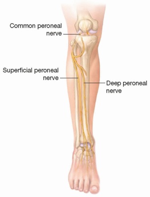
Image: The common peroneal nerve travels behind the knee before curving around the bony prominence on the outer side of the leg (the fibular neck). Because the nerve lies very close to the skin at this specific point, it is easily compressed by tight clothing, leg crossing, or external pressure.
Why the Peroneal Nerve Becomes Compressed
Several factors may increase pressure on the nerve.
Common causes include:
• Prolonged leg crossing
• Frequent squatting or kneeling
• Rapid weight loss reducing protective tissue around the nerve
• Direct trauma to the knee
• Fibular head fractures or knee injuries
• Repetitive knee stress in athletes (for example football players or runners), where muscle hypertrophy and repetitive mechanical pressure around the fibular head may contribute to nerve compression
• Compression during prolonged immobilization or surgery. In some patients, peroneal nerve dysfunction may develop after knee surgery, particularly after procedures involving major knee realignment, prolonged positioning, or postoperative swelling around the fibular head region.
• Tight casts or braces around the knee
• Diabetes or metabolic conditions affecting nerve vulnerability
In selected cases, ganglion cysts, schwannomas, or other local structural lesions near the fibular head may directly compress the peroneal nerve and contribute to progressive neurological symptoms.
In many patients, more than one factor contributes to nerve compression.
Symptoms of Peroneal Nerve Compression
Symptoms usually affect the foot and outer side of the lower leg.
Common symptoms include:
• Foot drop (difficulty lifting the foot)
• Weakness when lifting the toes or ankle. In more advanced cases with foot drop, patients may develop a characteristic “steppage gait,” lifting the knee higher than normal while walking in order to prevent the toes from dragging on the ground.
• Pain or tenderness on the outer side of the knee near the fibular head. Some patients experience burning pain, electric-shock sensations, or painful hypersensitivity along the outer side of the leg or on the top of the foot, especially when chronic nerve irritation leads to neuropathic pain mechanisms.
• Frequent tripping while walking
• Numbness on the outer side of the leg
• Reduced sensation on the top of the foot
• Difficulty walking on the heels
In more advanced cases, prolonged nerve compression may lead to muscle weakness and gait disturbances.
Symptoms may vary depending on which branch of the peroneal nerve is affected. Compression of the common peroneal nerve near the fibular head most often causes foot drop and weakness of ankle dorsiflexion, while superficial peroneal nerve involvement may mainly produce sensory symptoms on the outer leg and top of the foot. Compression of the deep peroneal nerve near the ankle may cause pain on the top of the foot or weakness of toe extension, or numbness between the first and second toes. Compression of the common peroneal nerve near the fibular head is by far the most common form of peroneal nerve entrapment and is the most frequent entrapment neuropathy of the lower extremity
How Peroneal Nerve Compression Is Diagnosed
Diagnosis begins with clinical examination and evaluation of symptoms.
During examination, the doctor may identify:
• Foot drop or weakness of ankle dorsiflexion
• Weakness of toe extension
• Reduced sensation over the outer leg and foot
• Tenderness around the fibular head. During examination, tapping over the peroneal nerve near the fibular head may reproduce tingling or electrical sensations radiating toward the foot (positive Tinel sign), supporting the suspicion of local nerve irritation or compression.
• Abnormal gait pattern
Additional tests may include:
• Nerve conduction studies (EMG) to evaluate nerve function
• Ultrasound to visualize nerve swelling, compression, scar tissue, ganglion cysts, local masses, or mechanical tethering affecting the nerve
• MRI of the knee region in selected cases
• Lumbar spine imaging if symptoms may originate from the spine
• Temporary anesthetic nerve blocks around the peroneal nerve in selected cases, which may help confirm whether the symptoms originate from local nerve compression
These tests help confirm the diagnosis and exclude other causes.
Conditions That Can Mimic Peroneal Neuropathy
Several disorders may produce similar symptoms.
These include:
• Lumbar disc herniation affecting the L5 nerve root
• Sciatic nerve injury
• Peripheral neuropathy
• Motor neuron disorders
• Local knee disorders
Careful clinical evaluation usually distinguishes these conditions.
Importance of Early Treatment
The duration of nerve compression is one of the most important prognostic factors. When severe weakness or foot drop persists for a prolonged period before decompression, the likelihood of complete neurological recovery may gradually decrease.
When Is Surgery Necessary in Peroneal Nerve Compression — Continue Conservative Treatment or Operate?
In many patients, the key question is whether foot drop and nerve function will recover with continued conservative treatment or whether surgical decompression of the common peroneal nerve is needed.
When symptoms are mild or improving, especially when strength gradually returns and walking becomes more stable, continued non-surgical treatment is usually appropriate.
Surgery becomes more likely when foot drop persists, when muscle weakness does not improve over time, or when findings suggest ongoing compression of the nerve at the fibular head.
The most important factor is timing — operating too early may not be necessary in milder cases, while delaying surgery in more severe or prolonged compression may reduce the chance of functional recovery of foot dorsiflexion.
Because this decision depends on symptom progression, severity of weakness, and clinical findings, different specialists may reasonably recommend either continued conservative treatment or surgical decompression based on how these factors are interpreted in an individual case.
Conservative Treatment
Many patients improve with non-surgical treatment.
Common approaches include:
• Avoiding leg crossing or prolonged squatting
• Activity modification
• Physical therapy and strengthening exercises
• Ankle-foot orthosis (AFO) for foot drop
• Medications for neuropathic pain
• Treatment of underlying metabolic disorders
• Ultrasound-guided hydrodissection in selected patients, which may be used to separate the nerve from surrounding scar tissue or adhesions by injecting fluid around the nerve to reduce mechanical irritation
• Peripheral nerve stimulation techniques in carefully selected chronic pain cases when conventional treatment options fail to provide sufficient symptom control
When symptoms are mild or moderate, these measures often lead to gradual improvement.
Surgical Treatment in Persistent Cases
Surgery may be considered when symptoms remain severe or progressive despite conservative treatment.
The most common surgical procedure is peroneal nerve decompression at the level of the fibular head.
During this operation, the surgeon releases the structures that compress the nerve as it passes around the neck of the fibula. This may include dividing tight fascial bands or surrounding tissues that narrow the space around the nerve. The goal of the procedure is to relieve pressure on the peroneal nerve, allowing the nerve to recover and gradually improve muscle strength and sensation in the foot.
Surgical treatment is usually recommended when there is:
• Persistent foot drop
• Progressive muscle weakness
• Electrodiagnostic confirmation of nerve compression
Possible Complications and Surgical Risks
Peroneal nerve decompression near the fibular head is generally considered a safe procedure. However, as with any surgery involving peripheral nerves, certain complications may occur, although they are relatively uncommon.
These may include:
Wound healing problems (dehiscence)
In some patients, healing of the surgical incision may be delayed or incomplete, particularly in the presence of diabetes, smoking, poor tissue healing capacity, or local postoperative tension around the knee.
Infection
Postoperative infection is uncommon but may require additional treatment if it occurs.
Persistent weakness or incomplete functional recovery
When nerve compression has already caused significant damage to the peroneal nerve, weakness of foot dorsiflexion or foot drop may persist even after technically successful decompression because nerve recovery capacity may already be limited.
Injury to sensory nerve branches
Small sensory branches around the fibular head region may occasionally be irritated during surgery, potentially leading to localized numbness, tingling, or scar sensitivity.
Scar-related discomfort
Some patients may experience pain, tenderness, or hypersensitivity around the surgical scar, especially during kneeling or pressure on the outer side of the knee.
Incomplete decompression or persistent compression
In selected cases, residual compression may remain around the nerve if all compressive structures are not fully released or if another site of nerve involvement coexists.
Most of these complications are uncommon, and in many patients symptoms gradually improve over time. It is important to distinguish these situations from more common causes of persistent symptoms, such as severe pre-existing nerve damage, lumbar nerve root involvement, or contributing factors affecting nerve recovery.
Why Symptoms May Persist After Peroneal Nerve Surgery
In some patients, weakness of foot dorsiflexion, numbness of the foot, gait instability, or sensory symptoms may persist even after technically successful decompression of the peroneal nerve near the fibular head. This does not necessarily mean that surgery was unsuccessful. In many cases, the procedure correctly relieves pressure on the nerve, but symptoms continue because the dominant mechanism responsible for weakness or nerve dysfunction has not been fully resolved or because additional contributing factors remain active.
Successful recovery depends not only on relieving nerve compression, but also on determining how long the nerve has been affected, whether another neurological condition contributes to symptoms, and whether irreversible nerve injury has already developed before treatment.
Unrecognized Alternative or Overlapping Diagnoses
Several disorders may mimic peroneal nerve compression or coexist with it. These conditions are outlined in the section “Conditions That Can Mimic Peroneal Neuropathy.”
If another neurological disorder is present — such as lumbar radiculopathy affecting the L5 nerve root or sciatic nerve involvement — decompression near the fibular head may not fully resolve symptoms.
Double Crush Syndrome and Multi-Level Nerve Involvement
In some patients, the same nerve pathway may be affected at more than one anatomical level. For example, lumbar nerve root compression may coexist with peroneal nerve compression near the knee.
If only one site of compression is treated, recovery may remain incomplete despite technically successful surgery.
Pre-existing Nerve Damage and Recovery Limitations
When foot drop or weakness has been present for a prolonged period, the peroneal nerve and affected muscles may already have undergone chronic structural and functional changes.
In such cases, improvement after decompression may remain slow or incomplete because nerve regeneration capacity and muscle recovery potential become limited over time.
Scar Tissue and Local Postoperative Factors
In some patients, scar tissue (fibrosis) may develop around the nerve after surgery. This may contribute to persistent irritation, altered nerve mobility, or local discomfort near the fibular head.
Although less common, postoperative swelling or mechanical sensitivity around the knee may also contribute to persistent symptoms.
Technical Factors Related to Surgical Outcome
In selected cases, persistent symptoms may be related to technical aspects of the decompression procedure itself.
Residual fascial bands, incomplete release of compressive structures, or persistent mechanical tension around the nerve may continue to affect nerve function even after surgery.
Although these situations are less common than severe pre-existing nerve injury or overlapping diagnoses, they should still be considered when recovery does not progress as expected.
Contributing Factors That May Maintain Symptoms After Surgery
Persistent symptoms are often influenced not only by the original nerve compression, but also by additional factors that impair nerve recovery or maintain nerve hypersensitivity.
These factors may include:
- Diabetes or metabolic disorders affecting nerve healing
- Rapid weight loss may reduce the protective soft tissue around the fibular head, increasing the vulnerability of the peroneal nerve to external pressure or mechanical irritation.
- Persistent mechanical irritation around the fibular head. Certain occupations or activities involving prolonged kneeling, squatting, repetitive knee flexion, or direct pressure on the outer side of the knee may increase the risk of chronic peroneal nerve compression.
- Lumbar spine disorders contributing to nerve vulnerability
- Central sensitization and chronic neuropathic pain mechanisms, where the nervous system itself becomes increasingly sensitive to pain signals over time.
- Delayed rehabilitation or prolonged gait abnormalities
Although these factors are rarely the primary cause, they may significantly influence long-term recovery and treatment response.
What Should Be Re-evaluated When Symptoms Persist
When symptoms continue after surgery, the most important step is not to repeat treatment blindly, but to reassess the underlying mechanism responsible for weakness or sensory symptoms.
This includes determining:
- whether the peroneal nerve near the fibular head remains the primary source of symptoms
- whether lumbar nerve root compression or another neurological condition is also involved
- whether severe pre-existing nerve injury limits recovery potential
- whether contributing factors continue to impair nerve healing or gait recovery
In many patients, different aspects of the condition have already been treated individually. However, lasting functional improvement often requires a comprehensive strategy based on a clearly defined mechanism of symptoms.
Online pain consultation for pain after surgery in detail
How the video consultation works — step by step
Answers to questions about the process and success of video consultations for pain after urgery
See the page “Possible Reasons for Poor Pain Treatment Effectiveness of Pain After Nerve Surgery” for an explanation of why conventional chronic pain treatments often fail—and what we do differently.
Prognosis and Recovery
The prognosis depends mainly on the duration and severity of nerve compression.
When diagnosed early, many patients improve with conservative treatment.
If compression persists for a long time, recovery may take longer.
After surgical decompression, improvement usually occurs gradually over several months as nerve function recovers.
When to Seek Specialist Evaluation
Medical evaluation is recommended if:
• Weakness when lifting the foot develops
• Frequent tripping or gait instability appears
• Numbness in the foot persists for several months
• Symptoms interfere with daily activities
• The diagnosis remains uncertain
Early specialist evaluation may help prevent permanent nerve damage.
Request Peroneal Nerve Compression Second Opinion — 24-Hour Review (Priority Option Available Within Hours)
Persistent weakness when lifting the foot, numbness on the outer side of the leg, or reduced sensation on the top of the foot may raise several important questions:
Is this really peroneal nerve compression?
Could the symptoms come from the lumbar spine or another nerve disorder?
Should treatment remain conservative or should surgical decompression be considered?
Why are the symptoms lasting longer than expected?
An independent neurosurgical second opinion may help clarify the cause of compression of the common peroneal nerve near the fibular head,
confirm whether the symptoms correspond to peroneal neuropathy or another neurological condition,
and determine whether conservative treatment, physical therapy, protective bracing, or surgical decompression
offers the best approach based on the duration of symptoms, neurological findings, and previous treatments.
- ✔ Send a brief message describing your symptoms, when they began, and whether you experience weakness when lifting the foot or frequent tripping while walking
- ✔ You will receive a reply within 24 hours explaining whether an online consultation is appropriate and which documentation is required
- ✔ Priority cases: progressive foot drop, worsening weakness of the foot, or rapidly increasing walking difficulty despite previous treatment — write PRIORITY in your first message
- ✔ Previous medical reports, EMG studies, lumbar spine imaging, or knee imaging can be reviewed
- ✔ During consultation we explain whether observation, physiotherapy, ankle-foot orthosis, or surgical decompression may be appropriate — including expected recovery timelines and up to 10 days of follow-up clarification
Consultation fees typically range from $180–250 depending on case complexity and documentation volume.
Secure payment by credit card, PayPal invoice (USD), or bank transfer.
This corresponds to typical international specialist telehealth neurosurgical second-opinion services.
Frequently Asked Questions About Peroneal Nerve Compression
What is peroneal nerve compression?
Peroneal nerve compression, also called peroneal neuropathy or peroneal nerve entrapment, occurs when the common peroneal nerve is compressed near the outer side of the knee, most often around the fibular head or fibular neck. At this level the nerve lies very close to the skin, which makes it vulnerable to external pressure, trauma, tight braces, leg crossing, squatting, kneeling, or postoperative swelling. The nerve controls lifting of the foot and toes and provides sensation to the outer lower leg and top of the foot. When compressed, it may cause foot drop, weakness of ankle dorsiflexion, numbness, tingling, burning pain, frequent tripping, and difficulty walking normally.
What are the most common symptoms of peroneal nerve compression?
The most common symptoms of peroneal nerve compression are weakness when lifting the foot, difficulty lifting the toes, frequent tripping, numbness on the outer side of the lower leg, and reduced sensation on the top of the foot. In more advanced cases, patients may develop foot drop and a characteristic steppage gait, lifting the knee higher than normal to prevent the toes from dragging on the ground. Some patients also have pain or tenderness near the fibular head on the outer side of the knee. Burning pain, electric-shock sensations, or hypersensitivity may appear when chronic nerve irritation produces neuropathic pain. Symptoms vary depending on whether the common, deep, or superficial peroneal branch is primarily affected.
What causes compression of the peroneal nerve?
Compression of the peroneal nerve most often occurs near the fibular head because the nerve is superficial and passes through a tight anatomical corridor. Common causes include prolonged leg crossing, squatting, kneeling, rapid weight loss, direct trauma to the knee, fibular head fractures, knee injuries, tight casts or braces, prolonged immobilization, and compression during or after surgery. Athletes may develop compression from repetitive knee stress, muscle hypertrophy, or mechanical pressure around the fibular head. Diabetes and other metabolic disorders can make the nerve more vulnerable. In selected cases, ganglion cysts, schwannomas, scar tissue, local masses, or other structural lesions directly compress the nerve. Often, several mechanical and biological factors act together.
Is peroneal nerve compression dangerous?
Peroneal nerve compression is not usually dangerous in a life-threatening sense, but it can become functionally serious because it may cause foot drop and walking difficulty. The common peroneal nerve controls muscles that lift the foot and toes. If compression is mild and improving, recovery may occur with avoiding pressure, physiotherapy, bracing, and treatment of contributing factors. The concern is persistent or progressive weakness. When foot drop lasts for a long time, the chance of complete recovery may decrease because the nerve and affected muscles can develop chronic structural and functional changes. Frequent tripping, worsening weakness, persistent numbness, or gait instability should prompt specialist evaluation, especially when symptoms do not improve over weeks or months.
Can peroneal nerve compression improve without surgery?
Yes. Peroneal nerve compression can improve without surgery when symptoms are mild, recent, or clearly improving. Conservative treatment usually focuses on removing pressure around the fibular head and protecting walking function. This may include avoiding leg crossing, prolonged squatting, kneeling, tight braces, or external pressure on the outer knee. Physical therapy, strengthening exercises, gait training, neuropathic pain medication, and treatment of metabolic disorders may help. An ankle-foot orthosis can prevent toe dragging and reduce falls in patients with foot drop. Some selected patients may benefit from ultrasound-guided hydrodissection to reduce mechanical irritation around the nerve. Surgery becomes more likely when weakness persists, foot drop does not improve, or tests show ongoing compression.
How is peroneal nerve compression diagnosed?
Peroneal nerve compression is diagnosed by combining symptoms, clinical examination, electrodiagnostic testing, and imaging when needed. The doctor checks ankle dorsiflexion, toe extension, sensation on the outer lower leg and top of the foot, tenderness around the fibular head, and walking pattern. Tapping over the nerve near the fibular head may reproduce tingling or electrical sensations toward the foot, supporting local nerve irritation. EMG and nerve conduction studies help confirm peroneal nerve dysfunction, localize the lesion, and estimate severity. Ultrasound can show nerve swelling, scar tissue, tethering, ganglion cysts, or local masses. MRI of the knee may be used in selected cases, while lumbar spine imaging is needed when L5 radiculopathy is possible.
What treatments are available for peroneal nerve compression?
Treatment for peroneal nerve compression depends on severity, duration, cause, and whether strength is improving. Conservative treatment includes avoiding pressure on the fibular head, stopping prolonged leg crossing or kneeling, activity modification, physical therapy, strengthening exercises, gait training, neuropathic pain medication, and treatment of diabetes or other metabolic factors. An ankle-foot orthosis can help stabilize walking when foot drop is present. In selected patients, ultrasound-guided hydrodissection may separate the nerve from scar tissue or adhesions and reduce mechanical irritation. Peripheral nerve stimulation may be considered in carefully selected chronic pain cases when standard treatments fail. Surgery is considered when weakness is severe, progressive, persistent, or when EMG and imaging suggest ongoing compression that is unlikely to recover spontaneously.
When is surgery recommended for peroneal nerve compression?
Surgery for peroneal nerve compression is recommended when symptoms are severe, progressive, or fail to improve with conservative treatment. The most important indication is persistent foot drop or weakness of ankle dorsiflexion, especially when EMG confirms peroneal nerve compression near the fibular head. Surgery is also considered when imaging shows a structural cause such as a ganglion cyst, schwannoma, local mass, scar tissue, or mechanical tethering. The operation usually involves decompression of the common peroneal nerve as it passes around the fibular neck. Tight fascial bands or surrounding tissues are released to reduce pressure. Timing matters because prolonged foot drop may reduce the chance of full recovery of dorsiflexion and walking function.
Can peroneal nerve compression cause permanent nerve damage?
Yes. Peroneal nerve compression can cause permanent nerve damage if weakness or foot drop persists for a long time. The duration and severity of compression are major prognostic factors. Early or mild compression may recover when pressure is removed and nerve function begins to return. However, if the nerve has been compressed long enough to cause significant axonal damage, recovery may be slow or incomplete. Persistent foot drop is especially important because muscles responsible for lifting the foot can weaken and gait may remain impaired. Delayed treatment may reduce the chance of full recovery, even after technically successful decompression. This is why progressive foot weakness, frequent tripping, or persistent dorsiflexion loss should be evaluated early.
Can symptoms persist after peroneal nerve decompression surgery?
Yes. Symptoms can persist after peroneal nerve decompression surgery, and this does not always mean that the operation failed. Surgery may correctly relieve pressure near the fibular head, but recovery can remain slow or incomplete if the nerve was already severely damaged before treatment. Foot drop present for a long time may recover only partially because the nerve and muscles have limited recovery capacity. Symptoms may also persist if another condition contributes, such as L5 lumbar radiculopathy, sciatic nerve injury, peripheral neuropathy, or double crush syndrome. Scar tissue, postoperative swelling, residual fascial bands, incomplete decompression, persistent mechanical irritation, metabolic disorders, or delayed rehabilitation can also limit recovery and require reassessment.
What are possible complications of peroneal nerve surgery?
Peroneal nerve decompression near the fibular head is generally considered safe, but complications can occur. Possible problems include delayed wound healing, infection, scar tenderness, localized numbness or tingling from irritation of small sensory branches, and discomfort during kneeling or pressure on the outer side of the knee. Some patients continue to have foot drop or weakness because the nerve had already suffered significant damage before surgery. Incomplete decompression or persistent compression may occur if all tight fascial bands or compressive structures are not fully released, or if another nerve level is involved. These issues must be distinguished from normal gradual nerve recovery, which can take months. Diabetes, smoking, and poor tissue healing may increase surgical risk.
What should be re-evaluated if symptoms continue after peroneal nerve surgery?
If symptoms continue after peroneal nerve surgery, the mechanism should be reassessed rather than repeating treatment blindly. The doctor should determine whether the peroneal nerve near the fibular head remains the main source, whether decompression was complete, whether scar tissue or residual fascial bands continue to irritate the nerve, and whether severe pre-existing nerve damage limits recovery. Other diagnoses must also be reconsidered, especially L5 lumbar radiculopathy, sciatic nerve injury, peripheral neuropathy, motor neuron disease, or local knee disorders. EMG, nerve conduction studies, ultrasound, knee MRI, and lumbar spine imaging may be needed. Contributing factors such as diabetes, rapid weight loss, mechanical pressure, delayed rehabilitation, gait abnormalities, and chronic neuropathic pain may also maintain symptoms.
Can I obtain an online consultation for peroneal nerve compression?
Yes. An online consultation can help when peroneal nerve compression is suspected, foot drop persists, or treatment decisions are unclear. Symptoms can be reviewed in detail, including weakness when lifting the foot, difficulty lifting the toes, frequent tripping, numbness on the outer leg or top of the foot, pain near the fibular head, and walking instability. EMG reports, nerve conduction studies, ultrasound, knee MRI, lumbar spine MRI, and previous surgical notes can also be reviewed. The consultation can clarify whether symptoms fit peroneal neuropathy, L5 radiculopathy, sciatic nerve involvement, or another condition. Progressive foot drop, worsening weakness, or rapidly increasing gait difficulty should be treated as a priority reason for specialist evaluation.
Why does peroneal nerve compression cause foot drop?
Peroneal nerve compression causes foot drop because the common peroneal nerve gives rise to the deep peroneal branch, which controls ankle and toe dorsiflexion. These muscles lift the foot during walking. When the nerve is compressed near the fibular head, signals to these muscles may be weakened or blocked. The patient may be unable to lift the foot properly, causing the toes to drag on the ground. To compensate, many patients develop a steppage gait, lifting the knee higher than normal during walking. Foot drop is clinically important because it indicates motor involvement, not only sensory irritation. Persistent foot drop requires careful evaluation because the chance of full recovery decreases when motor weakness remains untreated for too long.
How is peroneal nerve compression different from L5 radiculopathy?
Peroneal nerve compression and L5 radiculopathy can both cause foot weakness, so distinguishing them is essential. Peroneal nerve compression usually occurs near the fibular head and often causes weakness of foot and toe dorsiflexion, sensory symptoms on the outer lower leg and top of the foot, tenderness near the fibular head, and sometimes a positive Tinel sign at the knee. L5 radiculopathy originates from the lumbar spine and may include back pain, radiating leg pain, weakness in muscles not supplied only by the peroneal nerve, and different reflex or sensory patterns. EMG and nerve conduction studies help localize the lesion. Lumbar spine MRI is considered when symptoms may come from a disc herniation or nerve root compression.
Why is the peroneal nerve vulnerable near the fibular head?
The peroneal nerve is vulnerable near the fibular head because it wraps around the neck of the fibula and lies very close to the skin. At this point there is little soft tissue protection, especially after weight loss or in thin patients. External pressure from leg crossing, squatting, kneeling, braces, casts, surgical positioning, or direct trauma can compress the nerve against the bone. The nerve also passes through a tight anatomical corridor where fascial bands or surrounding tissues may increase pressure. Because of this superficial course, even simple mechanical factors can cause clinically important weakness or numbness. This anatomical vulnerability explains why common peroneal nerve compression at the fibular head is the most frequent entrapment neuropathy of the lower extremity.
Can rapid weight loss cause peroneal nerve compression?
Yes. Rapid weight loss can increase the risk of peroneal nerve compression because it reduces the protective soft tissue around the fibular head. The common peroneal nerve is already superficial at this location. When the cushioning layer becomes thinner, the nerve becomes more exposed to pressure from leg crossing, sleeping position, kneeling, squatting, braces, or hard surfaces. Weight loss alone may not be the only cause, but it can make ordinary mechanical pressure more harmful. Patients may develop numbness on the outer leg or top of the foot, weakness of foot dorsiflexion, or foot drop. Treatment focuses on avoiding pressure, protecting the nerve, physiotherapy, bracing when needed, and monitoring recovery. Persistent weakness requires specialist evaluation.
Can a ganglion cyst, schwannoma, or local mass cause peroneal nerve compression?
Yes. A ganglion cyst, schwannoma, local mass, scar tissue, or other structural lesion near the fibular head can directly compress the peroneal nerve. These causes are especially important when symptoms are progressive, focal, unexplained, or do not improve after removing common external pressure. Ultrasound can show nerve swelling, cysts, scar tissue, tethering, or local compression. MRI of the knee region may be used when a tumor, ganglion cyst, post-traumatic lesion, or deeper structural cause is suspected. If a mass or cyst is the main cause, conservative measures alone may not solve the problem. Surgical decompression may include removal of the lesion and release of the nerve. Correctly identifying a structural cause is essential before deciding treatment.
What is the difference between common, deep, and superficial peroneal nerve involvement?
The common peroneal nerve divides near the fibular head into the deep and superficial peroneal branches. Compression of the common peroneal nerve near the fibular head usually affects both motor and sensory functions and may cause foot drop, weakness of ankle dorsiflexion, toe extension weakness, numbness on the outer leg, and sensory loss on the top of the foot. Superficial peroneal nerve involvement may mainly cause sensory symptoms on the outer leg and dorsum of the foot, sometimes with weakness of foot eversion. Deep peroneal nerve involvement may cause toe extension weakness, pain on the top of the foot, or numbness between the first and second toes. This pattern helps localize the compression level.
What is the role of an ankle-foot orthosis in peroneal nerve compression with foot drop?
An ankle-foot orthosis, or AFO, helps patients with foot drop walk more safely while the peroneal nerve is recovering or while treatment decisions are being made. It holds the ankle in a better position and prevents the toes from dragging during walking. This can reduce tripping, falls, fatigue, and compensatory steppage gait. An AFO does not heal the nerve by itself, but it protects function and improves mobility during the recovery period. It may be used with physiotherapy, strengthening exercises, gait training, and treatment of the underlying compression. If foot drop is improving, the brace may be temporary. If weakness persists, further evaluation with EMG, ultrasound, MRI, or surgical consultation may be needed.
What is ultrasound-guided hydrodissection for peroneal nerve compression?
Ultrasound-guided hydrodissection is a minimally invasive procedure sometimes used in selected patients with peroneal nerve compression or irritation. Under ultrasound guidance, fluid is injected around the nerve to separate it from surrounding scar tissue, adhesions, or tight fascial planes. The goal is to reduce mechanical irritation and improve nerve mobility without open surgery. This approach may be considered when imaging suggests tethering or local soft-tissue irritation, and when symptoms are not severe enough to require immediate decompression. It is not a replacement for surgery when there is persistent foot drop, progressive weakness, a structural mass, or clear severe compression. Its role depends on symptoms, imaging findings, EMG results, and the treating specialist’s experience.
What is double crush syndrome in peroneal nerve compression?
Double crush syndrome means that the same nerve pathway is affected at more than one level. In peroneal nerve compression, local entrapment near the fibular head may coexist with L5 lumbar radiculopathy, sciatic nerve involvement, peripheral neuropathy, or metabolic nerve vulnerability. If only the peroneal nerve near the knee is treated, symptoms may improve only partially because another lesion continues to impair nerve function. Double crush is especially important when symptoms are more widespread than expected, when back pain or radiating leg pain is present, when EMG suggests more than one lesion, or when recovery after decompression is incomplete. Correct diagnosis requires evaluating both the local nerve and the proximal nerve pathway, not only the fibular head.
Why can recovery after peroneal nerve decompression take several months?
Recovery after peroneal nerve decompression can take several months because surgery removes pressure, but the nerve and muscles need time to recover. If compression was mild and short-lasting, improvement may be faster. If foot drop or weakness was present for a long time, the nerve may already have significant axonal damage, and muscle recovery may be limited. Nerve regeneration is slow, and functional improvement depends on how much of the nerve remains capable of recovery. Scar tissue, postoperative swelling, incomplete decompression, lumbar radiculopathy, peripheral neuropathy, diabetes, rapid weight loss, ongoing pressure near the fibular head, gait abnormalities, and delayed rehabilitation can also slow improvement. Gradual recovery can be normal, but lack of progress should prompt reassessment.