Dr Željko Kojadinović — NEUROHIRURGIJA I LEČENJE BOLA
Dr Zeljko Kojadinovic — Pain Treatment & Neurosurgery


Symptoms, MRI Diagnosis, Bleeding Risk, and Treatment Options
Author:
Dr. Zeljko Kojadinovic, MD, PhD
— Neurosurgeon and Pain Management Specialist
Specialized Experience:
30 years of clinical expertise in neurosurgery.
Last medically reviewed:
January 6, 2026
Who This Brain and CNS Cavernoma Page Is For
This page is for patients and families who have been told that an MRI shows a brain or spinal cord cavernoma — also called a cerebral or spinal cavernous malformation, or CCM — or who have seizures, neurological symptoms, or concerns about bleeding risk.
If recommendations differ, a bleed is mentioned, or you are unsure whether your brain or CNS cavernoma should be treated or monitored — or whether it is actually causing your epilepsy — an individualized neurosurgical second opinion can clarify the real risks and the safest next steps based on your MRI and symptoms.
When patients usually seek a second opinion for brain or CNS cavernoma
- MRI shows a brain or spinal cord cavernoma — also called a cerebral or spinal cavernous malformation, or CCM — and it is unclear whether the lesion is low-risk or requires treatment
- Seizures or seizure-like episodes are present, and it is uncertain whether the brain cavernoma is truly the cause
- New neurological symptoms develop in the presence of a brain or CNS cavernoma
- The imaging report mentions a “bleed,” “microbleeding,” or hemosiderin, and the clinical significance is unclear
- The cavernoma is located in a high-risk area such as the brainstem, spinal cord, deep brain, or eloquent brain, and medical recommendations differ
- Multiple cavernomas are present, raising the possibility of familial CCM, and a clear monitoring strategy is needed
In these situations, a focused telehealth review can clarify what the MRI findings truly mean, whether the brain or CNS cavernoma explains the symptoms, and which next step is most appropriate: Request Consultation
Brain and CNS Cavernoma — Quick Summary (Read This First)
- A brain or CNS cavernoma is a cluster of abnormal, dilated capillary spaces within the brain or spinal cord, often described as having a raspberry-like appearance. It is also called a cerebral or spinal cavernous malformation. It is a low-flow lesion that passively fills and drains blood through small capillaries connected to the surrounding normal circulation.
- Brain and CNS cavernomas are usually present from birth, although some may develop later or appear de novo, particularly in familial cases or after radiation. They are not caused by lifestyle, stress, trauma, or high blood pressure, and many remain silent for years.
- Unlike a brain AVM, a brain or CNS cavernoma has no large feeding arteries or draining veins that directly enter the lesion. Blood flow is low-pressure and occurs through small capillary-type channels.
- The main risks of a brain or CNS cavernoma are seizures and small internal bleeding. Most brain cavernomas cause irritation of nearby brain tissue from repeated micro-bleeds rather than massive hemorrhage. However, clinically significant bleeding is most concerning when a cavernoma is located in the brainstem, spinal cord, or near critical brain centers, where even small hemorrhages can cause noticeable neurological deficits.
- About 30–50% of brain or CNS cavernomas never cause symptoms and are discovered incidentally on MRI.
- MRI is the key test for brain and CNS cavernoma diagnosis. Cavernomas have a characteristic “popcorn” or “mulberry” appearance with a dark hemosiderin rim from prior micro-bleeding. This is usually sufficient for diagnosis, so biopsy is not required.
- Most brain or CNS cavernomas do not need surgery. Deep, eloquent, brainstem, spinal cord, or asymptomatic lesions are usually managed with observation and MRI follow-up.
- Surgery is mainly considered for symptomatic brain or CNS cavernomas — especially those causing drug-resistant epilepsy or repeated symptomatic bleeding and that are surgically accessible.
- The bleeding risk is low if a brain or CNS cavernoma has never bled, but higher if a prior symptomatic hemorrhage has occurred.
- Stereotactic radiosurgery for cavernoma is controversial and is usually reserved only for selected deep or brainstem cavernomas that have already bled and cannot be safely removed.
- In most cases, a brain or CNS cavernoma is not a contraindication to pregnancy, anticoagulant or antiplatelet therapy, air travel, driving, or participation in most sports. Individual recommendations may vary depending on lesion location, prior bleeding, and clinical symptoms. Many people live a normal life with a cavernoma and never require treatment.
- A spinal cavernoma is a cavernous malformation located inside the spinal cord, and it is less common than a brain cavernoma. Symptoms usually relate to spinal cord irritation or bleeding and may include limb weakness, numbness, sensory changes, walking difficulty, pain, or bladder and bowel disturbance. Because the spinal cord has very little free space, even a small symptomatic bleed can cause noticeable neurological symptoms, so spinal cavernomas require careful MRI-based evaluation and individualized treatment planning.
- Use the Contents box to jump to sections on bleeding risk, MRI diagnosis, epilepsy, or treatment options.
Most patients only need the Quick Summary plus the sections on Bleeding Risk and Treatment Options. The rest is for deeper understanding.
Contents
- Who this is for
- When to consult
- Quick summary
- What is cavernoma
- How common
- Single vs multiple
- Inherited form
- Where they occur
- How they affect
- Symptoms
- Without symptoms
- MRI diagnosis
- Treatment options
- No symptoms
- Epilepsy
- Headache
- Bleeding
- Brainstem cases
- MRI follow-up
- Bleeding risk
- Pregnancy & drugs
- Radiosurgery
- Surgical notes
- Why Specialist Opinions Differ
- Start consultation
- FAQs
- Patient resources
Definition (What Is a Brain or CNS Cavernoma?)
A brain or CNS cavernoma (CCM) is a low-flow vascular malformation composed of clusters of dilated, thin-walled capillary-type channels in the brain or spinal cord. Blood flow is present within a cavernoma and occurs slowly through these abnormal capillary-like spaces. Blood enters via small arterial capillaries and exits via venous capillaries, which connect to the normal arterial and venous circulation of the surrounding brain. Unlike AVMs, cavernomas do not have a direct arteriovenous shunt or large, identifiable feeding arteries and draining veins. Because there is no high-pressure arterial inflow, small leaks and repeated micro-bleeds are more common than massive hemorrhages. These bleeds typically leave a characteristic rim of hemosiderin (blood-breakdown products) in the surrounding brain tissue.
Because of its bumpy, purple-red appearance, it is often described as looking like a small raspberry or mulberry.
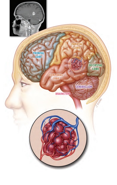
Image:Temporal lobe cavernoma composed of clusters of dilated, thin-walled capillary-type channels. The ‘bumpy’ purple surface is why these lesions are often compared to a raspberry or mulberry.
Incidence / Prevalence (How Common Is Brain Cavernoma?)
Cavernomas are not rare on modern MRI. Population studies suggest that about 0.4–0.8% of people have a cavernoma, and many are discovered incidentally when MRI is performed for unrelated reasons.
Multiplicity and Inheritance (Single vs Multiple CNS Cavernomas)
Sporadic Cavernoma
Most cavernomas are congenital, meaning they are present at birth, but some can develop later in life or appear de novo, particularly in familial cases or after brain radiation. In most cases, cavernomas are not inherited and occur sporadically.
Familial Cavernoma (Inherited Form)
In some cases, the condition is hereditary (genetic). This follows an autosomal dominant pattern, which means that if one parent carries a variant in the CCM1, CCM2, or CCM3 genes, there is a 50% chance of passing the condition to each child. Patients with this genetic form are much more likely to develop multiple cavernomas over their lifetime, and new lesions may appear over time.
However, the presence of multiple cavernomas alone does not automatically mean the condition is inherited, and clinical context and family history are important.
Genetic counseling may be considered in selected cases.
Locations (Brain vs Spinal Cord) — Where Do Cavernomas Occur?
Cavernomas can occur in:
- Supratentorial brain (70–80%): The most common site. These lesions are often associated with seizures.
- Cerebellum and brainstem (15–25%): Less common but clinically sensitive areas that control balance and vital functions.
- Spinal cord (1–5%): Uncommon; symptoms usually relate to limb weakness or sensory changes.
Although exact percentages vary between studies, the key clinical point is that brainstem and spinal cord cavernomas are more risk-sensitive, because even small bleeds can produce significant neurological symptoms. You can learn more about brain anatomy and its different regions on the following page.
How a Cavernoma Can Affect the Brain or Spinal Cord
Symptoms usually result from:
- Repeated micro-bleeds and hemosiderin irritation: Tiny leaks leave behind iron deposits (hemosiderin) in the surrounding brain tissue. Over time, this leads not only to chemical irritation but also to local inflammation and the formation of gliotic (scar-like) tissue around the lesion. This chronically altered brain environment can become electrically unstable and often results in seizures, especially when the cavernoma is located in the cerebral cortex.
- Symptomatic hemorrhage: A more significant bleed that causes a neurological deficit. This is especially concerning in eloquent brain regions (critical areas controlling speech, movement, or vision) or the brainstem and spinal cord, where even a small amount of pressure can impact vital functions.
Clinical Presentation (Symptoms) of Brain or CNS Cavernoma
Approximately 30–50% of cavernomas remain asymptomatic throughout life.
Common presentations include:
- Seizures (especially with supratentorial cavernomas) in ~25–40% of patients
- Focal neurological symptoms (weakness, numbness, speech or balance problems), in ~10–25% of patients, depending on location. These are caused by repeated microbleeding, local edema, and venous congestion around the cavernoma — even without a large hemorrhage
- Headache (nonspecific) in ~10–20%. In many cases, the headache is caused by another condition rather than the cavernoma itself, such as migraine or occipital neuralgia, which should be properly identified and treated.
- Symptomatic hemorrhage: Hemorrhage is less common as an initial presentation compared with seizures. In unselected populations, the average annual risk of bleeding is approximately 1% per patient-year. In incidentally discovered cavernomas, the annual risk is much lower (around 0.08-0.2% per year). However, after a prior symptomatic hemorrhage, the risk of rebleeding rises substantially, reaching approximately 15–30% over five years (2.7–6.5% annually), particularly in brainstem or spinal cord lesions.
Overall, many cavernomas present with seizures or are found incidentally, while major hemorrhage is less common.
How Often Are Brain or CNS Cavernomas Asymptomatic?
A large proportion of cavernomas are discovered incidentally, with approximately 30–50% of patients remaining asymptomatic.
In familial cavernoma, many carriers may have multiple lesions but no clinical symptoms for many years.
Diagnosis: How Is a Brain or CNS Cavernoma Identified?
MRI is the primary tool for diagnosis. In the vast majority of cases, an MRI scan alone is sufficient to confirm a cavernoma, making a brain biopsy unnecessary.
A typical cavernoma has a very distinct look on MRI:
- The „Popcorn“ Appearance: The center of the lesion has a bubbly, mixed-signal core (resembling popcorn or a mulberry) because it contains blood products at different stages of aging.
- The Dark Rim: The lesion is usually surrounded by a dark ring of hemosiderin (iron deposits from previous micro-bleeds).
- Special Sequences: We use highly sensitive „blood-detecting“ sequences (such as SWI or GRE) that act like a magnifying glass, allowing us to see even the smallest old micro-bleeds or additional lesions that might not show up on a standard scan.

Image: The arrow on the brain MRI indicates a cavernoma with a typical ‘popcorn’ appearance, surrounded by a dark halo (hemosiderin rim) caused by previous capillary bleeding
Does Angiography (DSA) Show a Cavernoma?
Usually no. Cavernomas are typically angiographically occult, meaning DSA is negative in most cases. Angiography is used only when other vascular lesions need to be excluded.
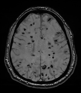
Image: MRI scan showing multiple cavernomas in both cerebral hemispheres. They appear dark because iron deposits from prior bleeding (hemosiderin) are highlighted as black on iron-sensitive MRI sequences.
Treatment Overview for Brain or CNS Cavernoma (What Are the Options?)
1) Asymptomatic Cavernoma (No Symptoms)
For an incidentally discovered cavernoma — especially if it is deep, eloquent, in the brainstem, or multiple — the usual approach is:
- No surgery
- Clinical follow-up and MRI surveillance, tailored to location and clinical history
However, in carefully selected cases, surgical removal may be considered if:
• The cavernoma is solitary
• Located in a non-eloquent, surgically accessible area
• The patient has a high-risk occupation (e.g., pilot, diver)
• Long-term anticoagulation is anticipated
• Or if the psychological burden of living with the lesion is significant
Such decisions require individualized risk–benefit analysis.
2) Cavernoma-Related Epilepsy
First-line treatment: The initial approach is typically anti-seizure medication to stabilize electrical activity in the brain.
Surgical Treatment for Epilepsy: Surgery is considered primarily for drug-resistant epilepsy. However, before proceeding, a doctor must first confirm that the epilepsy is indeed caused by the cavernoma and not by another underlying condition. You can learn more about epilepsy surgery on the following page
Identifying the Source (The „Guilty“ Lesion): When a patient has multiple cavernomas, it is vital to determine exactly which one — if any — is triggering the seizures. This is especially critical when lesions are located in different hemispheres or near eloquent (functional) brain regions. An experienced clinician identifies the true „trigger“ by analyzing several factors:
- Seizure Semiology: Studying the exact nature and „pattern“ of the seizures to determine their point of origin in the brain.
- Advanced EEG Monitoring: Using scalp electrodes (and occasionally internal ones) to map electrical activity and correlate it with the lesion.
- High-Resolution MRI & Functional Imaging: Correlating the location and characteristics of the cavernoma (such as the presence of a hemosiderin rim) with the brain’s functional maps.
Key Clinical Point: We must be certain of the source before considering surgery. In carefully selected patients, microsurgical resection of the specific „guilty“ lesion can significantly improve seizure control and, in many cases, lead to a seizure-free life.
3) Headache and Whether a Brain Cavernoma Is Truly the Cause
Headache is one of the most common reasons why brain MRI is performed, and a cavernoma is often discovered incidentally during this evaluation. However, in most cases, the headache is not caused by the cavernoma itself.
Unlike seizures or focal neurological deficits, which can be directly explained by repeated micro-bleeding and irritation of surrounding brain tissue, headache is a nonspecific symptom and is far more commonly related to other conditions such as migraine, tension-type headache, or occipital neuralgia.
The presence of a cavernoma on MRI does not automatically mean it is responsible for the patient’s pain. Misattributing headache to a cavernoma can lead to unnecessary concern or even inappropriate treatment decisions, including surgery that would not address the real cause of symptoms.
For this reason, it is essential to determine whether the headache pattern, neurological findings, and imaging features are truly consistent with a cavernoma-related mechanism — or whether another, more common and treatable cause of headache is present.
4) Hemorrhage (Bleeding) and When Surgery Is Considered in Brain or CNS Cavernoma
What Counts as “Bleeding”?
A clinically important event is a symptomatic hemorrhage — a bleed that produces new neurological symptoms and is confirmed on imaging. Emergency management follows the same core principles as other intracerebral hemorrhages; a practical step-by-step explanation is available on our Intracerebral Hemorrhage (ICH) page.
When Is a Brain or CNS Cavernoma Usually Operated On?
Microsurgery is most often considered when the cavernoma is:
- Symptomatic and surgically accessible (meaning it can be reached without damaging vital brain areas), or
- Associated with recurrent symptomatic hemorrhage, or
- Causing progressive neurological symptoms clearly attributable to the lesion
- In selected asymptomatic, low-risk, superficial lesions, when long-term anticoagulation, high-risk physical activities, or significant quality-of-life concerns are present, and surgical morbidity is expected to be minimal
Surgical treatment usually involves a craniotomy (surgical opening of the skull). This is followed by precise localization of the cavernoma using MRI-based neuronavigation in the operating room (GPS-like system for the brain) and complete removal of the lesion. Unlike many other brain lesions, accessing a cavernoma—especially when it is deep-seated—is often more challenging than separating it from the surrounding brain tissue and removing it once reached.
Read more about potential complications following craniotomy and open brain surgery on this page.
If surgery is performed for epilepsy, removal often includes not only the cavernoma itself but also the surrounding hemosiderin-stained and gliotic tissue, which may contribute to seizure generation. The extent of resection is carefully individualized and guided by anatomical location, functional mapping when necessary, and the goal of maximizing seizure control while preserving neurological function.
Brainstem Cavernomas (Special Situation)
Brainstem cavernomas are managed more conservatively because surgical morbidity is significantly higher compared with supratentorial lesions.
Surgery is typically considered only when:
• There is recurrent symptomatic hemorrhage, especially with progressive neurological deficits,
• A symptomatic lesion reaches or approaches the brainstem surface, making safe access feasible,
• A significant hematoma causes persistent or worsening neurological impairment and becomes surgically accessible.
Asymptomatic brainstem cavernomas are generally not operated on.
Beyond the specific characteristics of the cavernoma, the safety of the procedure is determined by the surgeon’s experience in accurately assessing risks and indications, their technical surgical skill in performing the operation safely, and the availability of advanced hospital equipment.
Follow-Up (Monitoring) of Brain or CNS Cavernoma
MRI follow-up depends on:
- Location (brainstem/spinal cord vs superficial cortex)
- Symptoms
- History of hemorrhage
- Sporadic vs familial disease
Stable, incidental lesions are monitored less frequently, while high-risk or recently symptomatic cavernomas require closer follow-up.
Pregnancy, Delivery, and Blood Thinners — Do They Increase Bleeding Risk in Brain or CNS Cavernoma?
Pregnancy and Vaginal Delivery
Prospective studies show that pregnancy does not increase hemorrhage risk in women with brain or spinal cavernomas, and vaginal delivery is generally safe in appropriate candidates.
Anticoagulants / Antiplatelet Therapy
Available observational cohort studies suggest no clear increase in intracranial hemorrhage risk in cavernoma patients receiving either anticoagulant therapy (such as warfarin or direct oral anticoagulants) or antiplatelet therapy (such as aspirin or clopidogrel) when these treatments are clinically indicated. However, the overall evidence remains limited and is largely based on non-randomized data, particularly in patients without prior symptomatic hemorrhage.
In contrast, the safety of thrombolytic therapy in patients with cavernomas is not well established, and available evidence is insufficient to confirm that it carries no additional risk.
For these reasons, antithrombotic therapy alone is not considered a routine indication for surgical removal of an otherwise asymptomatic cavernoma. Decisions should be individualized, taking into account lesion location, prior bleeding history, and the balance between natural history and surgical risk.
Air travel:
Flying is safe for patients with cerebral cavernomas because there is no evidence that cabin pressure changes increase bleeding risk.
Sports and physical activity:
Most patients with cavernomas can safely exercise and participate in sports; only high-impact or head-injury–prone activities (such as boxing, tackle football, or extreme sports) are usually discouraged in those with prior hemorrhage or brainstem lesions.
Stereotactic Radiosurgery (SRS) — Does It Work for Brain or CNS Cavernoma?
SRS for cavernomas remains controversial.
- It is not a standard alternative to surgery for superficial, operable lesions.
- For deep or eloquent cavernomas, particularly brainstem lesions with prior symptomatic hemorrhage, SRS may be considered when surgical risk is unacceptably high. Some observational studies suggest SRS may reduce re-bleeding rates in selected hemorrhagic brainstem cases, but evidence remains limited and controversial.
Surgical Note (For Patient Understanding)
When surgery is performed, the goal is complete removal of the cavernoma while minimizing injury to surrounding brain tissue. Previous micro-bleeding often creates a gliotic or hemosiderin plane that helps guide safe separating from the surrounding brain tissue and removing it once reached.
On this page, you can learn more about other neurosurgical cerebrovascular diseases.
Why Do Specialists Disagree About CNS Cavernoma Management?
This complexity explains why answers to these questions may differ between specialists. Below are the main reasons.
Is The Cavernoma Really The Cause Of Symptoms?
Seizures, headache, or neurological symptoms may be present, but not all are necessarily caused by the cavernoma. In many patients, especially when multiple lesions or common conditions like migraine are present, it is uncertain whether the cavernoma is the true source or an incidental finding.
How High Is The Real Risk Of Bleeding?
The risk of hemorrhage varies widely depending on prior bleeding, location, and individual factors. Some specialists emphasize the relatively low annual risk in incidental cases, while others focus on the significantly higher risk after a prior symptomatic hemorrhage, especially in brainstem or spinal cord lesions.
When Is Surgery Truly Necessary?
Surgical indication is not based on a single factor but on a balance between symptoms, location, and surgical risk. Some specialists recommend surgery in symptomatic or accessible lesions, while others prefer observation if symptoms are mild, stable, or not clearly attributable to the cavernoma.
Is Epilepsy Really Caused By The Brain Cavernoma?
In patients with seizures, it is not always clear whether the cavernoma is the epileptic focus. Especially in cases with multiple lesions or complex seizure patterns, additional evaluation is required before considering surgery, and opinions may differ on whether resection will improve seizure control.
Does Headache Justify Treatment?
Headache is common but rarely caused directly by a cavernoma. Misinterpreting headache as cavernoma-related can lead to unnecessary concern or even inappropriate treatment. In some cases, headache may actually be related to tension-type headache driven by anxiety after the MRI finding, while the cavernoma is incidental. When the lesion is superficial and surgically accessible, some specialists may still consider removal to eliminate uncertainty, while others prefer to first clarify whether the cavernoma truly explains the symptoms.
How Much Weight Should Be Given To Lesion Location?
Location significantly affects both risk and treatment decisions. Cavernomas in deep or eloquent areas (such as the brainstem or spinal cord) carry higher surgical risk, leading some specialists to avoid intervention, while others may still consider surgery after repeated symptomatic bleeding.
Why Do Some Specialists Recommend Surgery While Others Do Not?
Different specialists weigh the potential benefit and surgical risk differently based on their experience and clinical judgment. Some prioritize intervention when there is a reasonable chance of symptom relief or prevention of future bleeding, while others favor observation when expected benefit is uncertain or procedural risk is significant.
Request Cavernoma (Cerebral or Spinal Cavernous Malformation) Neurosurgery Consultation — 24-Hour Review or Priority Option (Usually Within 3 Hours)
When an MRI report reveals a cavernoma (also called a cavernous malformation or CCM), patients and families are often left with difficult questions about bleeding risk, seizures, neurological symptoms, and whether surgery or monitoring is the safest option.
An independent neurosurgical second opinion can help clarify how risky your specific cavernoma truly is, whether it should be treated or observed, and what outcome you can realistically expect based on its exact location, size, and MRI features.
- ✔ Send a short message describing the cavernoma diagnosis, symptoms (seizures, weakness, numbness, headache, bleeding), and what the MRI report says
- ✔ You’ll receive a reply within 24 hours explaining if and how we can help in your specific cavernoma situation
- ✔ Time-sensitive cases: if bleeding has occurred, neurological symptoms are worsening, or doctors are urgently recommending surgery, consultations can often be arranged within a few hours — write PRIORITY in your first message
- ✔ MRI images and hospital reports can be reviewed once initial contact is established
- ✔ During the consultation, we explain cavernoma type, bleeding and seizure risk, treatment options (observation, epilepsy management, or surgery), and realistic long-term outcomes — with up to 10 days of follow-up for brief questions
Consultation fees typically range from $180–250, depending on cavernoma complexity and MRI findings.
Secure payment by credit card, PayPal invoice (USD), or bank transfer.
This is within the usual range for international specialist telehealth second opinions in neurosurgery.
Frequently Asked Questions About Brain Cavernoma and CNS Cavernous Malformation
What is a brain cavernoma or cerebral cavernous malformation?
A brain cavernoma, also called a cerebral cavernous malformation, cavernous malformation, or CCM, is a low-flow vascular malformation made of clusters of dilated, thin-walled capillary-type channels. It is often described as having a raspberry, mulberry, or popcorn-like appearance. Unlike a brain AVM, a cavernoma does not have large feeding arteries or draining veins and does not create a high-pressure arteriovenous shunt. Blood flow inside a cavernous malformation is slow and low-pressure. Symptoms, when they occur, are usually related to repeated micro-bleeds, hemosiderin irritation, seizures, focal neurological symptoms, or occasionally a symptomatic hemorrhage.
Is a brain cavernoma or cavernous malformation the same as an AVM?
No. A cavernoma, or cavernous malformation, and a brain AVM are different vascular malformations. A brain AVM is a high-flow lesion where arteries connect directly to veins through a nidus, creating high-pressure blood flow and a different bleeding pattern. A cavernoma is a low-flow lesion made of abnormal capillary-type spaces. It usually does not show large feeding arteries or draining veins and is often angiographically occult. Cavernous malformations more commonly cause repeated small internal bleeds, hemosiderin deposition, irritation of nearby brain tissue, seizures, or focal symptoms. This distinction is important because diagnosis, bleeding risk, treatment options, and surgical strategy are different from AVM management.
Is a brain cavernoma or cerebral cavernous malformation something you are born with?
Many cavernomas, or cerebral cavernous malformations, are congenital, meaning they are present from birth, but some may appear later in life. This can happen especially in familial CCM syndromes or after previous brain radiation. Cavernous malformations are not caused by stress, trauma, lifestyle, high blood pressure, or infection. Many remain silent for years and are discovered only when MRI is performed for headache, seizures, neurological symptoms, or another reason. A single sporadic cavernoma is usually not inherited. Multiple cavernous malformations raise the possibility of a familial form, although the presence of more than one lesion does not automatically prove a genetic cause.
How common is a brain cavernoma or cerebral cavernous malformation?
Cavernomas, also called cerebral cavernous malformations when located in the brain, are not rare on modern MRI. Population studies suggest that approximately 0.4–0.8% of people may have a cavernoma. Many are discovered incidentally during MRI performed for unrelated symptoms, such as headache, dizziness, trauma evaluation, or nonspecific neurological complaints. A significant proportion of cavernous malformations remain asymptomatic throughout life. The clinical importance of a cavernoma depends less on the diagnosis itself and more on its location, whether it has bled, whether it is associated with seizures or neurological symptoms, and whether there are single or multiple lesions. Most incidental cavernomas do not require immediate surgery.
Can a brain cavernoma or CCM be inherited?
Yes, but most cavernomas are sporadic and are not inherited. Familial cerebral cavernous malformation, or familial CCM, can occur when a person carries a pathogenic variant in genes such as CCM1, CCM2, or CCM3. This form follows an autosomal dominant pattern, meaning there is a 50% chance of passing the condition to each child if one parent carries the variant. Familial cavernous malformation is more likely when multiple cavernomas are present, when new lesions appear over time, or when there is a family history of similar findings. Genetic counseling may be considered in selected cases, especially when multiple lesions or affected relatives are present.
What symptoms can a brain cavernoma or cavernous malformation cause?
A brain cavernoma or cerebral cavernous malformation may cause no symptoms at all, or it may present with seizures, headache, weakness, numbness, speech problems, visual symptoms, balance problems, or focal neurological deficits. Symptoms depend strongly on location. Supratentorial cortical cavernomas are more often associated with seizures. Brainstem, cerebellar, spinal cord, or deep cavernous malformations may cause neurological deficits even after a small bleed because these areas are anatomically sensitive. Some symptoms result from repeated micro-bleeds, hemosiderin irritation, local edema, or gliosis around the lesion. A cavernoma found on MRI does not automatically mean that all symptoms are caused by it.
Can a brain cavernoma or cerebral cavernous malformation cause seizures or epilepsy?
Yes. Seizures are one of the most common symptoms of supratentorial brain cavernomas, especially when the cerebral cavernous malformation is located near the cerebral cortex. Repeated micro-bleeds can leave hemosiderin and gliotic tissue around the cavernoma, creating an irritated area that may become electrically unstable. Treatment usually begins with anti-seizure medication. Surgery is considered mainly when epilepsy is drug-resistant or when the cavernous malformation is clearly the seizure source and can be removed safely. In patients with multiple cavernomas, it is especially important to identify the true “guilty” lesion through seizure pattern, EEG, MRI findings, and clinical correlation before considering surgery.
Can a brain cavernoma or cavernous malformation cause headaches?
Headache can occur in patients with a brain cavernoma or cerebral cavernous malformation, but it is a nonspecific symptom. In many cases, the cavernoma is found incidentally during MRI performed for headaches, while the true cause may be migraine, tension-type headache, occipital neuralgia, cervical pain, sinus-related headache, medication-overuse headache, or anxiety after the MRI finding. A cavernous malformation is more likely to be clinically relevant when headache is associated with new neurological symptoms, imaging evidence of recent bleeding, or location that plausibly explains the symptoms. Misattributing ordinary headache to a cavernoma can lead to unnecessary fear or inappropriate treatment decisions.
Can a brain cavernoma or cavernous malformation bleed or rupture?
Yes. A cavernoma, or cavernous malformation, can bleed, but its bleeding pattern is usually different from a ruptured aneurysm or AVM. Cavernous malformations are low-flow lesions, so repeated small internal bleeds and micro-bleeding are more common than massive hemorrhage. These small bleeds often leave hemosiderin around the lesion. A clinically important event is a symptomatic hemorrhage, meaning a bleed that causes new neurological symptoms and is confirmed on imaging. The risk of bleeding is usually low in incidentally discovered cavernomas, but it increases after a prior symptomatic hemorrhage, especially in brainstem or spinal cord cavernous malformations where even a small bleed can cause significant symptoms.
What symptoms suggest a brain cavernoma or cavernous malformation bleed?
Symptoms of a cavernoma or cavernous malformation bleed depend on the location of the lesion. A symptomatic hemorrhage may cause sudden or progressive weakness, numbness, speech difficulty, double vision, imbalance, vertigo, facial weakness, swallowing difficulty, new severe headache, seizure, or worsening of previous neurological symptoms. In the brainstem or spinal cord, even a small hemorrhage may cause important deficits because these regions are compact and functionally critical. Not every dark hemosiderin rim on MRI means there is a new dangerous bleed; hemosiderin can reflect prior micro-bleeding. The key issue is whether there are new symptoms and imaging evidence of a recent hemorrhage.
How is a brain cavernoma or cerebral cavernous malformation diagnosed?
MRI is the key test for diagnosing a cavernoma or cerebral cavernous malformation. In most cases, MRI alone is sufficient, and biopsy is not needed. A typical cavernoma has a mixed-signal “popcorn” or “mulberry” appearance, reflecting blood products at different stages of aging. It is often surrounded by a dark hemosiderin rim caused by previous micro-bleeding. Blood-sensitive MRI sequences such as SWI or GRE are especially useful because they detect hemosiderin and can reveal small or multiple CCM lesions that may not be obvious on standard sequences. MRI also helps determine location, relation to eloquent brain, and whether there are signs of recent bleeding.
What does a brain cavernoma or cerebral cavernous malformation look like on MRI?
On MRI, a brain cavernoma or cerebral cavernous malformation often has a characteristic “popcorn” or “mulberry” appearance. The center may look mixed or bubbly because it contains blood products of different ages. Around the lesion, MRI often shows a dark rim of hemosiderin, which represents iron-containing blood breakdown products from previous micro-bleeds. SWI and GRE sequences are particularly sensitive for detecting this hemosiderin signal and may show additional small cavernous malformations in patients with multiple lesions. This MRI appearance is usually distinctive enough to establish the diagnosis. The scan should also be interpreted in relation to symptoms, lesion location, and possible signs of recent hemorrhage.
What does hemosiderin mean around a brain cavernoma or cavernous malformation?
Hemosiderin is an iron-containing blood breakdown product that remains in tissue after previous bleeding or micro-bleeding. Around a cavernoma or cavernous malformation, it often appears as a dark rim on blood-sensitive MRI sequences such as SWI or GRE. This does not always mean there is a new or dangerous hemorrhage. It may reflect older micro-bleeds that occurred over time. However, hemosiderin can irritate nearby brain tissue and may contribute to seizures, especially when the cavernoma is located near the cerebral cortex. In epilepsy surgery, removal may sometimes include the cavernoma and selected surrounding hemosiderin-stained or gliotic tissue, depending on location and safety.
Does a brain cavernoma or cavernous malformation show on angiography or DSA?
Usually no. Cavernomas and cerebral cavernous malformations are typically angiographically occult, meaning they often do not appear on digital subtraction angiography (DSA). This is because they are low-flow vascular malformations without the large feeding arteries and draining veins seen in AVMs. MRI is therefore the main diagnostic test. DSA may be used only when another vascular lesion needs to be excluded, such as an AVM, dural arteriovenous fistula, or aneurysm, especially if the imaging pattern is unclear or bleeding has occurred. A negative angiogram does not rule out cavernoma when the MRI appearance is typical.
What is the difference between a brain cavernoma and a spinal cord cavernous malformation?
A brain cavernoma is located in the brain, while a spinal cord cavernous malformation is located inside the spinal cord. Both are low-flow cavernous malformations, but symptoms and treatment risk differ because of anatomy. Brain cavernomas may cause seizures, headaches, focal neurological symptoms, or hemorrhage depending on location. Spinal cord cavernomas more often cause limb weakness, numbness, sensory changes, pain, walking difficulty, or bladder-related symptoms if the spinal cord is affected. Spinal cord cavernous malformations are uncommon but clinically sensitive because there is limited space inside the cord. Even a small hemorrhage may produce significant neurological symptoms.
What makes a brainstem cavernoma or brainstem cavernous malformation different?
Brainstem cavernomas, or brainstem cavernous malformations, are managed more cautiously than many superficial cerebral cavernomas because the brainstem contains dense pathways controlling movement, sensation, eye movements, facial function, swallowing, balance, breathing, and consciousness. Even a small bleed in this compact region can cause significant neurological symptoms. Surgery in the brainstem carries higher risk and is usually considered only in selected situations, such as recurrent symptomatic hemorrhage, progressive neurological deficits, or when the lesion reaches or approaches a safe surgical surface. Asymptomatic brainstem cavernomas are generally not operated on. The decision depends on symptoms, bleeding history, accessibility, and surgical expertise.
Does every brain cavernoma or cavernous malformation need surgery?
No. Most cavernomas and cavernous malformations do not need surgery. An incidentally discovered cavernoma, especially if it is deep, in eloquent brain, in the brainstem, in the spinal cord, or one of multiple lesions, is often managed with observation and MRI follow-up. Surgery is considered only when the expected benefit is greater than the surgical risk. This is more likely when the cavernoma is symptomatic, surgically accessible, associated with recurrent symptomatic hemorrhage, causing progressive neurological symptoms, or clearly responsible for drug-resistant epilepsy. A cavernoma diagnosis alone is not enough to justify surgery; the decision must be individualized based on MRI and symptoms.
When is surgery recommended for a brain cavernoma or cavernous malformation?
Surgery is usually considered when a cavernoma or cavernous malformation is symptomatic and surgically accessible, when it causes recurrent symptomatic hemorrhage, when neurological symptoms are progressive and clearly attributable to the lesion, or when it is responsible for drug-resistant epilepsy. Superficial, non-eloquent cavernomas are generally more favorable for surgery than deep, brainstem, spinal cord, or eloquent-location lesions. If surgery is performed for epilepsy, the surgeon may remove not only the cavernoma but also selected surrounding hemosiderin-stained or gliotic tissue when safe. The goal is complete removal while minimizing injury to normal brain or spinal cord tissue.
Can a brain cavernoma or cavernous malformation be monitored instead of removed?
Yes. Many cavernomas and cavernous malformations are best monitored rather than removed. Observation is common for incidental lesions, asymptomatic cavernomas, deep or eloquent lesions, brainstem cavernomas without symptoms, spinal cord cavernomas without progression, and multiple cavernomas where no single lesion is clearly responsible for symptoms. Monitoring may include clinical follow-up and repeat MRI, with the interval depending on location, symptoms, hemorrhage history, and whether the lesion is sporadic or familial. Monitoring does not mean the diagnosis is ignored. It means that the expected surgical or procedural risk is higher than the likely benefit at that stage.
Can a brain cavernoma or cavernous malformation be cured?
A cavernoma or cavernous malformation can be considered cured locally if it is completely removed surgically and no residual lesion remains. This is most realistic for surgically accessible symptomatic cavernomas, especially superficial lesions. However, “cure” depends on the clinical situation. In familial CCM, new lesions may appear over time even if one lesion is removed. In epilepsy cases, seizure outcome depends on whether the removed cavernoma was truly the epileptic focus and whether surrounding hemosiderin or gliotic tissue also contributed to seizures. For deep, brainstem, spinal cord, or asymptomatic lesions, observation may be safer than attempting curative surgery.
Can radiosurgery be used for a brain cavernoma or cavernous malformation?
Stereotactic radiosurgery for cavernoma or cavernous malformation remains controversial. It is not a standard alternative to surgery for superficial, safely operable lesions. In selected deep or brainstem cavernomas that have already caused symptomatic hemorrhage and cannot be safely removed, radiosurgery may be discussed in specialized centers. Some observational studies suggest that radiosurgery may reduce rebleeding rates in selected hemorrhagic brainstem cases, but the evidence remains limited and debated. Radiosurgery also carries a risk of delayed radiation-related injury. For most cavernomas, the main options are observation, seizure management, or microsurgical removal when clearly indicated and technically safe.
Is pregnancy or vaginal delivery safe with a brain or spinal cord cavernoma?
In most cases, pregnancy does not appear to significantly increase hemorrhage risk in women with brain or spinal cavernomas, and vaginal delivery is generally safe in appropriate candidates. Individual recommendations may vary depending on lesion location, prior symptomatic hemorrhage, neurological status, and obstetric factors. A patient with a stable incidental cavernoma is usually managed differently from a patient with a recent brainstem hemorrhage or progressive neurological symptoms. Decisions about pregnancy monitoring, delivery mode, anesthesia, and anticoagulation should be individualized through coordination between neurology or neurosurgery, obstetrics, and anesthesia when relevant.
Can patients with a brain or spinal cord cavernoma take blood thinners?
Anticoagulant or antiplatelet therapy is not automatically forbidden in every patient with a cavernoma. Observational data suggest no clear increase in intracranial hemorrhage risk when these medications are clinically necessary, especially in patients without prior symptomatic hemorrhage, but the evidence is limited and decisions must be individualized. The balance depends on why blood thinners are needed, cavernoma location, prior bleeding history, neurological symptoms, and the risk of stopping the medication. Thrombolytic therapy is a separate issue and its safety in cavernoma patients is less certain. A cavernoma alone is not usually a routine indication for preventive surgery before antithrombotic treatment.
Is flying safe with a brain cavernoma or cerebral cavernous malformation?
In most patients with cerebral cavernoma or cerebral cavernous malformation, air travel is considered safe. There is no evidence that normal cabin pressure changes during commercial flights increase the risk of cavernoma bleeding. This is especially true for stable, incidental cavernomas without recent hemorrhage or progressive neurological symptoms. However, travel plans should be individualized if a patient recently had a symptomatic hemorrhage, has unstable neurological deficits, uncontrolled seizures, or a brainstem or spinal cord lesion with recent clinical change. The main practical concern is not the flight itself, but whether the patient is medically stable enough to travel and whether emergency care would be accessible if symptoms worsen.
Can patients with a brain or spinal cord cavernoma exercise or play sports?
Most patients with cavernomas or CCM can exercise and participate in ordinary physical activity. Walking, light fitness, swimming, cycling, and non-contact activities are usually acceptable when the patient is neurologically stable. High-impact sports or activities with a significant risk of head or spine trauma, such as boxing, tackle football, or extreme sports, are usually discouraged in patients with prior hemorrhage, brainstem lesions, spinal cord lesions, or uncontrolled seizures. Exercise recommendations should consider lesion location, bleeding history, seizure control, neurological deficits, and the type of activity. In many patients, avoiding unnecessary inactivity is also important for general health.
Why do specialists disagree about brain cavernoma or cavernous malformation management?
Specialists may disagree because cavernoma and cavernous malformation management depends on several uncertain balances: whether symptoms are truly caused by the lesion, how high the real bleeding risk is, whether epilepsy is generated by the lesion, whether surgery can be performed safely, and how much weight should be given to location. A superficial symptomatic cavernoma causing drug-resistant epilepsy may lead to a surgical recommendation. A deep, brainstem, spinal cord, or incidental cavernoma may lead to observation. Headache adds another layer of complexity because it is common and often unrelated to the cavernoma. Different specialists may weigh these risks differently based on experience and clinical judgment.
I have epilepsy and a brain cavernoma — do I need surgery?
No, not automatically. Having both epilepsy and a brain cavernoma does not by itself prove that surgery is necessary. The decision depends on whether the brain cavernoma is truly responsible for the seizures, whether appropriate anti-seizure medication controls them, and whether the lesion can be removed without causing an unacceptable neurological deficit.
Surgery is more likely to be considered when seizures continue despite adequate trials of appropriate medication, when the seizure pattern and EEG findings correspond to the location of the brain cavernoma, and when the lesion is surgically accessible. A superficial cortical brain cavernoma may present a very different risk–benefit balance from a deep lesion near speech, movement, memory, or visual pathways. You can read more about candidate selection and presurgical testing on our epilepsy surgery page.
How do doctors know whether a brain cavernoma is really causing my seizures?
Doctors cannot determine this from the MRI finding alone. A brain cavernoma is more likely to be the seizure source when it lies close to the cerebral cortex, the symptoms and pattern of the seizures correspond to that brain region, and EEG findings point to the same location. Hemosiderin and gliotic tissue around the brain cavernoma may further irritate the surrounding cortex and support a causal relationship.
The assessment is more difficult when there are multiple brain cavernomas, different seizure patterns, or disagreement between the MRI and EEG findings. Video-EEG monitoring, high-resolution epilepsy-protocol MRI, functional imaging, neuropsychological testing, and occasionally invasive EEG may then be required. The purpose is to identify the true epileptogenic lesion before surgery, because removing an incidental brain cavernoma may not improve seizure control. More details are available on our epilepsy surgery evaluation page.
If anti-seizure medication controls my seizures, should the brain cavernoma still be removed?
Usually not automatically. When seizures are well controlled, medication is tolerated, and the brain cavernoma has not caused symptomatic bleeding or progressive neurological deficits, continued medication and MRI monitoring may be safer than immediate surgery. The presence of a brain cavernoma on MRI alone is not a sufficient indication for removal.
Surgery may still be discussed in selected cases, depending on seizure severity, medication adverse effects, prior hemorrhage, lesion accessibility, occupation, driving limitations, and the patient’s long-term priorities. The expected benefit must be compared with the specific neurological risk of reaching and removing that brain cavernoma. General principles of drug-resistant epilepsy and lesionectomy are explained on our epilepsy surgery page.
How likely is brain cavernoma surgery to stop my seizures?
Brain cavernoma surgery can provide good seizure control in carefully selected patients, but it cannot guarantee permanent seizure freedom. The probability of success is higher when there is a single accessible brain cavernoma, the seizure pattern and EEG findings point to the same region, and the lesion can be completely removed together with relevant surrounding epileptogenic tissue when this is safe.
The outcome is less predictable when epilepsy has been present for a long time, several brain cavernomas are present, seizures arise from a wider cortical network, or surrounding hemosiderin-stained and gliotic tissue cannot be safely removed. Anti-seizure medication is generally continued after surgery and adjusted gradually according to seizure control. The expected outcome should therefore be estimated only after reviewing the MRI, EEG findings, seizure history, and surgical anatomy. Read more about lesionectomy and epilepsy surgery outcomes.
I have headaches and a brain cavernoma — how can doctors tell whether the brain cavernoma is really the cause?
In many patients, the brain cavernoma is not the cause of the headache. Brain cavernomas are often discovered incidentally when MRI is performed for migraine, tension-type headache, occipital neuralgia, cervical pain, or another common headache disorder. Simply finding a brain cavernoma on MRI does not establish that it is the source of pain.
A connection becomes more concerning when a new or unusually severe headache occurs together with new neurological symptoms, a seizure, or MRI evidence of a recent symptomatic hemorrhage. Doctors must compare the headache pattern, neurological findings, and actual MRI images rather than relying only on the radiology report. Removing a brain cavernoma solely because headache is present may not relieve the pain when another headache disorder is the true cause.
What are the specific risks of surgery for my brain cavernoma?
The risks of brain cavernoma surgery depend mainly on the lesion’s location, depth, size, previous bleeding, and relationship to critical brain structures. Possible complications include bleeding, brain swelling, infection, seizures, stroke, cerebrospinal fluid leakage, incomplete removal, and new neurological deficits involving movement, sensation, speech, vision, balance, memory, or cognition.
A superficial brain cavernoma in a non-eloquent part of the cerebral hemisphere may often be removed with relatively low neurological risk. A brain cavernoma in the brainstem, thalamus, basal ganglia, spinal cord, or eloquent cortex can carry a substantially greater risk because the surgical route passes close to critical pathways. The relevant comparison is therefore not surgery versus no risk, but the individualized surgical risk versus the natural risk of further bleeding, seizures, or neurological deterioration. See our detailed guide to craniotomy and brain surgery complications.
If I have multiple brain cavernomas, which one should be treated or removed?
Usually, not all brain cavernomas are removed. Treatment is directed toward the brain cavernoma that has caused symptomatic bleeding, progressive neurological impairment, or seizures and can be treated with an acceptable surgical risk. The remaining brain cavernomas are generally monitored unless they later become clinically relevant.
The largest brain cavernoma is not necessarily the responsible lesion. Doctors must correlate symptoms, neurological findings, seizure semiology, EEG results, and MRI features such as location and recent hemorrhage. When epilepsy is present, identifying the true “guilty” brain cavernoma is essential before considering surgery. Multiple brain cavernomas may also suggest familial cerebral cavernous malformation, particularly when there is a family history or new lesions appear over time. More information about evaluating multiple possible seizure-causing lesions is available on our epilepsy surgery and lesionectomy page.
Is monitoring safer than surgery for my brain cavernoma?
Monitoring is safer for many incidental, stable, deep, eloquent, brainstem, spinal cord, or multiple brain cavernomas, particularly when they have never caused symptomatic hemorrhage and are not clearly responsible for seizures or neurological deficits. Surgery becomes more reasonable when a brain cavernoma is surgically accessible and has caused recurrent symptomatic bleeding, progressive neurological impairment, or epilepsy that is not adequately controlled with medication.
The decision cannot be made from the size of the brain cavernoma or from the radiology report alone. Doctors must distinguish old micro-bleeding and hemosiderin from a recent symptomatic hemorrhage, determine whether the symptoms truly correspond to the lesion, and estimate the neurological risks of the surgical corridor. The safer strategy is the one expected to produce less overall harm for that individual patient, which may be observation in one case and surgery in another.
Can brain cavernoma or cavernous malformation MRI be reviewed through an online neurosurgical consultation?
Yes. Cavernoma or cerebral cavernous malformation MRI can often be reviewed through an online neurosurgical second opinion when MRI images, radiology reports, hospital records, and symptom history are available. The goal is to clarify whether the lesion is likely incidental or symptomatic, whether seizures, headache, neurological symptoms, or bleeding are truly related to the cavernoma, and whether observation, epilepsy management, surgery, or another strategy is most appropriate. This is especially useful when the cavernoma is located in the brainstem, spinal cord, or eloquent brain, when multiple cavernomas are present, or when different specialists have given different recommendations.
Additional Patient Resources About Cavernoma (Cerebral Cavernous Malformation / CCM)
If you want to read more from reputable organizations, these patient-friendly resources are a good starting point:
- Cavernoma Alliance UK — patient booklet / practical guide (PDF access via booklet page)
- Brain & Spine Foundation (UK) — Cavernoma (patient information leaflet)
- Mayo Clinic — Cavernous malformations (care & general info)
-
For deeper reading (open access, more technical):
Consensus Guidelines (open access) — diagnosis & clinical management of cavernous malformations (CCM)
Note: these sources are educational. Individual treatment decisions should always be made with a specialist who has reviewed your MRI and symptoms.
Structural vascular lesions (abnormal vessels) presented on the Cerebrovascular page
- Brain aneurysm (ruptured & unruptured)
- Brain AVM (arteriovenous malformation)
- Cavernous malformation (cavernoma)
- Dural arteriovenous fistula
- Developmental venous anomaly
- Small-vessel disease & spontaneous intracerebral hemorrhage