Dr Željko Kojadinović — NEUROHIRURGIJA I LEČENJE BOLA
Dr Zeljko Kojadinovic — Pain Treatment & Neurosurgery

Author:
Dr. Zeljko Kojadinovic, MD, PhD
— Neurosurgeon and Pain Management Specialist
Specialized Experience:
30 years of clinical expertise in neurosurgery
Last medically reviewed:
January 7, 2026
Spine vs. Spinal Cord — what is the difference?
Many patients confuse the spine and the spinal cord, but these are not the same structures.
- The spine (vertebral column) is a bony and joint-based structure that supports the body, allows movement, and protects neural elements.
- The spinal cord is part of the central nervous system and runs inside the spine, within the spinal canal.
A simple way to imagine this:
👉 the spine is the tunnel, and the spinal cord is the cable inside it.

Image: The spine is composed of vertebrae stacked one on top of the other. Inside, these bones form the spinal canal, which acts as a protective tunnel. The spinal cord and the nerve roots that branch off from it pass through this canal.
What is the spine made of?
The spine is composed of vertebrae, stacked one on top of another.
In total, there are 33–34 vertebrae, divided into five regions:
- Cervical spine – 7 vertebrae
- Thoracic spine – 12 vertebrae
- Lumbar spine – 5 vertebrae
- Sacral spine – 5 fused vertebrae
- Coccygeal spine – 3–4 small vertebrae
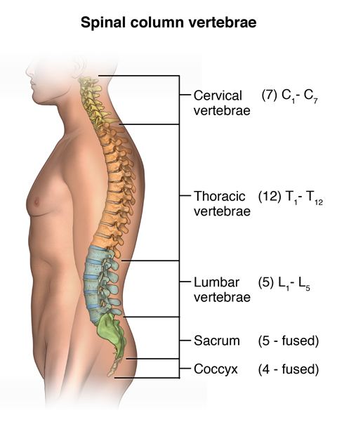
Image: Regions of the spine: cervical (neck), thoracic (mid-back), lumbar (lower back), sacral, and coccygeal (tailbone).
Main functions of the spine:
- supports body weight
- enables bending and rotation
- protects the spinal cord and nerve roots
Structure of a vertebra — terms patients often see in reports
Each vertebra consists of:
- Vertebral body – the weight-bearing front part
- Processes (Bony Projections) – bony projections for muscle and ligament attachment, and articulations (including spinous, transverse, and articular processes).
- Lamina – flat bony plates forming the posterior wall of the canal

Image: A vertebra and its parts: the vertebral body, the lamina, and the processes (spinous, articular, and transverse processes). Discs are located between vertebral bodies and consist of a soft central part (nucleus pulposus) and a strong outer ring (annulus fibrosus).
How are vertebrae connected?
Vertebrae are not fixed together — they are connected by flexible structures:
1. Intervertebral discs
Discs are located between vertebral bodies and act as shock absorbers.
Each disc consists of:
- a soft central part (nucleus pulposus)
- a strong outer ring (annulus fibrosus)
Discs allow movement and load distribution, but can cause problems when they bulge or herniate.
2. Facet joints
Facet joints are small joints located at the back of the spine.
They:
- guide spinal movement
- prevent excessive sliding
- can be a source of pain (facet joint syndrome)
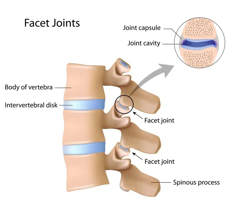
Image: The vertebrae are connected in the front by the intervertebral disc, located between the vertebral bodies. In the back, they are joined by the facet joints.
The spinal canal
The spinal canal is the space inside the spine that contains:
- the spinal cord
- nerve roots
- spinal meninges
Its boundaries:
- anteriorly – vertebral bodies and discs
- posteriorly – laminae and the ligamentum flavum (yellow ligament)
Thickening of ligaments or bony changes can narrow the canal, leading to spinal stenosis.
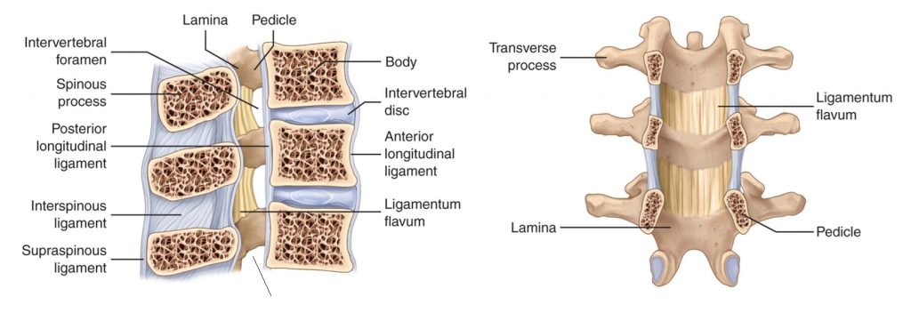
Image: The spinal canal and its boundaries. The front wall is formed by the back surfaces of the vertebral bodies and discs. The back wall is formed by the laminae and the yellow ligament (ligamentum flavum) situated between them (shown in the right image).
The spinal cord — function and extent
The spinal cord is part of the central nervous system.
Its function:
- transmits signals between the brain and the body
- enables movement, sensation, and reflexes
In adults, it extends:
- from the base of the brain
- down to approximately the L1–L2 vertebral level
Below this level, there is no spinal cord, only a bundle of nerve roots (cauda equina)

Image: The spinal cord inside the spinal canal. It extends from the first cervical vertebra down to the first or second lumbar vertebra. From that point downward, the canal contains the nerve roots for the legs that emerge from the spinal cord. This is why a lumbar disc herniation almost never results in compression of the spinal cord itself.
Nerve roots and spinal nerves
From the spinal cord emerge nerve roots:
- anterior (motor) roots
- posterior (sensory) roots, which contain dorsal root ganglia
These roots join together and exit the spinal canal through the:
Intervertebral (neural) foramina
These are small openings between adjacent vertebrae.
Disc bulges, herniations, bone spurs, or ligament thickening can compress the nerve here, causing radiating pain, numbness, or weakness.
Where do these nerves go?
After leaving the spine, spinal nerves form peripheral nerves that supply different body regions.
Cervical spine — nerves for the arms
The most important nerves include:
- Median nerve
- Ulnar nerve
- Radial nerve
- Axillary nerve
- Musculocutaneous nerve
These nerves control arm and hand movement, strength, and sensation.
Thoracic spine — nerves of the chest
Thoracic nerves continue as:
- Intercostal nerves
They run around the chest wall and are responsible for chest and upper abdominal sensation and movement.
Lumbar and sacral spine — nerves for the legs
The most important nerves include:
- Sciatic nerve
- Femoral nerve
- Obturator nerve
- Tibial nerve
- Common peroneal (fibular) nerve
These nerves control hip, leg, and foot movement and sensation.
This is why spinal problems often cause pain that radiates into the arm or leg, rather than staying only in the back.
Spinal cord coverings and the subarachnoid space
The spinal cord and nerve roots are surrounded by meninges (spinal coverings).
Between them lies the subarachnoid space, which:
- contains cerebrospinal fluid
- extends along the spinal cord
- continues around the proximal nerve roots until they receive their own dural sleeve
Changes in this space can be relevant in certain neurological and pain-related conditions.
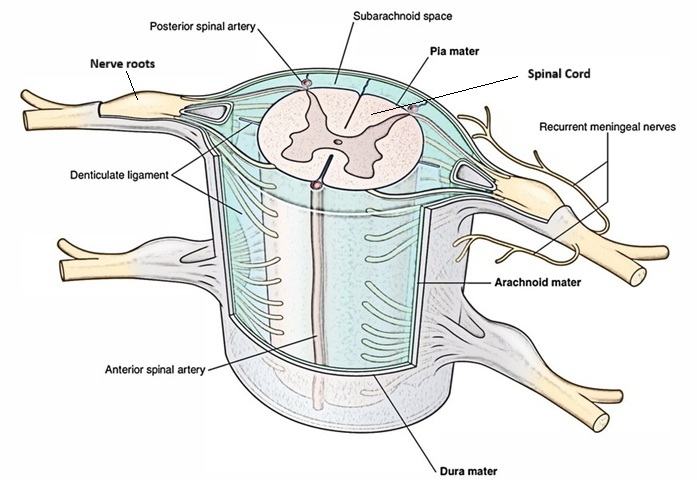
Image: The spinal cord and its nerve roots. The anterior (motor) and posterior (sensory) roots emerge from the spinal cord and join to form a single root. As they exit the spinal canal through openings between the vertebrae (foramina), they form the nerves. Both the spinal cord and the nerve roots are encased in protective layers called meninges (dura mater and arachnoid). Between the arachnoid layer and the nerve elements, clear cerebrospinal fluid (CSF) circulates.
Filum terminale is a thin fibrous structure that extends from the lower end of the spinal cord and anchors it to the sacrum. Its normal role is to stabilize the spinal cord while still allowing normal movement of the spine.
If the filum terminale is abnormally thick, short, or inelastic, it may place excessive tension on the spinal cord. This condition, known as tethered cord syndrome, can lead to neurological symptoms in children and, in some cases, pain and neurological complaints in adults, especially during spinal movement or prolonged standing.
This is a relatively uncommon condition and should always be evaluated in the appropriate clinical and imaging context.