Dr Željko Kojadinović — NEUROHIRURGIJA I LEČENJE BOLA
Dr Zeljko Kojadinovic — Pain Treatment & Neurosurgery

Author: Dr. Zeljko Kojadinovic, MD, PhD – Neurosurgeon and Pain Management Specialist
Last medically reviewed: June 02, 2026
Who this page is for
This page is for patients with hip pain that persists beyond 6–12 weeks even after a proper initial treatment trial (activity modification, medication, physical therapy), and where imaging (X-ray/MRI/ultrasound) shows no clear structural cause or only mild-to-moderate degenerative changes that don’t fully match the symptoms.
If you’re in this situation and want a focused evaluation to identify the dominant pain source (intra-articular hip/labrum or early OA, femoroacetabular impingement, greater trochanteric pain syndrome/gluteal tendinopathy, iliopsoas tendon/bursa, sacroiliac joint, referred lumbar radicular pain, or myofascial trigger points), you can request an online consultation with our specialist.
When patients usually seek a second opinion for hip pain
- Your MRI shows a labral tear or degenerative changes, but symptoms are inconsistent with imaging
- You have persistent groin or lateral hip pain despite physiotherapy and injections
- You are being advised to consider hip arthroscopy or hip replacement and want to understand if it is truly necessary
- Pain radiates to the thigh or knee and the true pain source remains unclear
A structured online consultation can clarify whether the pain is intra-articular, periarticular, or referred from the spine — and help determine the safest and most effective next step.
Hip pain is one of the most common musculoskeletal complaints in adults. Because the hip is a deep, load-bearing ball-and-socket joint surrounded by powerful muscles and tendons, pain may arise from local tissues or be referred from the lumbar spine or sacroiliac joint. Pinpointing the true generator is essential before planning treatment.
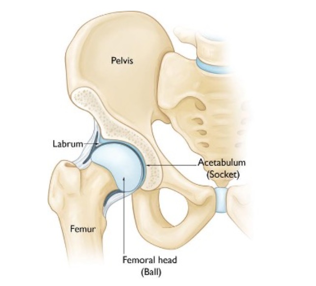
Image: Hip anatomy. The labrum is a ring of cartilage that surrounds the hip socket and helps keep the joint stable.
Common Causes of Hip Pain
Hip pain can be lateral, anterior, or deep groin pain — and each location points to a different dominant pain generator.
Frequent local sources include:
- Greater trochanteric pain syndrome (GTPS) — causes up to 20–25% of chronic hip pain in adults, more common in middle-aged women. It usually reflects gluteus medius or minimus tendinopathy; isolated “bursitis” alone accounts for less than 10% of cases.
- Femoroacetabular impingement (FAI)– extra bone bump occurs when extra bone develops on the femoral neck or around the hip socket. This causes friction during movement, gradually damaging the cartilage and labrum. It is found in about 10–15% of adults under 45 with persistent groin pain, especially active individuals or athletes.
- Labral tear — seen in roughly 10–20% of younger patients with mechanical hip symptoms; frequently overlaps with FAI or mild dysplasia. The labrum is a ring of tough cartilage that lines the rim of the hip socket and helps keep the joint stable, like a rubber seal. When this cartilage ring tears or frays, it can cause sharp or catching pain deep in the groin, sometimes with clicking, locking, or a sense that the hip may give way.
- Hip osteoarthritis (OA) — In osteoarthritis, the smooth cartilage that normally covers the ball and socket of the hip joint gradually wears away. As this protective layer becomes thinner, the underlying bone is exposed and small bone spurs (osteophytes) may form along the edges of the joint. These changes can cause deep, aching pain and stiffness, especially when standing up, walking, or climbing stairs. However, X-rays often show such wear-and-tear changes even in people who feel no pain — up to 15–20% of adults over 60 have visible arthritis, but fewer than half of them (≈8%) actually experience hip pain. This means that imaging findings do not always match the real pain source, because not every worn joint surface produces inflammation or nerve irritation.
- Iliopsoas or IT-band–related syndromes — found in about 5–10% of active adults. Pain or snapping in the front or side of the hip may result from bursal inflammation, tendon irritation (tendinitis), or muscle spasm caused by overuse or imbalance between hip flexors and stabilizers.
- Referred pain — lumbar spine or sacroiliac joint pathology is responsible for 10–15% of patients presenting with hip or groin pain.
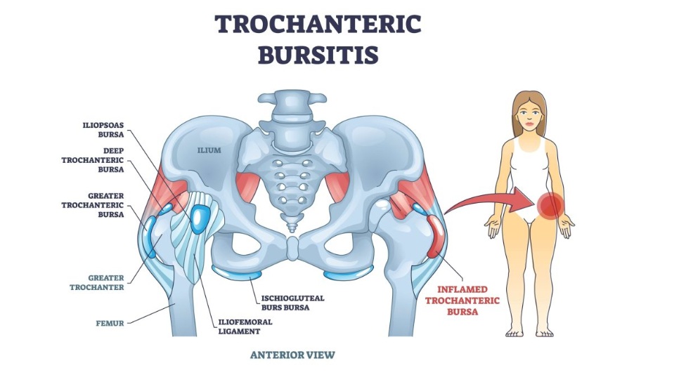
Image: Greater trochanteric pain syndrome (GTPS), showing hip bursae and gluteus minimus and medius tendons.
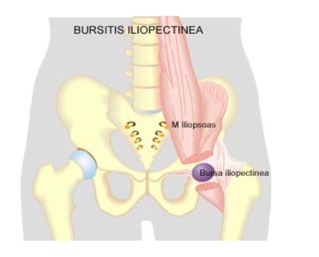
Image: Iliopsoas or IT-band–related syndromes
Symptoms of Hip Pain
Symptoms vary depending on the underlying cause of hip pain.
In greater trochanteric pain syndrome (GTPS), discomfort is usually felt on the outer side of the hip, sometimes radiating down the thigh. Pain worsens when lying on the affected side, climbing stairs, or walking uphill. Local tenderness over the bony prominence (greater trochanter) is typical, and patients often describe difficulty sleeping on that side.
In femoroacetabular impingement (FAI), pain is felt deep in the groin or front of the hip, often triggered by flexion or rotation — for example, when getting out of a car or tying shoes. Some patients notice a catching or clicking sensation in the hip caused by labral irritation.
When there is a labral tear, symptoms resemble those of FAI but may include sharp mechanical pain, occasional locking, or giving way of the hip. Discomfort typically appears during twisting or pivoting movements.
In hip osteoarthritis (OA), stiffness after sitting, pain during the first steps in the morning, and reduced range of motion are most common. Pain may spread toward the thigh or knee. Many patients report a dull ache that worsens with prolonged walking or standing.
With iliopsoas or IT-band–related syndromes, pain appears in the front or outer part of the hip, often accompanied by snapping, catching inside the hip, or a sense of tightness. Overuse, poor posture, or imbalance between hip flexors and stabilizing muscles can contribute.
Finally, referred pain from the lumbar spine or sacroiliac joint may mimic hip pain but is often associated with low back stiffness, numbness, or tingling in the leg. The hip itself moves freely, but pain increases with back movements or prolonged sitting.
Diagnostic Evaluation — Why Imaging Alone Is Not Enough
In many patients, plain films are often normal or show incidental age-related change that does not match the symptoms. Accurate diagnosis leans on:
- Focused physical exam (site-specific palpation, provocation tests, gait).
- Targeted ultrasound for tendons/bursae and snapping phenomena (dynamic exam).
- MRI when intra-articular pathology is suspected (labrum, cartilage) or when symptoms persist after guided therapy.
- Small diagnostic injections (e.g., peritrochanteric, intra-articular) to confirm the pain generator.
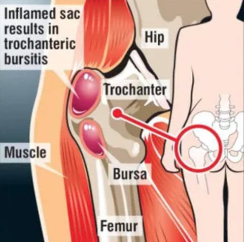
Image: Hip Anatomy and Trochanteric Bursitis
Treatment of Hip Pain
Most hip pain improves without surgery when the dominant source is identified and treated:
- Medication & local therapy — short courses of anti-inflammatory medication relieve acute inflammation and enable rehabilitation. When pain is well localized, ultrasound-guided injections of long-acting corticosteroids or platelet-rich plasma (PRP) can be placed precisely around the affected structure — such as the gluteal tendons, iliopsoas tendon sheath, or trochanteric bursa. These injections often provide substantial relief and prevent the need for surgery.
- Physiotherapy — progressive rehabilitation aims to restore hip control, gluteal strength, and pelvic stability. Activity modification (avoiding repetitive climbing, deep flexion, or prolonged standing) helps unload irritated structures. Strengthening the hip abductors and core muscles improves gait balance and long-term function.
- Procedure-level care — used when standard conservative therapy does not provide sufficient relief. Includes ultrasound-guided lavage or aspiration for recurrent trochanteric bursitis, hydrodistension for early capsular stiffness, and diagnostic intra-articular injections to differentiate joint from periarticular pain. When intra-articular pathology is confirmed, hip arthroscopy may treat femoroacetabular impingement (FAI) by removing bony overgrowth (cam/pincer deformity) and repairing or trimming the labrum. These minimally invasive procedures are performed only after full non-surgical care and restore hip motion while preserving joint integrity.
- Surgery — reserved for refractory structural pathology confirmed by imaging and diagnostic blocks. Arthroscopic labral repair or FAI correction is indicated when pain and mechanical symptoms persist despite image-guided and rehabilitative therapy. Cartilage restoration or microfracture may be used in younger patients with focal chondral lesions. Total hip replacement (arthroplasty) is indicated for advanced osteoarthritis (coxarthrosis) with major cartilage loss, stiffness, and severe movement restriction.
- ➤ Only about 10–15% of all patients with chronic hip pain eventually require surgical treatment, while the vast majority improve with targeted conservative and image-guided therapy.
Online pain consultation for regional pain in detail
Schematic explanation of the video consultation for regional pain
Answers to questions about the process and success of video consultations for regional pain
There are several common reasons for poor therapeutic outcomes in the treatment of chronic pain, which are often seen in patients with regional pain.
Artificial intelligence can also support the process by analyzing complex pain syndromes in fibromyalgia, but clinical expertise remains essential.
Why Hip Pain May Persist Despite Treatment
This is a common situation in patients who have already tried medication, injections, or physiotherapy — yet continue to experience pain.
In many cases, treatment does not fail because available methods are ineffective, but because the true pain generator has not been precisely identified. Imaging findings such as labral tears, early osteoarthritis, or degenerative changes may be present, but they do not always correspond to the actual source of pain.
Even when the primary structure is treated, symptoms may persist if additional contributing factors — such as altered gait mechanics, pelvic imbalance, muscle dysfunction, or systemic influences — continue to maintain irritation. Without recognizing and addressing all active mechanisms, treatment often leads to only partial or temporary relief.
Treatment of Contributing Factors in Hip Pain
Effective treatment of hip pain always begins with identifying the primary pain generator — the specific structure responsible for symptoms (such as intra-articular pathology including labrum or early osteoarthritis, greater trochanteric pain syndrome, iliopsoas tendon or bursa, or referred pain from the lumbar spine or sacroiliac joint).
However, in many patients, pain persists not only because of the local problem, but because additional contributing factors are not recognized or adequately addressed. These factors rarely act as the sole cause of pain, but they can maintain irritation, delay recovery, and reduce the effectiveness of otherwise appropriate treatment.
For that reason, successful management of hip pain requires not only treating the primary structure, but also understanding the broader mechanical and systemic context.
What contributing factors may play a role in hip pain?
- Repetitive loading and mechanical overload — Prolonged walking, running, stair climbing, or standing can continuously stress the hip joint and surrounding tendons. Improved by correcting load and activity patterns.
- Gait and pelvic mechanics — Altered walking pattern, pelvic tilt, or poor control of hip stabilizers increases stress on the joint and periarticular structures. Identified clinically and corrected through targeted rehabilitation.
- Muscle imbalance and reduced hip stability — Weak gluteal muscles and poor coordination lead to overload of structures such as the labrum or trochanteric region.
- Metabolic factors, pro-inflammatory diet and low-grade inflammation — Obesity, insulin resistance, and chronic inflammation increase joint load and pain sensitivity while slowing recovery.
- Nutritional deficiencies — Low vitamin D, vitamin B12, and other micronutrients may impair tissue repair and prolong symptoms.
- Sleep disturbances and night pain cycle — Pain worsens at night, especially when lying on the affected side, while poor sleep further increases pain sensitivity.
- Central sensitization — The nervous system becomes more sensitive over time, amplifying pain even when structural damage is limited.
- Reduced activity and deconditioning — Avoidance of movement leads to weakness, altered gait, and further overload of the hip.
- Medications and previous treatments — Certain medications such as statins, long-term use of pain medications, corticosteroids, or medications that cause excessive sedation may contribute to muscle or tendon-related pain, reduce tissue quality, or alter pain perception. Previous treatments, including repeated injections or incomplete rehabilitation, may also maintain symptoms if the underlying mechanism was not fully addressed.
- Other medical conditions and comorbidities — Conditions such as autoimmune diseases, thyroid disorders, diabetes, or chronic inflammatory states may increase pain sensitivity, affect tendon and joint structures, and reduce the response to otherwise appropriate treatment.
- Vitamin-related factors — Both deficiencies and excesses of certain vitamins (particularly vitamin B6) may contribute to nerve-related symptoms, burning pain, or altered sensitivity around the hip.
- Tissue quality and degeneration — Age-related changes, reduced blood supply, or repeated micro-injuries may impair tendon healing and increase vulnerability to persistent pain, even when mechanical factors are partially corrected.
Why this matters in practice
In many cases, treatment fails because the primary pain generator is not correctly identified, and therapy is directed only at contributing factors such as posture correction, exercise, or dietary changes. Conversely, even when the main structural cause is treated, failure to recognize and address contributing factors often leads to only partial or temporary improvement.
The most effective approach is a carefully selected combination of treatment that addresses both the primary pain generator and the contributing mechanical, functional, and systemic factors. In contrast, an inadequate or incomplete combination — even when it includes individually effective methods — is a common reason for suboptimal or short-lasting results.
This approach significantly increases the likelihood of long-term improvement and reduces the need for repeated injections or surgery.
In practice, many patients try to address contributing factors on their own — for example by improving posture, starting exercise programs, following an anti-inflammatory diet, using supplements (vitamin D, magnesium, glucosamine, chondroitin), or applying methods such as kinesio taping.
While these approaches can be helpful, they rarely lead to lasting improvement if the primary pain generator is not clearly identified and treated. On the other hand, even well-targeted medical treatment may fail if all contributing factors are not recognized and corrected.
Many patients reading this recognize that they have already tried one part of this approach — but not the complete strategy. This is one of the most common reasons why otherwise well-treated hip pain becomes chronic.
Prognosis and Long-Term Outlook in Hip Pain
Once the true pain source is identified and treated, most patients achieve meaningful, often complete relief and a return to full activity without surgery. Sustained pain control over several weeks typically predicts durable functional recovery.
Request Persistent Hip Pain Second Opinion — 24-Hour Review (Priority Option Available Within Hours)
Persistent hip pain despite physiotherapy, injections, medication, arthroscopy, or previous treatment
often raises important questions:
Is the pain really coming from inside the hip joint?
Do MRI, X-ray, or ultrasound findings truly explain the symptoms?
Is hip arthroscopy or hip replacement actually the right next step?
Could the pain come from gluteal tendons, trochanteric region, iliopsoas tendon or bursa, sacroiliac joint, lumbar spine, myofascial trigger points, or another referred source?
An independent specialist second opinion may help clarify whether the dominant pain source is
intra-articular, periarticular, or referred, whether the proposed procedure is medically justified,
and whether more targeted treatment — such as medication adjustment, or correction of contributing mechanical and systemic factors, ultrasound-guided evaluation, diagnostic injections, focused rehabilitation or
image-guided treatment, — may be more appropriate before arthroscopy or joint replacement.
- ✔ Send a brief message describing your hip pain location, how long it has lasted, what makes it worse, and which treatments have already been tried
- ✔ You will receive a reply within 24 hours explaining whether an online consultation is appropriate and which documentation is required
- ✔ Priority cases: severe persistent pain after hip arthroscopy, failed injections, conflicting specialist recommendations, rapidly worsening function, or uncertainty before proposed hip surgery or replacement — write PRIORITY in your first message
- ✔ MRI, X-ray, ultrasound reports, injection history, operative notes, physiotherapy summaries, and previous specialist opinions can be reviewed
- ✔ During consultation we analyze whether the dominant pain generator is inside the hip joint, around the hip joint, or referred from the lumbar spine or sacroiliac region, and which specific structures are most likely responsible
- ✔ We explain which treatment direction best matches the suspected dominant pain generator — including medication adjustment, correction of contributing factors, targeted rehabilitation, or diagnostic injections when the responsible structure is uncertain. If hip arthroscopy or replacement is being considered, we clarify whether the pain pattern supports that decision before discussing it with your local treating team — with up to 10 days of follow-up clarification.
Consultation fees typically range from $180–250 depending on case complexity and documentation volume.
Secure payment by credit card, PayPal invoice (USD), or bank transfer.
Based on our medical report, reimbursement may be possible if your insurance plan allows it.
This corresponds to typical international specialist telehealth second-opinion services for complex pain and treatment-decision review.
FAQ About Persistent Hip Pain
Why can persistent hip pain come from around the hip joint rather than inside the hip joint?
Persistent hip pain does not always come from the cartilage, labrum, or osteoarthritis inside the hip joint. The hip is a deep joint surrounded by powerful muscles, tendons, bursae, fascia, and nerves, and these surrounding structures can become the dominant pain generator. Pain may come from greater trochanteric pain syndrome, gluteal tendinopathy, iliopsoas tendon or bursa irritation, iliotibial band-related overload, or myofascial trigger points. These problems may cause severe symptoms even when X-ray or MRI shows only mild degenerative change. Pain on the outer hip, pain when lying on one side, pain with stairs, or local tenderness often suggests a periarticular source. Correct localization is essential before injections, arthroscopy, or hip replacement are considered.
What is the difference between intra-articular, periarticular and referred hip pain?
Intra-articular hip pain comes from inside the hip joint, such as the labrum, cartilage, early osteoarthritis, femoroacetabular impingement, or joint inflammation. It is often felt deep in the groin and may worsen with hip flexion, rotation, walking, or getting out of a car. Periarticular hip pain comes from structures around the joint, such as gluteal tendons, trochanteric bursa, iliopsoas tendon or bursa, iliotibial band, or surrounding muscles. This pain is often lateral, anterior, or movement-specific and may be tender to touch. Referred hip pain comes from another region, most often the lumbar spine or sacroiliac joint. The hip joint itself may move relatively well, while pain changes with back position, sitting, gait, or nerve irritation.
Can hip X-ray show osteoarthritis or degenerative changes that are not the true cause of pain?
Yes. Hip X-ray can show osteoarthritis, joint-space narrowing, osteophytes, sclerosis, or age-related degenerative change, but these findings do not always prove the source of pain. Many adults have visible hip wear on X-ray without significant symptoms, while others have disabling hip or groin pain with only mild imaging changes. X-ray is important for detecting advanced arthritis, deformity, fracture, or severe cartilage loss, but it does not show all pain-generating structures around the hip. Gluteal tendinopathy, trochanteric pain syndrome, iliopsoas irritation, sacroiliac pain, lumbar radicular pain, or myofascial overload may explain symptoms better than the X-ray. Treatment decisions should connect imaging with pain location, movement triggers, examination, and sometimes diagnostic injections.
Can hip MRI show labral tears, cartilage changes or arthritis findings that do not explain the pain?
Yes. Hip MRI can show labral tears, cartilage changes, femoroacetabular impingement-related findings, tendinopathy, bursitis, or early osteoarthritis, but not every MRI finding is clinically responsible for pain. Labral abnormalities and degenerative changes may be present without being the dominant pain generator. MRI is very useful when intra-articular pathology is suspected or when symptoms persist after guided treatment, but it must be interpreted together with the patient’s pain pattern. Deep groin pain with catching or pain during flexion and rotation may fit intra-articular disease. Lateral tenderness or pain when lying on one side may fit trochanteric or gluteal tendon pathology. MRI shows what is visible; clinical reasoning determines what is actually painful.
How can a doctor identify the real pain generator in persistent hip pain?
Identifying the real pain generator in persistent hip pain requires more than reading an MRI or X-ray report. The doctor must map whether pain is deep in the groin, lateral over the trochanter, anterior near the iliopsoas, posterior near the sacroiliac region, or radiating from the lumbar spine. Provocation tests, gait, range of motion, tenderness to palpation, pain during rotation, stair climbing, lying on the side, or prolonged sitting help separate joint, tendon, bursa, spine, and sacroiliac sources. Ultrasound can assess tendons, bursae, snapping phenomena, and guide injections. MRI helps when labrum, cartilage, FAI, or deeper structural pathology is suspected. When several findings coexist, targeted diagnostic injections can confirm which structure is most likely responsible.
Can hip pain come from gluteal tendons or greater trochanteric pain syndrome rather than the hip joint itself?
Yes. A common source of persistent lateral hip pain is greater trochanteric pain syndrome, which usually reflects gluteus medius or minimus tendinopathy rather than simple isolated bursitis. Patients often feel pain on the outer side of the hip, sometimes spreading down the thigh. It may worsen when lying on the affected side, climbing stairs, walking uphill, or standing for long periods. The hip joint may still move relatively well, and X-ray may show only mild or incidental arthritis. This matters because treating the hip joint itself may not solve pain if the dominant generator is the gluteal tendon or trochanteric region. Diagnosis depends on local tenderness, movement pattern, ultrasound or MRI findings, and response to targeted treatment.
Can lateral hip pain come from trochanteric bursitis, gluteal tendinopathy or iliotibial band irritation?
Yes. Lateral hip pain often comes from periarticular structures rather than the hip joint itself. Trochanteric bursitis, gluteal tendinopathy, and iliotibial band irritation can all cause pain over the outer hip. Many patients describe difficulty sleeping on the painful side, pain with stairs, pain while walking uphill, or tenderness directly over the greater trochanter. In modern practice, “trochanteric bursitis” is often only part of the problem; gluteus medius or minimus tendon irritation is frequently the more important pain generator. Iliotibial band tension and altered pelvic mechanics can maintain irritation. Treatment is different from treating intra-articular hip disease and may involve load modification, gluteal strengthening, ultrasound-guided injections, tendon-focused rehabilitation, and correction of gait or pelvic mechanics.
Can groin pain come from femoroacetabular impingement (FAI), labral tear or iliopsoas irritation?
Yes. Groin pain may come from several different hip-region structures. Femoroacetabular impingement can cause friction between the femoral neck and hip socket, irritating cartilage and the labrum, especially during flexion and rotation. A labral tear may produce sharp deep groin pain, catching, clicking, locking, or a feeling that the hip may give way. Iliopsoas tendon or bursa irritation can also cause anterior hip or groin pain, sometimes with snapping or tightness. These conditions can overlap, and imaging may show more than one abnormality. The key is to determine which finding matches the actual pain pattern. Pain when getting out of a car, tying shoes, pivoting, or rotating the leg may suggest intra-articular or iliopsoas-related mechanisms that need targeted evaluation.
Can hip pain come from the lumbar spine or sacroiliac joint rather than the hip itself?
Yes. Pain felt as hip, groin, buttock, thigh, or lateral hip pain may sometimes be referred from the lumbar spine or sacroiliac joint. Lumbar nerve root irritation can mimic hip pain, especially when symptoms are associated with low back stiffness, numbness, tingling, burning, or pain that changes with sitting, bending, or spinal movement. Sacroiliac joint dysfunction can cause buttock, posterior hip, groin, or thigh pain and may alter gait, increasing load on the hip region. This is especially important when hip imaging is normal or shows mild changes that do not match symptoms. A careful assessment must evaluate the hip, lumbar spine, sacroiliac region, gait, and neurological pattern together before assuming the pain comes from the hip joint itself.
Why can hip pain radiate to the thigh or knee?
Hip pain can radiate to the thigh or knee because the hip joint and surrounding structures share nerve pathways with regions lower in the leg. Hip osteoarthritis, labral irritation, femoroacetabular impingement, iliopsoas problems, or referred pain from the lumbar spine can all create symptoms that are felt beyond the hip. Some patients with true hip disease report pain mainly in the groin, anterior thigh, or even knee, which can delay diagnosis if the knee is examined in isolation. Conversely, lumbar radicular pain can be felt around the hip or thigh while the hip joint itself moves normally. This is why radiation of pain must be interpreted together with hip range of motion, back symptoms, neurological signs, imaging, and response to diagnostic injections.
Why can hip pain persist despite physiotherapy, injections or medication?
Hip pain can persist despite physiotherapy, injections, or medication when the dominant pain generator has not been identified precisely. Treatment may focus on a labral tear, early arthritis, or degenerative imaging finding, while the main pain source is gluteal tendinopathy, trochanteric pain syndrome, iliopsoas irritation, sacroiliac pain, lumbar referred pain, or myofascial overload. Injections may help temporarily if they reduce inflammation, but pain returns when gait mechanics, pelvic imbalance, weak gluteal control, repetitive loading, poor sleep, metabolic factors, or central sensitization continue to maintain irritation. Medication may reduce symptoms but does not identify the cause. Persistent hip pain therefore requires reassessment of both the primary pain generator and the mechanical, functional, and systemic contributing factors.
Why can hip pain persist after hip arthroscopy or previous hip surgery?
Hip pain can persist after hip arthroscopy or previous hip surgery when the operation treated a structural finding that was not the dominant pain source, or when several pain mechanisms were present at the same time. Arthroscopy may address femoroacetabular impingement or labral pathology, but pain may continue if gluteal tendinopathy, trochanteric pain syndrome, iliopsoas irritation, sacroiliac pain, lumbar referred pain, scar-related symptoms, or central sensitization are maintaining the problem. Pain can also persist if rehabilitation does not restore hip control, gluteal strength, pelvic stability, and gait mechanics. In some cases, true surgical problems must be excluded, but when imaging and surgical findings do not explain ongoing pain, the wider regional pain generator must be reassessed.
Why is hip arthroscopy not always useful for chronic hip pain?
Hip arthroscopy is not always useful for chronic hip pain because not every labral tear, FAI-related finding, or mild structural abnormality is the true source of symptoms. Arthroscopy is most appropriate when the patient has persistent mechanical symptoms, groin pain, compatible examination findings, and imaging that clearly matches intra-articular pathology after adequate non-surgical treatment. It is much less likely to help if the dominant pain generator is greater trochanteric pain syndrome, gluteal tendinopathy, iliopsoas irritation, sacroiliac dysfunction, lumbar referred pain, myofascial pain, metabolic or systemic contributors, or central sensitization. Before arthroscopy, the key question is not only whether an abnormality exists on MRI, but whether that abnormality explains the patient’s pain and functional limitation.
Why is hip replacement not always the best solution for hip pain?
Hip replacement is not always the best solution because it treats advanced intra-articular hip osteoarthritis, not every cause of hip-region pain. Total hip replacement is usually considered when there is major cartilage loss, severe stiffness, reduced movement, significant functional limitation, and symptoms that clearly match advanced joint degeneration. If pain mainly comes from gluteal tendinopathy, trochanteric pain syndrome, iliopsoas irritation, sacroiliac dysfunction, lumbar radiculopathy, myofascial pain, central sensitization, or systemic contributing factors, replacing the hip joint may not fully solve symptoms. X-ray or MRI changes alone are not enough. The decision should be based on correlation between imaging, examination, pain location, walking limitation, response to diagnostic injections, and whether the hip joint is truly the dominant pain generator.
Why can pain persist even after technically successful hip replacement?
Pain can persist after technically successful hip replacement when the implant is well positioned but pain is maintained by another structure or mechanism. Some patients had periarticular or referred pain before surgery that was not primarily caused by the hip joint. Examples include gluteal tendinopathy, trochanteric pain syndrome, iliopsoas irritation, lumbar spine pathology, sacroiliac pain, myofascial pain, or central sensitization. Postoperative scar sensitivity, muscle weakness, gait changes, soft-tissue irritation, or altered pelvic mechanics may also maintain symptoms. True orthopedic complications such as infection, loosening, instability, malposition, fracture, or leg-length issues must always be excluded first. If these are not present, persistent pain requires reassessment of regional soft tissues, spine or sacroiliac referral, nervous system sensitivity, and contributing factors.
What complications should be excluded when hip pain persists after surgery or hip replacement?
When hip pain persists after arthroscopy or hip replacement, true orthopedic complications must first be excluded. Depending on the procedure, these may include infection, implant loosening, instability, malposition, fracture, leg-length discrepancy, heterotopic ossification, persistent impingement, residual labral or cartilage pathology, iliopsoas irritation, scar-related pain, or postoperative stiffness. After replacement, blood tests and imaging may be needed if infection or loosening is suspected. After arthroscopy, persistent mechanical symptoms may require reassessment of intra-articular pathology and rehabilitation progress. However, if these complications are excluded, pain may come from gluteal tendons, trochanteric region, sacroiliac joint, lumbar spine, myofascial structures, small sensory nerves, central sensitization, or systemic contributing factors. This distinction is essential before repeat surgery or revision is considered.
Can central sensitization make hip pain persist even when structural damage is limited?
Yes. Central sensitization can make hip pain persist or feel disproportionate even when structural damage is limited. In this situation, the nervous system becomes more sensitive and amplifies pain signals from the hip, pelvis, spine, or surrounding tissues. The pain is real, but the intensity may no longer reflect only the visible tissue damage on MRI or X-ray. Central sensitization is more likely when pain has lasted for months, sleep is poor, multiple treatments have failed, symptoms spread, or ordinary movement becomes painful. Treatment must still identify and reduce the primary peripheral pain generator, but it also needs to address pain amplification, sleep, graded activity, mechanical overload, medication strategy, and contributing systemic factors. Ignoring sensitization can make otherwise reasonable local treatment only partially effective.
Can metabolic or systemic factors make persistent hip pain harder to treat?
Yes. Metabolic and systemic factors can make persistent hip pain harder to treat, even when a local hip diagnosis is present. Obesity and insulin resistance can increase mechanical load and low-grade inflammation. Diabetes, thyroid disease, autoimmune conditions, chronic inflammatory states, poor sleep, and pro-inflammatory diet may increase pain sensitivity and reduce tissue recovery. Low vitamin D or B12 may impair healing, while excess vitamin B6 can contribute to nerve-related sensory symptoms in some patients. Medications, repeated injections, incomplete rehabilitation, deconditioning, and long-term pain cycles may also maintain symptoms. These factors rarely act as the only cause of hip pain, but they can reduce the effect of otherwise appropriate treatment. Durable improvement often requires addressing the primary pain generator and the broader contributing context.
When are diagnostic injections useful in persistent hip pain?
Diagnostic injections are useful when persistent hip pain has several possible sources or when imaging findings do not clearly match symptoms. A small amount of local anesthetic can be injected into a suspected pain generator, such as the hip joint, peritrochanteric region, iliopsoas bursa, sacroiliac region, or another targeted structure. If the patient’s typical pain improves temporarily, that structure is more likely to be clinically important. This can help separate intra-articular hip pain from gluteal tendon pain, trochanteric pain syndrome, iliopsoas irritation, sacroiliac pain, or referred lumbar pain. Diagnostic injections are not a complete treatment plan by themselves, but they can prevent poorly targeted procedures and help decide whether rehabilitation, image-guided treatment, arthroscopy, or replacement is reasonable.
Can an online video consultation help identify the source of persistent hip pain?
An online video consultation can help identify the likely source of persistent hip pain when the situation is non-emergent and the patient already has imaging, treatment history, or conflicting recommendations. MRI, X-ray, ultrasound, injection, physiotherapy, or surgery reports can be reviewed together with the exact pain location and symptom pattern. Video assessment can evaluate gait, movement triggers, pain during hip flexion or rotation, difficulty with stairs, pain when lying on one side, and possible lumbar or sacroiliac contribution. It cannot replace urgent in-person care or every physical test, but it can often clarify whether the dominant pain generator is intra-articular, periarticular, referred, or maintained by contributing factors. It can also guide what should be checked locally next.
Additional Patient Resources — Hip Pain
-
NHS — Hip pain in adults
Common causes, self-care options, and when to seek medical attention. -
AAOS OrthoInfo — Osteoarthritis of the Hip
Symptoms, diagnosis, and treatment of degenerative hip changes. -
AAOS OrthoInfo — Femoroacetabular Impingement (FAI)
FAI and labral damage as common causes of groin pain. -
AAOS OrthoInfo — Hip Bursitis / Lateral Hip Pain
Pain on the outer side of the hip (peritrochanteric region) and its treatment. -
RSNA RadiologyInfo — MRI of the Hip
The role of MRI in evaluating the labrum, cartilage, and surrounding soft tissues. -
AAOS — Hip Conditioning Program
Safe strengthening and flexibility exercises after pain improvement. -
AAPM&R KnowledgeNow — Lateral Hip / Greater Trochanteric Region
Overview of peritrochanteric pain syndromes — professional yet easy to understand.