Dr Željko Kojadinović — NEUROHIRURGIJA I LEČENJE BOLA
Dr Zeljko Kojadinovic — Pain Treatment & Neurosurgery

Author:
Dr. Zeljko Kojadinovic, MD, PhD
— Neurosurgeon and Pain Management Specialist
Specialized Experience:
30 years of clinical expertise in neurosurgery
Last medically reviewed:
January 8, 2026
What is the skull?
The skull is the bony structure that forms the head. Its primary role is to protect the brain and sensory organs, support the face, and provide passageways for nerves and blood vessels connecting the brain with the rest of the body.
The adult human skull consists of 22 bones. These bones are tightly joined together and, in normal conditions, do not move.

Image: Bones of the skull — Facial bones and Neurocranium (Cranium)
How the skull connects to the face and the spine
The skull is anatomically divided into two major parts:
- the neurocranium, which encloses and protects the brain
- the facial skeleton (viscerocranium)
The skull articulates with the first cervical vertebra (atlas) at the foramen magnum, a large opening at the base of the skull. Through this opening, the brainstem continues as the spinal cord, allowing head mobility while preserving protection of vital neural structures. The foramen magnum is a critical anatomical landmark; any displacement of brain tissue through this opening (herniation) due to high pressure can be life-threatening.
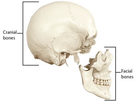
Image: Bones of the skull — Cranial and Facial regions

Image: Skull bones are joined by sutures. These joints appear as interlocking, jagged edges where the bones of the skull weave together.
What the skull is used for
The skull has several essential functions:
- protection of the brain and brainstem
- protection of cranial nerves and major blood vessels
- formation of the eye sockets, nasal cavity, and oral cavity
- housing of hearing and balance organs
- providing attachment points for muscles of the head and neck
Calvaria and skull base
From an anatomical and clinical perspective, the skull is divided into:
- the calvaria (skull vault)
- the skull base
The calvaria (skull vault)
The calvaria forms the roof and sides of the cranial cavity. It is composed mainly of flat bones.
Some bones of the calvaria are relatively simple in shape, such as the parietal bones, which form much of the lateral skull vault. Other bones are more complex in structure and function. A key example is the temporal bone, which participates both in the skull vault and the skull base.

Image: The calvaria (skull vault)
The skull base
The skull base is a complex, irregular structure that supports the brain from below. It separates the brain from the nasal cavity, sinuses, and upper neck structures.
The skull base is traditionally divided into three cranial fossae (basin-like depressions that support the different parts of the brain):
- anterior cranial fossa
- middle cranial fossa
- posterior cranial fossa
These fossae accommodate different parts of the brain and are separated and reinforced by dural folds.

Image: The skull base and its three fossae (anterior, middle, and posterior). Percentages indicate the frequency of fractures in each region.
Dural partitions and intracranial compartments
Inside the skull, the brain is not suspended freely. Instead, it is held in place by strong dural membranes. These membranes act as internal partitions, creating separate compartments that prevent the brain from shifting during head movement.
Important dural partitions include:
- falx cerebri, which separates the two cerebral hemispheres
- tentorium cerebelli, which separates the cerebrum from the cerebellum
- falx cerebelli, a smaller fold in the posterior fossa
These partitions help stabilize the brain and influence how pressure, bleeding, or swelling spreads inside the skull.

Image: The falx cerebri and tentorium cerebelli — dural partitions (membranes) that divide the cranial cavity.
These dural partitions are not just anatomical features — they determine how bleeding and swelling spread inside the skull. For example, epidural hematomas typically remain limited by suture lines, while subdural hematomas can spread across large areas of the brain surface. Herniation patterns are also strongly influenced by the tentorium and falx.
Openings of the skull base (foramina)
The skull base contains numerous openings (foramina) that allow nerves and blood vessels to pass between the brain and the rest of the body.
Examples include:
- foramen magnum — passage of the brainstem, spinal cord, and vertebral arteries
- optic canal — passage of the optic nerve and ophthalmic artery
- jugular foramen — passage of major venous drainage and lower cranial nerves
- carotid canal — passage of the internal carotid artery
Because of these openings, fractures or lesions of the skull base can affect nerves, blood flow, or cerebrospinal fluid dynamics.

Image: Superior view of the left skull base (left) and inferior view of the right skull base (right). Shown are the brainstem and the numerous nerves, arteries, and veins passing through the foramina and canals of the skull base.
Sinuses of the skull
Some skull bones contain air-filled cavities called paranasal sinuses. These include the frontal, ethmoid, sphenoid, and maxillary sinuses.
The sinuses are not fully developed at birth. They gradually form and enlarge during childhood and adolescence (most reach near-adult size during the teenage years).
Sinuses reduce skull weight, influence voice resonance, and are closely related to the skull base and orbital structures.
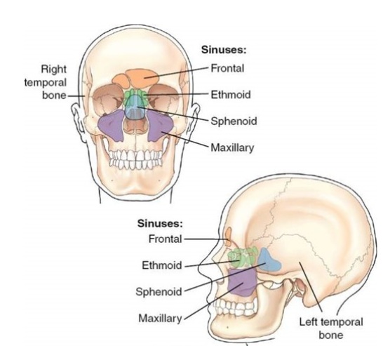
Image: Paranasal sinuses – frontal, ethmoid, sphenoid, and maxillary sinuses.
Ear structures and mastoid air cells
The skull also contains important structures related to hearing and balance.
The inner and middle ear are housed within the temporal bone, especially in its dense and complex petrous pyramid, which also contains the facial nerve canal. This region represents one of the most anatomically intricate areas of the skull.
Behind the ear, the temporal bone contains mastoid air cells — a system of small air-filled spaces that communicate with the middle ear. These structures are clinically important because infections or fractures in this area can spread to adjacent intracranial compartments.

Image: Internal and middle ear — nerves and arteries within the complex anatomy of the petrous part of the temporal bone.
Why skull anatomy matters clinically
Understanding skull anatomy is essential when imaging reports or clinical notes mention:
- skull fractures
- skull base injuries
- Traumatic Brain Injury
- epidural or subdural hematomas
- cranial nerve deficits
- cerebrospinal fluid leaks
- sinus or ear-related complications
It also provides a necessary foundation for understanding surgical access to the brain, such as a craniotomy.
The skull is not just a rigid shell. Its structure, compartments, and openings strongly influence how disease, trauma, and pressure affect the brain.