Dr Željko Kojadinović — NEUROHIRURGIJA I LEČENJE BOLA
Dr Zeljko Kojadinovic — Pain Treatment & Neurosurgery

Author: Dr. Zeljko Kojadinovic, MD, PhD – Neurosurgeon and Pain Management Specialist
Last medically reviewed: June 7, 2026
Who this page is for
This page is for patients with chronic or recurrent knee pain that persists for months — even after proper conservative therapy or surgery. MRI can show structural changes that appear significant but do not actually explain the pain.
In many such cases, the real problem lies in extra-articular pain sources — such as irritated tendons, bursae, or small nerve branches around the knee — which are not clearly visible on imaging and are not resolved by surgery. If surgery and RFA have not relieved your pain, it is essential to determine whether total knee replacement (TKA) would actually address the problem — or if a less aggressive, targeted treatment is actually the correct option.
These hidden causes can often be recognized during a detailed online consultation through symptom mapping, movement analysis, and review of prior reports. You can request an online consultation with our specialist to determine whether your pain may come from such treatable sources.
If you are unsure whether an online consultation can help after all previous tests and treatment, read why this consultation is different.
When patients usually seek a second opinion for persistent knee pain
- Your MRI shows structural changes (cartilage wear, meniscal tear, tendinopathy), but the findings do not clearly explain the severity of your pain
- You continue to have pain despite physiotherapy, injections, RFA, or even prior surgery
- You are being advised to consider arthroscopy or joint replacement and want to understand whether surgery is truly necessary
- Your pain is localized to a specific area (inner side, outer side, front, or back of the joint), but imaging appears “normal”
- Different doctors have given different recommendations about whether to operate, inject, or simply wait
A focused telehealth review can help determine whether the pain source is intra-articular or extra-articular, whether surgery is medically justified, and what targeted treatment options may provide real improvement.
Contents
- Epidemiology and Misconceptions
- Distribution of Pain Sources
- Intra-articular sources
- Extra-articular Causes
- Symptom Patterns Overview
- Symptoms from Intraarticular Causes
- Symptoms Mimicking Joint Pain
- Diagnostic Approach
- Treatment of Knee Pain
- Treatment of Contributing Factors in Knee Pain
- Prognosis and Long-Term Outlook
- Knee Pain FAQ
- Additional Patient Resources
Epidemiology and Common Misconceptions
Knee pain is among the top three musculoskeletal complaints in adults, affecting up to 25% of people over 50.
However, radiographic osteoarthritis (OA) is found in far more individuals than those who report pain — only about half of those with X-ray evidence of OA actually have symptoms.
Conversely, many patients with disabling pain have normal or near-normal imaging because the source is extra-articular (bursae, tendons, or muscle attachments).
This mismatch explains why so many ineffective surgeries are performed based solely on imaging.
Distribution of Pain Sources
Knee pain may originate from structures located inside the joint capsule (intra-articular) or from soft tissues outside the capsule (extra-articular). In clinical practice, roughly 50–60% of painful knees are intra-articular in origin, and 40–50% are extra-articular. Distinguishing between the two is crucial, since their mechanisms, treatment, and prognosis differ completely.
Intra-articular sources
Pain arising from inside the joint capsule reflects irritation or injury of structures such as:
- Articular cartilage, which undergoes degenerative wear in osteoarthritis, leading to loss of the smooth gliding surface between the femur and tibia.
- Menisci, two crescent-shaped fibrocartilaginous pads that act as shock absorbers between the femur and tibia. Degeneration or tearing disrupts joint congruence and causes mechanical irritation of the joint lining.
- Synovial membrane, which lines the inner surface of the joint capsule, is a thin, vascular tissue that produces the lubricating synovial fluid necessary for smooth cartilage movement. When irritated by cartilage wear debris, crystal deposits (such as urate or calcium pyrophosphate), or chemical by-products of degeneration, it becomes inflamed — a condition known as synovitis. The inflamed membrane thickens and secretes excess fluid, leading to joint effusion, swelling, and stiffness even when X-rays appear normal.
- Cruciate ligaments (anterior and posterior), which stabilize forward and backward translation (sliding motion) of the tibia beneath the femur. Partial or complete rupture leads to abnormal joint motion and secondary inflammation.
- Less frequent intra-articular pain generators include plica irritation (redundant synovial folds that become inflamed) and loose bodies of cartilage or bone moving freely inside the joint space.
Overall, intra-articular pain reflects structural damage, inflammation, or mechanical instability within the joint itself.
Extra-articular sources
Extra-articular pain often comes from surrounding soft tissues rather than the joint itself. Pain from outside the capsule arises from the supporting tendons, ligaments, bursae, and fat pads that stabilize or move the joint:
- Collateral ligaments (medial and lateral) provide side-to-side stability and may be strained or inflamed after overload or trauma.
- Tendons of the quadriceps and patellar mechanisms may develop microtears and degeneration (tendinopathy) from repetitive stress.
- Bursae — small fluid-filled sacs that reduce friction — can become inflamed (bursitis). Common sites include the prepatellar bursa (in front of the kneecap), the infrapatellar bursa (below it), and the pes anserine bursa (inner side of the upper shin), which often coexists with arthritis.
- Collateral ligament injuries cause pain and tenderness on either the inner (medial) or outer (lateral) side of the knee, often after sudden twisting or side impact. Mild sprains produce sharp pain when the knee bends sideways, while more significant injuries can lead to swelling and a feeling of instability.
- Hoffa’s fat pad, located just beneath the patellar tendon, can become inflamed or fibrotic after repetitive compression or direct trauma.
- Iliotibial (IT) band friction at the lateral femoral condyle produces irritation of the underlying tissue due to repetitive flexion and extension.
- In the posterior (popliteal) region, extra-articular pain may originate from strain of the gastrocnemius or hamstring tendons, rupture of a Baker’s cyst, or entrapment of the tibial or peroneal nerve.
Vascular problems such as popliteal artery entrapment or superficial thrombophlebitis can occasionally mimic musculoskeletal pain.
In some patients, apparent knee pain is referred from the hip or lumbar spine, despite normal imaging of the knee itself.
Understanding whether the lesion is intra- or extra-articular is essential: true intra-articular damage (e.g., meniscal tear, cruciate injury, advanced OA) may require surgical intervention, while most extra-articular conditions respond well to conservative or image-guided treatment.
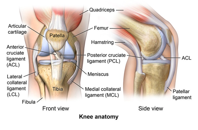
Image- Complex knee anatomy
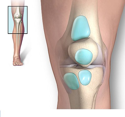
Image: Inflamed knee bursae. While treatment is generally successful, they can be a source of severe knee pain and mobility issues for years before a proper diagnosis.
Symptoms of Knee Pain
Clinical presentation depends on the underlying anatomical source.
Symptoms from Intraarticular Causes
Pain is usually deep and diffuse, felt “inside the joint,” and aggravated by weight bearing, climbing stairs, or prolonged walking.
- In osteoarthritis, pain is dull and mechanical, with stiffness, swelling, and crepitus (crackling) during motion.
- Meniscal tears cause sharp, localized joint-line pain (inner or outer), sometimes with catching, locking, or a sense of giving way.
- Synovitis or chondral lesions lead to persistent swelling, warmth, and a feeling of fullness even when X-rays are normal.
- Cruciate injuries cause deep pain and a sense of instability, especially when pivoting or descending stairs.
- Occasional stabbing pain may occur when a plica fold or loose body gets trapped during movement.
Symptoms from Extraarticular Causes
Pain is typically well localized, reproducible by palpation or specific movement, and related to the tendon, bursa, or ligament involved.
- Pes anserine bursitis causes tenderness on the inner upper shin, sometimes radiating upward.
- IT-band friction produces lateral knee pain that worsens with repetitive bending.
- Patellar or quadriceps tendinopathy leads to pain above or below the kneecap, aggravated by climbing, running, or squatting.
- Hoffa’s fat pad inflammation produces burning or pressure-like anterior pain beneath the kneecap.
- Posterior knee pain (popliteal area) may stem from muscle strain, Baker’s cyst, or nerve irritation, often described as deep tightness behind the knee.
- Referred pain from the hip or lumbar spine mimics knee pain but is associated with normal knee motion and imaging.
In summary, intra-articular pain tends to be deep, aching, and diffuse, while extra-articular pain is superficial, focal, and movement-related. Correct localization of the pain generator is the key to effective treatment.
Diagnostic Approach — Why Imaging Alone Is Not Enough
Plain radiographs detect joint-space narrowing, osteophytes, and sclerosis — but these are not reliable predictors of pain.
Accurate diagnosis integrates:
- Focused physical exam: palpation of specific tender zones, patellar tracking, gait, and dynamic pain tests.
- Ultrasound: reveals soft-tissue causes (bursitis, tendinopathy, fluid) and allows guided diagnostic injections.
- MRI: indicated when intra-articular pathology (such as a meniscal tear or cartilage defect) is suspected, or when conservative therapy fails. It may also suggest extra-articular abnormalities, but it cannot reliably differentiate the true pain generator — unlike a focused clinical examination, ultrasound assessment, or navigated diagnostic blocks. MRI can show structural changes that appear significant but do not actually explain the pain.
- Diagnostic blocks: selective injections (pes anserine, IT-band, intra-articular) to confirm the pain generator.
Treatment of Knee Pain
Most patients improve without surgery once the true pain source is identified and treated.
Medication & local therapy — Short courses of NSAIDs for acute inflammation. When pain is localized, ultrasound-guided injections of long-acting corticosteroids or platelet-rich plasma (PRP) can be placed precisely into the affected bursa, tendon sheath, or joint space.
Physiotherapy — A structured program aimed at restoring quadriceps and gluteal control, correcting load distribution, and improving flexibility.
Key principles include gradual strengthening, avoidance of deep squats or stairs during flares, and activation of hip abductors to offload the knee.
Interventional Pain Knee Procedures
- For extra-articular pain: ultrasound-guided lavage or aspiration of inflamed bursae, or peritendinous hydrodissection.
- For intra-articular pathology: diagnostic or therapeutic intra-articular injections, viscosupplementation, or radiofrequency ablation (RFA) of genicular nerves when standard therapy fails.
Surgery —
- Arthroscopy is not recommended for degenerative knee disease without mechanical locking; large meta-analyses show minimal benefit beyond physiotherapy. Partial meniscectomy or chondroplasty is reserved for true mechanical lesions verified clinically and on MRI.
- Total knee arthroplasty (TKA) is indicated for advanced OA with major cartilage loss and severe limitation of movement.
- Genicular nerve RFA provides pain relief for about 6–12 months in patients who are poor surgical candidates or awaiting arthroplasty. The procedure interrupts sensory fibers transmitting pain from the joint capsule and subchondral bone, primarily through the superior medial, superior lateral, and inferior medial genicular nerves.
However, it does not act on extra-articular pain sources — such as tendons, bursae, or small nerve branches around the knee — which often remain symptomatic even after a technically successful RFA. If you have undergone genicular denervation but your knee pain persists, it may originate from these extra-articular structures, which are not influenced by RFA and can often be identified through a targeted clinical or ultrasound-guided evaluation.
Only about 10–15% of all chronic knee pain cases ultimately require surgical or nerve-ablative procedures.
The remaining majority can be successfully managed with a combination of conservative therapy (activity modification, medications, targeted physical therapy) and image-guided management — which includes ultrasound-guided diagnosis and precise local treatments such as perineural injections, hydrodissection, or tendon and bursal procedures that directly target the actual pain source.
Online pain consultation for regional pain in detail
Schematic explanation of the video consultation for regional pain
Answers to questions about the process and success of video consultations for regional pain
There are several common reasons for poor therapeutic outcomes in the treatment of chronic pain, which are often seen in patients with regional pain.
Artificial intelligence can also support the process by analyzing complex pain syndromes in fibromyalgia, but clinical expertise remains essential.
Why Knee Pain Does Not Improve Despite Treatment
Many patients continue to experience knee pain despite undergoing standard treatments such as medication, physiotherapy, or injections.
In a large number of cases, this is not because treatment options are ineffective, but because the dominant pain source has not been clearly identified. Imaging findings such as cartilage degeneration, meniscal changes, or mild osteoarthritis are common, but they often do not fully explain the intensity or pattern of symptoms.
Pain may persist when treatment is directed at structural findings rather than the true functional pain generator, or when contributing factors — such as altered biomechanics, muscle imbalance, joint overload, or systemic influences — are not addressed. As a result, even well-applied therapies may fail to achieve lasting improvement.
Treatment of Contributing Factors in Knee Pain
Effective treatment of knee pain always begins with identifying the primary pain generator — the specific structure responsible for symptoms (such as intra-articular pathology including cartilage or meniscus, pes anserine bursa, patellar or quadriceps tendon, iliotibial band, or small nerve branches around the knee).
However, in many patients, pain persists not only because of the local problem, but because additional contributing factors are not recognized or adequately addressed. These factors rarely act as the sole cause of pain, but they can maintain irritation, delay recovery, and reduce the effectiveness of otherwise appropriate treatment.
For that reason, successful management of knee pain requires not only treating the primary structure, but also understanding the broader mechanical and systemic context.
What contributing factors may play a role in knee pain?
- Repetitive loading and mechanical overload — Walking, stairs, squatting, or prolonged standing can continuously stress the same structures, especially in degenerative conditions.
- Lower limb alignment and biomechanics — Varus/valgus alignment, altered gait, or poor load distribution increase stress on specific compartments of the knee.
- Muscle imbalance and reduced joint stability — Weak quadriceps, gluteal muscles, or poor neuromuscular control lead to overload of tendons, bursae, or joint surfaces.
- Hip and core dysfunction — Poor hip control (especially abductors) shifts load toward the knee and contributes to persistent symptoms.
- Metabolic factors, pro-inflammatory diet and low-grade inflammation — Obesity, insulin resistance, and chronic inflammation increase joint stress and pain sensitivity.
- Nutritional deficiencies — Low vitamin D, B12, or other micronutrients may impair tissue recovery.
- Sleep disturbances and chronic pain cycle — Poor sleep increases pain sensitivity and delays healing.
- Central sensitization — The nervous system amplifies pain signals even when structural damage is limited.
- Reduced activity and deconditioning — Avoidance of movement leads to weakness and further joint overload.
- Medications and previous treatments — Certain medications such as statins, long-term use of pain medications, corticosteroids, or medications that cause excessive sedation may contribute to muscle or tendon-related pain, reduce tissue quality, or alter pain perception. Previous treatments, including repeated injections or incomplete rehabilitation, may also maintain symptoms if the underlying mechanism was not fully addressed.
- Other medical conditions and comorbidities — Conditions such as autoimmune diseases, thyroid disorders, diabetes, or chronic inflammatory states may increase pain sensitivity, affect tendon and joint structures, and reduce the response to otherwise appropriate treatment.
- Vitamin-related factors — Both deficiencies and excesses of certain vitamins (particularly vitamin B6) may contribute to nerve-related symptoms, burning pain, or altered sensitivity around the knee.
- Tissue quality and degeneration — Age-related changes, reduced blood supply, or repeated micro-injuries may impair tendon healing and increase vulnerability to persistent pain, even when mechanical factors are partially corrected.
Why this matters in practice
In many cases, treatment fails because the primary pain generator is not correctly identified, and therapy is directed only at contributing factors such as exercise, posture, or lifestyle changes. Conversely, even when the main structural cause is treated, failure to recognize and address contributing factors often leads to only partial or temporary improvement.
The most effective approach is a carefully selected combination of treatment that addresses both the primary pain generator and the contributing mechanical, functional, and systemic factors. In contrast, an inadequate or incomplete combination — even when it includes individually effective methods — is a common reason for suboptimal or short-lasting results.
This approach significantly increases the likelihood of long-term improvement and reduces the need for repeated injections or surgery.
In practice, many patients try to address contributing factors on their own — for example through exercise programs, posture correction, anti-inflammatory diet, supplements (vitamin D, magnesium, glucosamine, chondroitin), or supportive methods.
While these approaches can be helpful, they rarely lead to lasting improvement if the primary pain generator is not clearly identified and treated. On the other hand, even well-targeted medical treatment may fail if all contributing factors are not recognized and corrected.
Many patients reading this recognize that they have already tried one part of this approach — but not the complete strategy. This is one of the most common reasons why otherwise well-treated knee pain becomes chronic.
Prognosis and Long-Term Outlook
When the pain source is accurately localized and treated, most patients experience substantial and sustained improvement.
Persistent relief after a guided injection or successful physiotherapy usually predicts durable recovery and reduced need for surgery.
Why an Online Consultation Can Help When Knee Pain Persists Despite Treatment
A video consultation for persistent knee pain can help identify the exact source of your pain — one or more pain generators — as well as the factors that trigger and maintain it. Knee MRI or X-ray findings can show cartilage wear, meniscal changes, arthritis, or tendon abnormalities, but these findings do not always explain the severity or location of pain. This is achieved through a detailed conversation and review of your MRI scans, X-rays, ultrasound reports, previous treatments, injections, RFA or surgery reports when available. During the session, you are instructed to perform specific knee movements, walking or stair-related tests, regional pressure tests, and pain-location mapping to see what increases, reduces, or changes your pain. This helps identify which knee pain source is active. Many extra-articular pain generators and sustaining factors cannot be clearly identified from imaging alone.
This may sound like examinations you have already had. It is not — because what matters most is not the test itself, but who interprets it. Only a specialist with deep knowledge of knee pain anatomy knows which questions to ask, how to distinguish pain from inside the joint from pain around the joint, where to instruct you to press, which movements to test, and how to decide whether the pain pattern matches MRI findings, RFA targets, arthroscopy, or knee replacement. This is not just another opinion.
You will also receive advice on which additional factors that trigger and sustain knee pain should be investigated — such as altered gait mechanics, lower-limb alignment, muscle imbalance, hip or lumbar spine contribution, repetitive loading, local inflammation, tendon or bursa irritation, small nerve-branch irritation, metabolic factors, vitamin deficiencies, side effects of other medications, and other overlooked contributors. In many patients who have already visited several specialists, these factors have still not been fully investigated.
Once the main knee pain mechanism is identified, treatment follows: targeted treatment of the dominant pain source, combined with correction of contributing mechanical, functional, and systemic factors. This may include medication adjustment, focused rehabilitation, activity modification, ultrasound-guided evaluation, diagnostic blocks, or image-guided treatment when needed. When selecting or adjusting medications, we take into account whether patients are older, sensitive to side effects, or have other health conditions, and we use a safe combination for the shortest necessary duration to avoid medication overload. All recommendations are explained during the conversation and are also given in a written medical report.
Many patients assume that because physiotherapy, injections, RFA, arthroscopy, denervation, or even previous surgery have not helped, knee replacement or another procedure is now the only option. In many cases, this is not true — previous treatment may have targeted imaging findings rather than the true pain generator. During the consultation, we help assess whether genicular nerve denervation, arthroscopy, or knee replacement is medically justified, whether the pain pattern actually supports that procedure, and what should be checked before such a decision. We also help when pain persists after these interventions, by reassessing whether the remaining pain comes from inside the joint, around the joint, small nerve branches, the hip, lumbar spine, altered mechanics, or another overlooked source.
Request Persistent Knee Pain Second Opinion — 24-Hour Review (Priority Option Available Within Hours)
Persistent knee pain despite physiotherapy, injections, RFA, arthroscopy, denervation, or previous surgery
often raises important questions:
Is the pain really coming from inside the knee joint?
Do MRI or X-ray findings truly explain the symptoms?
Is arthroscopy, genicular nerve denervation, or knee replacement actually the right next step?
Could the pain come from tendons, bursae, fat pad irritation, small nerve branches, the hip, lumbar spine, sacroiliac region, or another extra-articular source?
An independent specialist second opinion may help clarify whether the dominant pain source is
intra-articular or extra-articular, whether the proposed procedure is medically justified,
whether more targeted treatment — such as medication adjustment, correction of contributing mechanical and systemic factors, ultrasound-guided evaluation, diagnostic injections, focused rehabilitation, or image-guided treatment — may be more appropriate before arthroscopy or joint replacement.
- ✔ Send a brief message describing your knee pain location, how long it has lasted, what makes it worse, and which treatments have already been tried
- ✔ You will receive a reply within 24 hours explaining whether an online consultation is appropriate and which documentation is required
- ✔ Priority cases: severe persistent pain after arthroscopy, knee denervation, or knee replacement; conflicting specialist recommendations; rapidly worsening function; or uncertainty before a proposed operation — write PRIORITY in your first message
- ✔ MRI, X-ray, ultrasound reports, operative notes, injection history, RFA or denervation reports, and physiotherapy summaries can be reviewed
- ✔ During consultation we analyze whether the dominant pain generator is inside the knee joint or around the joint, which specific structures are most likely responsible, and whether contributing factors may be maintaining the pain.
- ✔ We explain which treatment direction best matches the suspected dominant pain generator — including medication adjustment, correction of contributing factors, targeted rehabilitation, or diagnostic blocks when the responsible structure is uncertain. If denervation, arthroscopy, or knee replacement is being considered, we clarify whether the pain pattern supports that decision before discussing it with your local treating team — with up to 10 days of follow-up clarification.
Consultation fees typically range from $180–250 depending on case complexity and documentation volume.
Secure payment by credit card, PayPal invoice (USD), or bank transfer.
Based on our medical report, reimbursement may be possible if your insurance plan allows it.
This corresponds to typical international specialist telehealth second-opinion services for complex pain and treatment-decision review.
FAQ About Persistent Knee Pain
Why can persistent knee pain come from around the joint rather than inside the knee joint?
Persistent knee pain does not always come from the joint surface, cartilage, meniscus, or arthritis inside the knee. In many patients, the painful structure is outside the joint capsule: tendons, bursae, ligaments, fat pads, muscle attachments, or small nerve branches around the knee. These extra-articular structures can produce severe and very localized pain, even when X-ray or MRI findings look mild or do not explain the symptoms. This is why the exact pain location, tenderness to palpation, movement triggers, gait, and response to targeted injections can be more informative than imaging alone. If treatment is directed only at a visible MRI finding while the true pain generator is outside the joint, physiotherapy, injections, RFA, arthroscopy, or even surgery may fail.
What is the difference between intra-articular, extra-articular and periarticular knee pain?
Intra-articular knee pain comes from structures inside the joint capsule, such as cartilage, meniscus, synovial membrane, cruciate ligaments, loose bodies, or advanced osteoarthritis. It is often deeper, more diffuse, and related to weight bearing, swelling, stiffness, locking, or instability. Extra-articular pain comes from structures outside the joint capsule, such as bursae, tendons, ligaments, iliotibial band, Hoffa’s fat pad, muscle attachments, or nearby nerves. This pain is often more localized and reproducible by pressing on a specific tender point or performing a specific movement. Periarticular pain is a broader term for pain around the joint. The distinction matters because intra-articular disease may sometimes require joint-directed treatment, while many extra-articular causes respond better to targeted injections, ultrasound-guided procedures, rehabilitation, or nerve-oriented treatment.
Can knee X-ray show degenerative changes that are not the true cause of pain?
Yes. Knee X-rays often show degenerative changes such as joint-space narrowing, osteophytes, sclerosis, or osteoarthritis, especially in adults over 50. However, these findings do not automatically prove that the visible degeneration is the true source of pain. Many people have radiographic osteoarthritis without significant symptoms, while others have disabling pain with normal or near-normal X-rays. X-ray is useful for identifying advanced joint degeneration, deformity, fractures, and severe cartilage loss, but it cannot reliably identify soft-tissue pain generators such as bursitis, tendinopathy, fat pad irritation, small nerve branch pain, or referred pain from the hip or spine. For this reason, treatment decisions should not be based only on “degeneration” seen on X-ray. The imaging must match the pain pattern and physical findings.
Can knee MRI show meniscus, cartilage or arthritis findings that do not explain the pain?
Yes. Knee MRI can show meniscal degeneration, cartilage wear, chondral defects, tendinopathy, synovitis, bone marrow changes, or mild osteoarthritis, but not every finding is the true cause of pain. MRI is excellent for showing anatomy, yet it does not automatically prove which structure is generating symptoms. A meniscal tear, cartilage change, or degenerative finding may be incidental, especially if the pain location, movement triggers, swelling pattern, or examination do not match that structure. MRI also may not clearly show some extra-articular pain sources, small nerve branches, subtle bursitis, dynamic irritation, or referred pain. Therefore, MRI should be interpreted together with a focused examination, symptom mapping, ultrasound assessment, and sometimes diagnostic blocks. The key question is not “what does MRI show?” but “which finding explains this patient’s pain?”
How can a doctor identify the real pain generator in persistent knee pain?
Identifying the real pain generator in persistent knee pain requires more than reading an MRI report. The doctor must map the exact pain location, determine whether the pain is deep or superficial, check which movements provoke it, and compare symptoms with anatomy. Palpation of specific tender zones, patellar tracking, gait analysis, dynamic tests, and comparison with hip, lumbar spine, and sacroiliac sources are important. Ultrasound can show bursitis, tendinopathy, fluid, tendon sheath irritation, or soft-tissue pain sources and can guide precise injections. Diagnostic blocks are especially useful when several possible pain generators exist. If a selective injection temporarily relieves the patient’s typical pain, it strongly suggests that the blocked structure is clinically relevant. This process helps avoid treating MRI findings that are not actually painful.
Can knee pain come from small nerve branches rather than cartilage, meniscus or arthritis?
Yes. Knee pain can come from small sensory nerve branches around the knee rather than from cartilage, meniscus, or arthritis. These nerves supply the skin, joint capsule, periosteum, ligaments, and soft tissues around the knee. When irritated by trauma, surgery, scar tissue, inflammation, repeated injections, mechanical overload, or local entrapment, they may produce burning, stabbing, stinging, electric, hypersensitive, or sharply localized pain. This type of pain may not be well explained by MRI or X-ray. It may also persist after arthroscopy, knee replacement, or genicular nerve ablation if the treated structure was not the dominant pain source. Recognizing nerve-related knee pain requires careful symptom mapping, sensory examination, knowledge of regional neuroanatomy, and sometimes diagnostic blocks or ultrasound-guided perineural treatment.
Can knee pain come from the hip, lumbar spine or sacroiliac region?
Yes. Pain felt in the knee can sometimes be referred from the hip, lumbar spine, sacroiliac region, or nerves supplying the leg. Hip disorders may refer pain toward the thigh or knee, while lumbar nerve root irritation can mimic knee pain or produce associated sensory symptoms. Sacroiliac or pelvic mechanics can also change gait and load distribution, indirectly worsening knee symptoms. Referred pain is especially important when knee imaging is normal or shows mild findings that do not match the severity of pain. It should also be considered when knee motion is relatively preserved, when pain changes with back or hip position, or when symptoms extend beyond the knee. A correct diagnosis requires checking the entire kinetic chain, not only the knee joint.
Can pain behind the knee come from nerves, tendons or a Baker’s cyst rather than the joint itself?
Yes. Pain behind the knee, in the popliteal region, has a different differential diagnosis from pain inside the joint line. It may come from a Baker’s cyst, gastrocnemius or hamstring tendon strain, posterior capsule irritation, bursitis, vascular problems, or irritation of nerves near the posterior knee. A Baker’s cyst may produce fullness, tightness, or pain behind the knee, especially when bending or walking. Tendon and muscle injuries are often movement-related and reproducible by resisted testing. Nerve irritation may cause burning, radiating, tingling, or electric sensations. Vascular conditions are less common but must be considered when swelling, calf pain, color change, or circulatory symptoms are present. Because posterior knee pain has many possible causes, imaging alone is not enough; localization and clinical pattern are essential.
Why can knee pain persist despite physiotherapy, injections or medication?
Knee pain can persist despite physiotherapy, injections, or medication when treatment is not directed at the true pain generator. For example, therapy may focus on arthritis seen on X-ray while the dominant pain comes from pes anserine bursitis, Hoffa’s fat pad, patellar tendinopathy, small nerve branches, referred pain, or altered biomechanics. Injections may help temporarily if they reduce inflammation, but pain returns when load distribution, muscle imbalance, repetitive stress, or systemic factors are not corrected. Physiotherapy may also fail if the program is too generic or aggravates the painful structure during a flare. Medication can reduce symptoms but usually does not identify the cause. Persistent pain requires reassessment of the primary structure, contributing mechanical factors, metabolic influences, sleep, central sensitization, and previous treatment choices.
Why can knee pain persist after arthroscopy or knee surgery?
Knee pain can persist after arthroscopy or knee surgery when the procedure corrected a visible structural finding but not the dominant pain source. Degenerative meniscal tears, cartilage wear, or mild intra-articular changes may be present on MRI, but they may not be the main cause of symptoms. Pain may instead arise from bursae, tendons, fat pad irritation, scar tissue, small sensory nerve branches, altered biomechanics, or referred pain from the hip or spine. Arthroscopy is generally not recommended for degenerative knee disease without true mechanical locking, because the benefit may be limited when pain is not caused by a treatable mechanical lesion. Persistent pain after surgery should not be interpreted automatically as surgical failure; the real question is whether the original pain generator was correctly identified before surgery.
Why can knee pain persist after genicular nerve ablation or knee denervation?
Knee pain can persist after genicular nerve ablation or knee denervation because these procedures mainly target sensory fibers transmitting pain from the joint capsule and subchondral bone. They do not treat all possible pain generators around the knee. If the dominant pain source is extra-articular — such as pes anserine bursitis, patellar or quadriceps tendinopathy, iliotibial band irritation, Hoffa’s fat pad inflammation, scar-related pain, or small superficial nerve branch irritation — genicular denervation may not help enough. RFA may also fail when central sensitization, poor biomechanics, muscle imbalance, inflammatory state, or referred pain from the lumbar spine or hip maintains symptoms. A diagnostic block before denervation can improve patient selection, but persistent pain after RFA usually means the pain generator needs to be reassessed.
Can central sensitization make knee pain persist even when structural damage is limited?
Yes. Central sensitization can make knee pain persist or feel disproportionate even when structural damage is limited. In this situation, the nervous system becomes more sensitive and amplifies pain signals from the knee and surrounding tissues. A patient may feel severe pain from mild tissue irritation, pressure, movement, or previously non-painful stimuli. Central sensitization does not mean the pain is imaginary. It means that the local pain generator and the nervous system response must both be considered. It is more likely when pain has lasted for months, sleep is poor, stress is high, multiple treatments have failed, or pain spreads beyond the original structure. Treatment then requires a combined approach: identifying the primary pain source, reducing peripheral irritation, improving sleep and function, and addressing nervous system hypersensitivity.
Can metabolic or systemic factors make persistent knee pain harder to treat?
Yes. Metabolic and systemic factors can make persistent knee pain harder to treat, even when the local diagnosis is correct. Obesity increases mechanical load across the knee and may also contribute to low-grade inflammation. Insulin resistance, diabetes, thyroid disorders, autoimmune disease, chronic inflammatory states, poor sleep, and pro-inflammatory diet can increase pain sensitivity and reduce tissue recovery. Nutritional problems such as low vitamin D or B12 may impair healing, while excessive vitamin B6 can contribute to nerve-related symptoms in some patients. Certain medications, repeated injections, incomplete rehabilitation, deconditioning, and chronic pain cycles may also maintain symptoms. These factors are rarely the only cause of knee pain, but they can reduce the effect of otherwise appropriate treatment. Durable improvement often requires addressing both the primary pain generator and these contributing factors.
When are diagnostic blocks useful in persistent knee pain?
Diagnostic blocks are useful in persistent knee pain when imaging shows several possible abnormalities, when symptoms do not match MRI findings, or when it is unclear whether pain is intra-articular, extra-articular, nerve-related, or referred. A diagnostic block uses a local anesthetic injection around a suspected pain generator, such as the knee joint, pes anserine bursa, iliotibial band region, tendon sheath, genicular nerve region, or small sensory nerve branch. If the patient’s typical pain temporarily improves, the blocked structure is likely clinically relevant. This can prevent unnecessary surgery, poorly targeted injections, or denervation procedures that would not address the true source. Diagnostic blocks are most useful when combined with clinical examination, ultrasound guidance, movement testing, and careful interpretation of the patient’s usual pain pattern.
Can a diagnostic block show whether genicular nerve denervation may help?
A diagnostic genicular nerve block can help estimate whether genicular nerve denervation may relieve knee pain, but it is not perfect. The block temporarily anesthetizes sensory nerve pathways that transmit pain from parts of the knee joint capsule and subchondral bone. If the patient’s typical deep joint pain improves significantly for the expected anesthetic duration, genicular RFA may be more likely to help. However, a positive block does not guarantee long-term success, and a negative or incomplete response may suggest that the dominant pain comes from extra-articular structures, small superficial nerve branches, tendons, bursae, fat pad irritation, referred pain, or central sensitization. The block should therefore be interpreted in context. It is a tool for patient selection, not a complete diagnosis by itself.
Why is knee arthroscopy not always useful for chronic knee pain?
Knee arthroscopy is not always useful for chronic knee pain because many chronic pain cases are degenerative, extra-articular, inflammatory, or biomechanical rather than caused by a surgically correctable mechanical lesion. Arthroscopy may help when there is true mechanical locking, loose bodies, or a clearly symptomatic mechanical meniscal tear that matches the patient’s history and examination. However, for degenerative knee disease without true locking, evidence shows limited benefit beyond physiotherapy and conservative management. If pain comes from bursitis, tendinopathy, fat pad irritation, small nerve branches, central sensitization, or referred pain from the hip or spine, arthroscopy will not address the main problem. Before arthroscopy, the key question should be whether the MRI finding is truly responsible for the patient’s symptoms.
Why is knee replacement not always the best solution for knee pain?
Knee replacement is not always the best solution for knee pain because it treats advanced intra-articular joint degeneration, not every pain source around the knee. Total knee arthroplasty is most appropriate when there is advanced osteoarthritis with major cartilage loss, significant functional limitation, and symptoms that match the joint degeneration. If the dominant pain comes from tendons, bursae, fat pad irritation, small nerve branches, referred lumbar or hip pain, central sensitization, or metabolic/systemic pain amplification, replacing the joint may not fully relieve symptoms. MRI or X-ray degeneration alone is not enough. The pain pattern, examination, movement limitations, response to injections, and functional disability must support the decision. Before replacement, it is important to confirm that the joint itself is the main pain generator.
Why can pain persist even after technically successful knee replacement?
Pain can persist after technically successful knee replacement when the implant is well positioned but the original or new pain generator is not fully addressed. Some patients had extra-articular pain before surgery, such as tendon, bursa, fat pad, nerve branch, hip, spine, or central sensitization-related pain, and the replacement did not target that source. Others develop postoperative scar sensitivity, soft-tissue irritation, altered biomechanics, muscle weakness, stiffness, or neuropathic pain around the incision or joint. Persistent pain after knee replacement also requires exclusion of mechanical problems, loosening, infection, instability, malalignment, or patellar tracking issues. However, when those are absent, evaluation must move beyond the implant and assess soft tissues, nerves, referred pain, systemic factors, and chronic pain mechanisms.
What complications should be excluded when knee pain persists after surgery or knee replacement?
When knee pain persists after arthroscopy or knee replacement, true surgical or orthopedic complications must first be excluded. These include infection, implant loosening, malalignment, instability, patellar tracking problems, stiffness or arthrofibrosis, scar-related pain, recurrent meniscal or cartilage pathology, progression of arthritis in untreated compartments, or a new injury. Pain around the incision may also come from irritation or injury of small skin sensory nerves. These problems usually require in-person orthopedic assessment, examination, blood tests when infection is suspected, and appropriate imaging. If these complications are excluded, persistent pain may come from extra-articular structures, small nerve branches, referred pain from the hip or spine, central sensitization, or contributing mechanical and systemic factors. This distinction is essential before repeat surgery, denervation, or replacement revision is considered.
What should be checked before deciding on knee surgery, arthroscopy, denervation or replacement?
Before deciding on knee surgery, arthroscopy, denervation, or replacement, the doctor should confirm that the proposed procedure targets the true pain generator. This means checking whether pain is intra-articular, extra-articular, nerve-related, referred from the hip or spine, or maintained by central sensitization and systemic factors. Imaging should match the pain location, physical examination, movement triggers, swelling pattern, and functional limitation. If the pain source is unclear, ultrasound assessment or diagnostic blocks may help identify whether the joint, bursa, tendon, fat pad, genicular nerves, or smaller sensory branches are responsible. It is also important to review previous injections, physiotherapy, RFA, arthroscopy, systemic conditions, medications, metabolic factors, and rehabilitation. A technically correct procedure can still fail if it treats the wrong structure.
Can an online video consultation help identify the source of persistent knee pain?
An online video consultation can help identify the likely source of persistent knee pain when the problem is non-emergent and the patient has already had imaging, treatment, or conflicting opinions. The consultation can review MRI and X-ray reports, previous injections, surgery, RFA, physiotherapy, symptom history, and exact pain location. During video assessment, movement analysis, pain mapping, gait observation, and provocation patterns may suggest whether pain is intra-articular, extra-articular, nerve-related, referred from the hip or spine, or influenced by contributing factors. Video consultation cannot replace urgent in-person care or all physical tests, but it can often clarify whether the current treatment plan matches the likely pain generator. It can also help decide whether further ultrasound evaluation, diagnostic blocks, targeted treatment, or surgical reassessment is reasonable.
Additional Patient Resources — Knee Pain
-
NHS — Knee pain in adults
Common causes, self-care, red flags, and when to seek medical help. -
AAOS OrthoInfo — Osteoarthritis of the Knee
Symptoms, diagnosis, and treatment options for knee osteoarthritis. -
AAOS OrthoInfo — Meniscus Tears
Meniscal pain, when arthroscopy helps, and non-surgical care. -
RSNA RadiologyInfo — MRI of the Knee
What a knee MRI shows and when it is indicated. -
AAOS — Knee Conditioning Program
Safe strengthening and flexibility exercises to support recovery.