Dr Željko Kojadinović — NEUROHIRURGIJA I LEČENJE BOLA
Dr Zeljko Kojadinovic — Pain Treatment & Neurosurgery


Causes, Symptoms, Diagnosis, Bleeding Risk, and Treatment Options
Author:
Dr. Zeljko Kojadinovic, MD, PhD
— Consultant Neurosurgeon
Specialized Experience:
30 years of clinical expertise in neurosurgery.
Last medically reviewed:
June 6, 2026
Who This Brain AVM Page Is For
This Brain Arteriovenous Malformation (AVM) resource is designed primarily for patients and families who have been told that an MRI, CT, or angiography scan shows a brain AVM —
or who have experienced a brain hemorrhage, seizures, or unexplained neurological symptoms and are trying to understand what the diagnosis really means.
This page explains the biology of AVMs, their symptoms, and the decision-making process behind modern treatment options like embolization, radiosurgery, and microsurgical removal.
If you are receiving different recommendations or feel uncertain about your specific treatment path, an individualized
neurosurgical second opinion
can help clarify the risks and benefits based on your unique imaging and clinical situation.
When patients usually seek a second opinion for brain AVM (arteriovenous malformation)
- You have been told that an MRI, CT, or angiography shows a brain AVM, but the real bleeding risk is unclear
- You had a brain hemorrhage or neurological symptoms and an AVM was found on imaging
- You are being advised to consider embolization, radiosurgery, or open brain surgery and want to understand the true risks
- Different doctors give different recommendations about whether your AVM should be treated or monitored
If this reflects your situation, a focused telehealth review can clarify how dangerous the AVM truly is, what your realistic treatment options are, and what is — and is not — medically indicated in your case: Request Consultation
Brain Arteriovenous Malformation (AVM) — Quick Summary (Read This First)
- A brain AVM is an abnormal tangle of arteries and veins inside the brain. Blood flows directly from arteries into veins without normal capillaries, creating dangerous high pressure.
- This page focuses on brain AVM, not AVMs in the lungs, uterus, bowel, skin, or limbs. Vascular malformations outside the brain have different risks, imaging tests, specialists, and treatment principles.
- Brain AVMs are present from birth, although they may remain silent for decades. They are not caused by lifestyle, stress, or infection, but they can remain silent for many years before causing symptoms.
- The main danger is bleeding into the brain. The fragile vessels inside a brain AVM can rupture, causing stroke, coma, or sudden neurological damage.
- Seizures, headaches, or weakness may be warning signs. Some brain AVMs are discovered only after a brain hemorrhage occurs.
- MRI and CT can show the AVM and its location in the brain, but digital subtraction angiography (DSA) is the most precise test for mapping the feeding arteries, the nidus, and the draining veins, which is essential for treatment planning.
- Not every brain AVM requires immediate treatment. Management decisions depend on the risk of bleeding—influenced by size, location, venous drainage, and prior hemorrhage. In many cases, especially with large or deep AVMs in older patients, careful monitoring and symptom-based care are preferred over invasive attempts to eliminate the lesion.
- Treatment focuses on closing the brain AVM nidus. This may involve embolization (glue or Onyx), radiosurgery, open surgery, or a combination.
- Blocking only the feeding arteries of brain AVM is not enough. The nidus itself must be treated to truly eliminate the AVM.
- Many brain AVMs can be safely cured or controlled when managed by an experienced neurovascular team using modern techniques.
- Use the Contents box to jump to sections on bleeding risk, embolization, radiosurgery, or surgery.
Most families only need the Quick Summary plus the sections on Bleeding Risk and Treatment Options. The rest is for deeper understanding.
Contents
- Who This Brain AVM Page Is For
- Quick Summary
- What is a Brain AVM?
- AVM location
- Is Brain AVM Congenital?
- How Common are AVMs?
- Is Brain AVM Inherited?
- When Do AVMs Become Symptomatic?
- Symptoms Without Bleeding
- 1) Epileptic Seizures
- 2) Chronic Headache
- 3) Neurological Deficits
- 4) Cranial Nerve Compression
- What Happens When an AVM Bleeds?
- Bleeding Risk
- AVM Classification (Spetzler–Martin)
- How AVMs are Diagnosed
- CT Scan
- CTA (CT Angiography)
- MRI / MRA
- Digital Subtraction Angiography (DSA)
- How Brain AVMs are Treated
- Treatment If the AVM Has Bled
- Treatment If the AVM Has Not Bled
- Microsurgical Removal
- Stereotactic Radiosurgery
- Endovascular Embolization
- Large & Complex AVMs
- When Is „No Treatment“ the Best Option?
- Why Specialist Opinions Differ?
- Request Consultation
- AVM and Epilepsy
- Conclusion
- FAQs
- Patient Resources
What is a Brain AVM?
A brain arteriovenous malformation (AVM) is an abnormal connection between arteries and veins inside the brain.
Normally, blood flows:
artery → capillaries → vein
In an AVM, the capillary bed is missing.
Blood flows directly from high-pressure arteries into veins, forming a tangled cluster of vessels called a nidus. Veins are naturally designed for low-pressure blood; the high pressure from arteries causes them to stretch and weaken, similar to a balloon being overinflated.
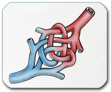
Image: Arteriovenous malformation. The red part represents the arterial component, and the blue part represents the venous component of the malformation.
This causes:
- abnormally high pressure in veins,
- fragile vessel walls
- turbulent flow
- risk of rupture
- irritation of surrounding brain tissue
An AVM is therefore not a tumor — it is a high-flow vascular shunt (an abnormal „short circuit“ where blood flows directly from arteries into veins, bypassing the protective capillary bed) that carries a risk of rupture and disrupts normal blood flow within the brain.

Image: Frontal lobe brain AVM shown as a tangle of abnormal blood vessels (nidus). The red arrow indicates the feeding artery, while the blue arrow points to the draining vein of the malformation. An associated aneurysm on the feeding artery is marked with a green arrow. The yellow arrow indicates an intra-nidal aneurysm within the arterial part of the malformation.
Brain AVM Location
Brain arteriovenous malformations can occur anywhere in the brain, but their location significantly influences symptoms, hemorrhage risk, and treatment strategy.
Approximately 80–90% of AVMs are located in the supratentorial compartment, meaning within the cerebral hemispheres. These include:
- Cortical AVMs (involving the surface of the brain)
- Subcortical AVMs
- Deep AVMs affecting structures such as the basal ganglia or thalamus
The remaining 10–20% are infratentorial, involving the cerebellum or brainstem.
Location strongly affects clinical presentation:
- Cortical AVMs, particularly in the temporal and frontal lobes, are more commonly associated with epileptic seizures.
- Deep AVMs and those with exclusively deep venous drainage carry a higher risk of hemorrhage.
- Posterior fossa AVMs (cerebellum and brainstem) are less likely to present with seizures but may lead to more severe neurological deficits if bleeding occurs due to the limited space in this region.
Understanding the anatomical location of an AVM is essential for estimating individual risk and selecting the most appropriate management strategy.
Is Brain AVM Congenital?
Yes.
Brain AVMs are developmental (congenital) vascular malformations — they form during early fetal development, even though symptoms may not appear for decades.
They are not caused by lifestyle, blood pressure, or trauma.
How Common are AVMs?
Brain AVMs are rare:
- Estimated prevalence: ~10–20 per 100,000 people
- Many people never know they have one until imaging is done
Is Brain AVM Inherited?
Most AVMs are sporadic, meaning they occur by chance during development. They are not inherited, so you did not receive this from your parents, and you cannot pass it on to your children.
A slightly higher risk exists among first-degree relatives, but:
- the absolute risk remains low
- routine screening of family members is not recommended unless a known genetic syndrome is present (such as HHT — a condition causing abnormal blood vessel formation throughout the body).
When Do AVMs Become Symptomatic?
Although AVMs exist from birth, most become clinically evident between ages 15 and 45.
It is estimated that:
- Approximately 15–20% of AVMs remain „silent“ (asymptomatic) throughout a person’s life and are discovered only incidentally or during autopsy.
- Data across different series may vary, but approximately 80–85% of AVMs will eventually cause symptoms, with intracranial hemorrhage and seizures being the most common presentations.
They are discovered:
- After a hemorrhage (approx. 50% of cases): Often the first sign of an AVM’s presence.
- After seizures begin (approx. 20–30% of cases): Due to the impact of the malformation on the surrounding brain tissue.
- Incidentally on MRI (increasingly common): Found during scans for unrelated headaches or minor head injuries.
It is important to understand the fundamental differences between a brain AVM and a cerebral aneurysm, as they represent distinct vascular conditions with very different clinical behaviors. While both carry a risk of rupture, a ruptured aneurysm is statistically much more lethal, with mortality rates reaching 40–50% due to high-pressure bleeding into the subarachnoid space. In contrast, an AVM rupture has a significantly lower mortality rate of approximately 10–15%, as the bleeding is typically contained within the brain tissue (intracerebral) and often occurs at lower pressures. In AVM, outcomes after a bleed vary widely depending on the location, volume, and initial clinical grade of the hemorrhage. Furthermore, while an aneurysm is generally a single weak point or „blister“ on an artery, an AVM is a complex network that can also cause seizures or progressive neurological deficits by disrupting normal circulation over many years, even without bleeding.
What Are the Symptoms of an AVM if It Has Not Bled?
An AVM can cause neurological problems even if it has never ruptured.
1) Epileptic Seizures
Seizures are the second most common symptom of a brain AVM, occurring in approximately 20–30% of cases. They often serve as the first warning sign that leads to diagnosis.
Population-based studies suggest that the 5-year risk of experiencing a first seizure in patients with an untreated AVM is approximately 8%, and higher (up to ~20%) in those who have already suffered a hemorrhage.
If a first seizure occurs, the probability of developing epilepsy (recurrent unprovoked seizures) within the following five years is approximately 50–60%.
Seizures are significantly more common in AVMs involving the cerebral cortex, particularly in the temporal and frontal lobes. Deep-seated or cerebellar AVMs are much less likely to present with seizures and more often manifest with hemorrhage.
The „short circuit“ of the AVM causes chronic irritation to the surrounding brain cortex through several mechanisms:
- Steal phenomenon: Abnormal blood flow „steals“ oxygen from healthy tissue.
- Microscopic bleeding & Iron deposition: Small, undetected leaks leave iron deposits (hemosiderin) that are highly irritating to neurons.
- Gliosis: The formation of scar tissue around the malformation.
Some patients may experience seizures for years before the AVM is finally discovered on an MRI scan.
2) Chronic Headache Or Migraine-like Pain
Chronic or migraine-like headaches occur in up to 15–40% of patients with an AVM. While common, they are considered „non-specific“ because they can mimic standard migraines.
The pain is typically not caused by the AVM tissue itself, but by:
- Venous Congestion: The high-pressure flow causes „overcrowding“ in the veins, increasing local pressure.
- Vascular Stretching: The abnormal vessels stretch the pain-sensitive linings of the brain (dura mater).
- Meningeal Irritation: Minor, asymptomatic „micro-bleeds“ can irritate the brain’s surface.
For many patients, any persistent or recurring headache naturally causes anxiety that the AVM is the cause. While localized pain on the same side as the AVM is clinically significant, it is important to remember that AVM patients can also suffer from common, unrelated tension headaches or migraines. Distinguishing whether a headache is a direct symptom of the vascular shunt or an independent condition is a key part of the neurological evaluation.
3) Neurological Deficits
Less commonly, AVMs cause:
- weakness
- numbness
- vision problems
- speech problems
Mechanisms include:
- chronic venous hypertension
- “steal phenomenon” (blood diverted away from the normal brain), causing micro-ischemia in brain tissue around AVM
4) Cranial Nerve Compression (rare)
In unusual anatomical locations, enlarged draining veins may compress cranial nerves (for example causing trigeminal neuralgia or visual disturbances), but this is uncommon.
What Happens When an AVM Bleeds?
AVM rupture causes intracranial hemorrhage.
Most commonly:
- intracerebral hemorrhage (ICH): Bleeding directly into the brain tissue itself (the most common type for AVMs).
Sometimes: - Subarachnoid hemorrhage (SAH): Bleeding into the fluid-filled space surrounding the brain.
- Intraventricular hemorrhage (IVH): Bleeding into the brain’s ventricles (the internal chambers where spinal fluid is produced).
- Combinations of these: In extensive ruptures, blood may spread across multiple compartments.
Symptoms may include:
- sudden severe headache
- vomiting
- weakness or paralysis
- speech or vision loss
- loss of consciousness or coma
- seizures
Unlike aneurysm rupture, AVM hemorrhage:
- does not cause classic vasospasm
- has a different rebleeding pattern
- is dominated by the mass effect of the clot and brain injury
What Is the Risk That an AVM Will Bleed?
The risk depends on anatomy and prior rupture.
If the AVM Has Never Bled
Annual hemorrhage risk:
~1–3% per year
If the AVM Has Already Bled
Annual hemorrhage risk increases to:
~4–6% per year
Factors that increase bleeding risk:
- prior hemorrhage
- deep venous drainage
- deep brain location
- associated aneurysms on feeding arteries
- high-flow shunts
How Brain AVMs are Classified and How It Guides Treatment (Spetzler–Martin System)
This system scores three features:
| Feature | Points |
|---|---|
| Size of AVM (Small < 3 cm, Medium 3–6 cm, Large > 6 cm) | 1–3 |
| Location in eloquent brain (motor, speech, vision, brainstem, etc.) | 0 or 1 |
| Deep venous drainage | 0 or 1 |
Total = Grade I to V
The Spetzler–Martin grading system estimates the surgical risk of AVM resection based on nidus size, eloquence of adjacent brain, and venous drainage pattern. While it was developed to predict microsurgical risk, it also indirectly guides overall treatment strategy. Prior hemorrhage significantly influences decision-making but does not override anatomical risk.
Unruptured: Generally favorable for microsurgical resection, with high cure rates and low permanent morbidity in experienced centers. However, anatomical suitability alone does not mandate surgery. The decision also depends on patient age, symptom profile (for example, drug-resistant epilepsy or progressive neurological deficit), estimated lifetime hemorrhage risk, and individual comorbidities. In younger patients with long life expectancy, definitive elimination of hemorrhage risk is often considered reasonable. In older asymptomatic patients, careful observation may be equally appropriate.
Radiosurgery or selective endovascular strategies may be appropriate in certain patients, particularly when the lesion is located in eloquent or deep regions, when surgical risk is increased due to comorbidities, or when the patient prefers a less invasive approach.
Ruptured: Definitive surgical removal is usually strongly considered after stabilization, as it eliminates the risk of recurrent hemorrhage. Alternative or adjunctive treatments (such as embolization of associated aneurysms or radiosurgery in selected cases) may be incorporated depending on angioarchitecture and overall risk profile.
Grade III
Unruptured: Heterogeneous group. Some lesions are appropriate for surgery, others for radiosurgery or staged multimodal treatment. Individual anatomical features and center expertise are critical.
Ruptured: Hemorrhage increases the likelihood of active treatment, but management remains individualized.
Grade IV–V
Unruptured: Very high treatment risk. Large or deep-seated AVMs in this category are often not suitable for single-step complete removal. In many cases, management focuses on risk reduction rather than cure. Options may include staged embolization, targeted treatment of high-risk components, limited or staged radiosurgery in selected anatomy, or careful observation with symptom control.
Ruptured: Even after hemorrhage, complete resection remains high risk. Management may prioritize life-saving measures and selective stabilization rather than aggressive curative intent.
Important: In large high-grade AVMs, the natural history risk may be lower than the procedural risk. Hemorrhage increases urgency, but it does not automatically convert a high-grade AVM into a safe surgical candidate. Final decisions must balance natural history risk, patient age, neurological status, and center-specific expertise.
How AVMs are Diagnosed
CT Scan
Used in emergencies to detect bleeding.
CTA (CT Angiography)
Shows large vessels and may reveal the AVM.
MRI / MRA
Provides:
- brain damage from prior bleeds
- microhemorrhages
- seizure-related scarring
- AVM location and size
Digital Subtraction Angiography (DSA)
This is the gold standard. DSA is a minimally invasive imaging technique performed under local anesthesia, often with light conscious sedation to ensure the patient is comfortable and relaxed.
A neuro-radiologist inserts a thin catheter, typically through a puncture in the femoral artery (in the groin) or the radial artery (in the wrist). The catheter is then carefully navigated through the major blood vessels up to the arteries supplying the brain. Once positioned, a contrast agent is injected. This substance allows high-resolution, real-time X-ray images to visualize the blood flow, which clearly outlines the arteries and identifies AVMs, aneurysms, or other vascular pathologies.
It shows:
- feeding arteries
- nidus
- draining veins
- flow patterns
- associated aneurysms
If hemorrhage occurred, DSA helps determine:
- whether the AVM itself ruptured
- or whether an associated aneurysm was the bleeding source
This distinction is crucial for treatment planning.
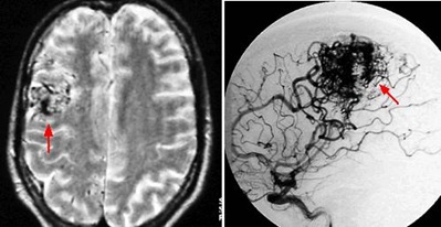
Image: The red arrow on the left shows a brain AVM on an MRI scan. The red arrow on the right shows the same AVM case on a DSA.
How Brain AVMs are Treated
The treatment of a brain AVM primarily depends on whether it has bled or not.
Treatment If the Brain AVM Has Bled
Management involves treating the hemorrhage first, followed by the treatment of the AVM itself.
1. Emergency care of hemorrhage
If there is a large clot compressing the brain, it may need urgent surgical removal. Management of an AVM-induced intracerebral hemorrhage (ICH) follows the same emergency principles as other hemorrhagic strokes.
Key differences in AVM bleeding:
• Aneurysm priority: If a brain AVM is associated with an aneurysm, the first priority is to treat the lesion that actually caused the bleeding — especially if the hemorrhage came from the aneurysm. In selected cases, both the aneurysm and the AVM can be treated during the same procedure.
• Higher need for surgery: AVM hemorrhages are often more superficial or atypically located compared to hypertensive bleeds. Consequently, they more frequently require surgical evacuation to relieve pressure from a significant clot.
2. AVM strategy when bled
After stabilization, the team decides on the best path forward:
• Can the AVM be safely removed during the same procedure as the clot evacuation, or should it be delayed?
• Should it be embolized first?
• Should radiosurgery be used later?
This is often multimodal treatment.
Treatment If the Brain AVM Has Not Bled
Step 1 – Treat symptoms
- Seizures → antiseizure medications
- Headache → evaluate for migraine, cervical, sinus, medication-overuse, and other types of headache.
Step 2 – Decide on AVM elimination: either removing the nidus or permanently stopping the blood flow through it.
Not every AVM should be treated.
The ARUBA trial showed that, in the short term, conservative management of unruptured AVMs was safer than intervention. However, this remains controversial, and modern specialized centers use careful patient selection. The decision is always individualized; for instance, a young patient with a 50-year life expectancy faces a vastly different cumulative risk of hemorrhage than an older patient.
The primary methods to eliminate blood flow through an AVM include: surgical resection, stereotactic radiosurgery, endovascular embolization, or a combined multimodality treatment approach. Partial treatment does not solve a problem — residual AVM continues to carry the same bleeding risk, so the goal is complete obliteration when treatment is chosen.
The decision on whether to treat an AVM and which modality to use depends on:
- SM (Spetzler–Martin system) grade- size, location and type of venous drainage
- bleeding risk
- patient age
- expected lifetime risk
- treatment risks
Surgical Removal of AVM (Microsurgery)
Surgery is performed only when complete removal can be achieved safely.
During surgery:
- craniotomy is performed to approach AVM
- the surgery is performed under microsurgical magnification
- feeding arteries of AVM are identified and closed first
- the AVM gradually collapses and it is carefully separated from the brain.
- draining veins of AVM are preserved until the very end
- only after all arteries are disconnected is the main vein coagulated and cut
- the nidus is removed
Cutting the vein too early would cause catastrophic bleeding.
Intraoperative angiography may be used to confirm complete removal.
Read more about potential complications following craniotomy and open brain surgery on this page.
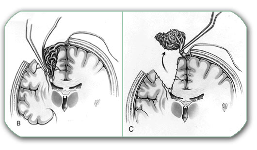
Image: Surgical resection of a brain AVM. During the procedure, the feeding arteries of the AVM are identified and closed first, causing the malformation to gradually collapse. The draining veins are strictly preserved until the very end. Only after all arterial feeders have been disconnected is the main vein cut, allowing for the safe removal of the nidus.
Stereotactic Radiosurgery (Gamma Knife, CyberKnife, LINAC)
Used mainly for:
- small AVMs (usually ≤3 cm)
- deep or eloquent locations (areas of the brain controlling speech, movement, or vision)
- residual AVM after embolization or surgery
How it works: High-dose radiation targets the nidus, causing progressive damage to the vessel lining. Over time, this leads to progressive scarring and eventual closure (obliteration) of the malformation.
Dosing and Sessions:
- Dose: Typically, a single high dose of 15 to 25 Gy (Grays) is delivered precisely to the nidus.
- Sessions: Usually performed as a single-session treatment. However, for slightly larger or complex AVMs, staged-volume radiosurgery (dividing the AVM into parts treated months apart) or fractionated sessions may be used to protect the surrounding healthy brain tissue.
The “Waiting Period”
The closure is not immediate; it typically takes 2–3 years for the AVM to fully disappear. During that time:
• The AVM is still present: The malformation does not vanish immediately and remains part of the brain’s circulation.
• The risk of bleeding continues: The risk of rupture remains at its baseline rate until the closure is complete.
• Regular follow-up is required: Periodic MRI/MRA scans are necessary to monitor the progress, with a final digital subtraction angiography (DSA) to confirm total obliteration.
Repeat Radiosurgery
If complete obliteration is not achieved after the initial radiosurgical treatment (typically assessed 2–3 years later with MRI and confirmatory DSA), repeat radiosurgery may be considered in selected patients. This approach is most appropriate for small residual nidus volumes. The decision depends on residual size, location, prior radiation dose, patient age, and cumulative risk of radiation-induced injury. While repeat treatment can increase overall obliteration rates, it must be carefully balanced against the risk of delayed radiation-related complications.
Endovascular embolization (gluing)
During endovascular embolization, a microcatheter is advanced through a feeding artery (the standard transarterial approach) as close as possible to the AVM nidus. The catheter insertion technique is the same as that used for DSA. A liquid embolic agent (such as Onyx or n-BCA “medical glue”) is then injected with the aim of penetrating and filling the nidus itself, so that the abnormal arteriovenous shunt is closed from within. The goal is not simply to occlude proximal feeding arteries, because isolated feeder occlusion may recruit collateral supply and usually does not cure the AVM. While selective feeder embolization may be used as part of surgical preparation, targeted rupture control, or palliative flow reduction, only nidus-directed embolization is considered curative for brain AVMs.
In selected cases, embolization is performed as part of a multimodality plan or as a primary intervention:
- (1) Pre-operative embolization: Performed to reduce intraoperative bleeding and surgical complexity by occluding deep or difficult-to-reach feeders.
- (2) Pre-radiosurgery embolization: Used to reduce the nidus size or treat high-risk compartments, making the malformation more suitable for targeted radiation.
- (3) Targeted or palliative embolization: Aimed at securing a focal “weak point” (such as an intranidal aneurysm or a ruptured compartment) when immediate complete cure is not feasible.
- (4) Stand-alone treatment: Employed in selected small AVMs where complete obliteration is achievable through filling of the nidus alone.
It is rarely curative alone for complex AVMs. Complete cure requires elimination of the nidus, not just proximal flow reduction.
Risks include:
- stroke
- hemorrhage
- vessel perforation
- venous thrombosis
These risks are minimized through careful patient selection, high-resolution angiography, and experienced neurointerventional teams.
Transvenous approach (special technique)
The transvenous approach (embolization through the draining vein rather than the feeding arteries) is an emerging technique used only in highly selected AVMs, typically those with:
• small, compact nidus
• single or favorable venous drainage
• anatomy suitable for controlled retrograde filling
Special anti-reflux strategies (such as “pressure-cooker” techniques) are used to allow safe retrograde penetration of the nidus while protecting the normal veins. This approach is not standard embolization and is reserved for centers with advanced expertise.
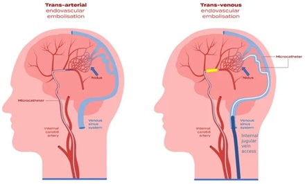
Image: Endovascular embolization techniques. The left side shows the common transarterial approach, while the right side shows the transvenous approach.
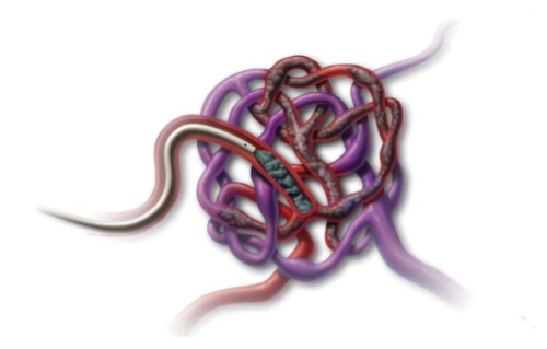
Image: Endovascular embolization of an AVM. A microcatheter delivers a liquid embolic agent („medical glue“) directly into the nidus to close the abnormal shunt from within.
In Conclusion: When Is „No Treatment“ the Best Option?
Some AVMs are best left untreated and monitored through regular imaging. This is typically the case for:
- Spetzler-Martin Grade IV & V: Large malformations (often > 6 cm) where the risk of surgery outweighs the risk of natural history.
- Deep-seated or eloquent locations: When the AVM is in a region where any intervention could cause a major neurological deficit.
- Asymptomatic lesions: AVMs that have never bled, especially in older patients where the cumulative lifetime risk of hemorrhage is lower than the risk of intervention.
Why Do Medical Opinions Differ in Brain AVM Treatment?
Differences in medical opinion are common because treatment decisions depend on how bleeding risk, AVM anatomy, patient age, and treatment risk are balanced against each other, rather than on a single finding.
Is The AVM Dangerous Enough To Require Treatment?
Some specialists recommend treatment based on the presence of an AVM alone, while others consider intervention only if there is significant lifetime hemorrhage risk, especially in younger patients or after prior bleeding.
Should An Unruptured AVM Be Treated Or Observed?
Some follow the ARUBA-based conservative approach, recommending observation for unruptured AVMs, while others advocate treatment in selected patients with favorable anatomy and long life expectancy, where cumulative bleeding risk becomes relevant.
Which Treatment Option Is The Safest And Most Effective?
There is no single best method, as some recommend microsurgery for low-grade AVMs, others favor radiosurgery for deep or eloquent locations, while embolization is often used as part of a combined strategy rather than a standalone cure.
Can Partial Treatment Reduce Risk Or Is Complete Obliteration Necessary?
Some believe that reducing flow or treating high-risk components is sufficient, while most specialists emphasize that only complete elimination of the nidus removes the bleeding risk, and partial treatment may leave the patient exposed.
When Is “No Treatment” Actually The Best Option?
In large, deep, or high-grade AVMs, different specialists and centers may assess treatment risk differently based on their experience and expertise. Some consider intervention justified when they believe the risk can be controlled, while others recommend observation when the estimated procedural risk exceeds the natural history risk of the AVM.
Request Brain AVM (Arteriovenous Malformation) Neurosurgery Consultation — 24-Hour Review or Priority Option (Usually Within 3 Hours)
When an MRI, CT, or angiography report reveals a brain arteriovenous malformation (AVM), families are often suddenly faced with difficult and high-stakes decisions about bleeding risk, intervention, or long-term monitoring.
An independent neurosurgical second opinion can help clarify how dangerous the AVM truly is, whether it is treatable, and which combination of embolization, radiosurgery, or surgery offers the safest path forward.
- ✔ Send a short message describing the diagnosis, symptoms (headache, seizures, weakness, bleeding), and what the MRI, CT, or angiography report says about the AVM
- ✔ You’ll receive a reply within 24 hours explaining if and how we can help in your specific AVM situation
- ✔ Time-sensitive cases: if bleeding has occurred, neurological symptoms are worsening, or doctors are urgently recommending intervention, consultations can often be arranged within a few hours — write PRIORITY in your first message
- ✔ MRI, CT, angiography images, and hospital reports can be reviewed once initial contact is established
- ✔ During the consultation, we explain AVM type, bleeding risk, treatment options (embolization, radiosurgery, surgery), and realistic outcomes — with up to 10 days of follow-up for brief questions
Consultation fees typically range from $180–250, depending on AVM complexity and imaging findings.
Secure payment by credit card, PayPal invoice (USD), or bank transfer.
This is within the usual range for international specialist telehealth second opinions in neurovascular and neurosurgical care.
AVM and Epilepsy
In patients with unruptured brain AVMs, seizures are the presenting symptom in approximately 25–40% of cases, making AVMs a leading cause of secondary (structural) epilepsy in young adults.
Treatment Strategy for AVM-Related Epilepsy:
- Medication First: Initially, seizures are managed with anti-seizure medications.
- When to Consider Intervention:
- Refractory Epilepsy: If seizures cannot be controlled with medication, AVM elimination (resection, radiosurgery) or specialized epilepsy surgery may be considered to reduce seizure frequency.
- Preventive Elimination (Grade I–II): In younger patients with favorable anatomy (Spetzler–Martin Grade I or II), complete AVM removal is often recommended even if seizures are well-controlled. This is done to eliminate the cumulative, lifelong risk of hemorrhage and to potentially cure the epilepsy by removing the structural cause.
Clinical Take-Home Message
A brain AVM is a congenital vascular short-circuit that can cause:
- bleeding
- seizures
- neurological deficits
Some AVMs must be treated urgently.
Others should never be touched.
Correct management requires:
- precise angiographic analysis
- understanding of natural history
- and realistic weighing of risk vs benefit.
This is exactly where expert second-opinion consultation makes the biggest difference.
Frequently Asked Questions About Brain AVM (Arteriovenous Malformations)
What is a brain AVM?
A brain arteriovenous malformation (AVM) is an abnormal connection between arteries and veins inside the brain. Normally, blood flows from arteries into tiny capillaries and then into veins. In a brain AVM, the capillary bed is missing, so blood passes directly from high-pressure arteries into veins. This forms a tangled cluster of abnormal vessels called a nidus. Because veins are not designed for arterial pressure, they can stretch, weaken, and become prone to rupture. A brain AVM is not a tumor. It is a high-flow vascular shunt that can cause bleeding, seizures, headaches, or neurological symptoms depending on its size, location, and blood flow pattern.
Is a brain AVM something you are born with?
Yes. Brain AVMs are developmental or congenital vascular malformations, meaning they form during early fetal development. However, symptoms may not appear for many years or even decades. A person may be born with a brain AVM and discover it only after a seizure, headache investigation, neurological symptom, or brain hemorrhage. Brain AVMs are not caused by stress, lifestyle, infection, high blood pressure, or trauma. Most are sporadic, meaning they occur by chance and are not inherited from parents. Routine screening of family members is usually not recommended unless there is a known genetic syndrome, such as hereditary hemorrhagic telangiectasia.
Is a brain AVM the same as a brain aneurysm?
No. A brain AVM and a brain aneurysm are different vascular conditions. An aneurysm is usually a single weak, balloon-like dilation on an artery. A brain AVM is a complex network of abnormal arteries and veins connected through a nidus. Both conditions can rupture and cause bleeding, but their behavior is different. A ruptured aneurysm often causes subarachnoid hemorrhage and carries very high early mortality. AVM bleeding more commonly causes intracerebral hemorrhage inside the brain tissue. Some AVMs may also have associated aneurysms on feeding arteries or inside the nidus, and these weak points can influence bleeding risk and treatment planning.
Is a brain AVM a tumor?
No. A brain AVM is not a brain tumor. It is a vascular malformation, meaning an abnormal formation of blood vessels. Unlike tumors, AVMs are not made of uncontrolled growing tumor cells. Their danger comes from abnormal blood flow, fragile vessel walls, high pressure inside draining veins, and the risk of rupture. A brain AVM may appear as a mass-like cluster on imaging, and it can irritate or affect nearby brain tissue, but it is not cancer. Treatment decisions are therefore based on bleeding risk, seizures, location, venous drainage, size, and procedural risk rather than on tumor behavior.
Why is a brain AVM dangerous and what makes it bleed?
A brain AVM is dangerous because it creates a high-flow shortcut between arteries and veins without the normal protective capillary bed. Arterial pressure enters veins that are normally designed for lower pressure. Over time, this can stretch and weaken vessel walls, create turbulent flow, and increase the risk of rupture. Bleeding risk is influenced by several factors, including prior hemorrhage, deep brain location, deep venous drainage, associated aneurysms, and high-flow shunts. Some AVMs remain stable for years, while others bleed unexpectedly. This is why individual risk cannot be judged only by the presence of an AVM; it requires careful analysis of anatomy, symptoms, and angiographic features.
Can a brain AVM rupture or cause a brain bleed?
Yes. A brain AVM can rupture and cause intracranial hemorrhage. The most common bleeding pattern is intracerebral hemorrhage, meaning bleeding directly into the brain tissue. In some cases, bleeding can also enter the ventricles or the subarachnoid space. Symptoms may include sudden severe headache, vomiting, weakness, speech difficulty, vision loss, seizure, loss of consciousness, or coma. After a bleed, treatment first focuses on stabilizing the patient and managing the hemorrhage. Later, the neurovascular team decides whether the AVM should be removed, embolized, treated with radiosurgery, or managed with a staged or combined approach.
What symptoms can a brain AVM cause?
A brain AVM may cause symptoms even before it bleeds. Common presentations include seizures, headaches, weakness, numbness, speech problems, visual disturbances, or other focal neurological symptoms. Seizures are more common when the AVM involves the cerebral cortex, especially frontal or temporal regions. Headaches may occur but are non-specific, because many patients with AVMs can also have unrelated migraine or tension-type headaches. Some AVMs are discovered incidentally on MRI performed for another reason. Others are diagnosed only after hemorrhage. The exact symptom pattern depends on AVM location, surrounding brain irritation, venous congestion, steal phenomenon, and whether bleeding has occurred.
Can a brain AVM cause seizures?
Yes. Seizures are one of the most common ways an unruptured brain AVM becomes symptomatic. AVM-related seizures are more likely when the malformation is located in or near the cerebral cortex, especially in the frontal or temporal lobes. Several mechanisms may contribute: irritation of surrounding brain tissue, chronic abnormal blood flow, microscopic bleeding, hemosiderin deposition, gliosis, and steal phenomenon. Initial treatment often includes anti-seizure medication. If seizures remain uncontrolled or if the AVM has favorable anatomy and significant lifetime bleeding risk, AVM elimination by surgery, radiosurgery, or a combined strategy may be considered. The seizure plan and AVM treatment plan must be individualized together.
How is a brain AVM diagnosed?
Brain AVM diagnosis usually begins with CT, CTA, MRI, or MRA, depending on whether the patient presents with bleeding, seizures, headaches, or incidental imaging findings. CT is often used in emergencies to detect hemorrhage. MRI can show the AVM, surrounding brain changes, prior microbleeds, or seizure-related scarring. CTA and MRA can show larger vessels and the general vascular structure. However, digital subtraction angiography (DSA) remains the gold standard for detailed AVM evaluation. DSA shows feeding arteries, the nidus, draining veins, flow patterns, and associated aneurysms. This level of detail is essential for estimating risk and planning treatment.
What is the difference between MRI/MRA and DSA for brain AVM?
MRI and MRA are very useful for detecting a brain AVM, showing its location, estimating size, and evaluating surrounding brain tissue. They can reveal prior bleeding, microhemorrhage, scarring, or structural changes. However, they do not always show the full flow dynamics of the malformation. DSA, or digital subtraction angiography, is more precise because it shows the AVM in real time as contrast flows through feeding arteries, the nidus, and draining veins. This is especially important when planning embolization, surgery, radiosurgery, or deciding whether an associated aneurysm or AVM compartment caused a hemorrhage. MRI helps identify the lesion; DSA maps it for decision-making.
What is the “nidus” of an AVM?
The nidus is the central tangled core of the AVM where arteries connect directly to veins without a normal capillary bed. It is the key part of the malformation. Treating only the feeding arteries is usually not enough, because the nidus may recruit new collateral blood supply and the AVM can remain active. True cure requires complete elimination or closure of the nidus, either by microsurgical removal, successful radiosurgical obliteration, curative embolization in selected cases, or a combined treatment strategy. The nidus is also where some dangerous weak points, such as intranidal aneurysms, may be found. Understanding the nidus is essential for treatment planning.
What does AVM life expectancy depend on?
Life expectancy with a brain AVM depends on the individual risk of bleeding, whether the AVM has already ruptured, the patient’s age, AVM location, venous drainage, associated aneurysms, neurological condition, and treatment risk. A small superficial AVM in a young patient may create a significant lifetime hemorrhage risk because many decades remain ahead. In contrast, a large deep high-grade AVM in an older asymptomatic patient may carry a procedural risk higher than the natural history risk. Life expectancy is therefore not determined by the diagnosis alone. It depends on balancing natural history risk against the risks and benefits of observation, embolization, radiosurgery, surgery, or combined treatment.
Can a brain AVM be cured?
A brain AVM can be considered cured only when the nidus is completely eliminated and there is no remaining abnormal arteriovenous shunt. This can sometimes be achieved with microsurgical removal, radiosurgery after delayed obliteration, embolization in selected small AVMs, or a combination of treatments. Partial treatment may reduce flow or treat a dangerous weak point, but it does not necessarily remove the long-term bleeding risk if a residual nidus remains. Cure is more realistic for small, superficial, low-grade AVMs in non-eloquent regions. Large, deep, or high-grade AVMs may be too risky to eliminate completely, and observation or targeted risk reduction may be safer.
Does every brain AVM need treatment?
No. Not every brain AVM requires immediate treatment. Management depends on whether the AVM has bled, the estimated future hemorrhage risk, patient age, symptoms, AVM size, eloquence of the surrounding brain, venous drainage pattern, and procedural risk. Some small, superficial AVMs in younger patients may be reasonable candidates for curative treatment. Other AVMs, especially large, deep, high-grade, or asymptomatic lesions in older patients, may be safer to monitor. Treatment is not chosen simply because an AVM exists. The key question is whether eliminating the AVM is safer than living with it under observation and symptom control.
When is observation or no treatment the best option for a brain AVM?
Observation may be the best option when the risk of treatment is higher than the expected natural history risk of the AVM. This is often considered for large Spetzler–Martin grade IV or V AVMs, deep-seated malformations, lesions in eloquent brain areas, asymptomatic AVMs in older patients, or cases where complete cure is unlikely without major neurological risk. Observation does not mean ignoring the problem. It usually includes clinical follow-up, imaging when appropriate, seizure control if needed, and reassessment if symptoms change or bleeding occurs. In high-grade AVMs, the safest plan may be careful monitoring rather than aggressive intervention.
What does the Spetzler–Martin grade mean for brain AVM treatment?
The Spetzler–Martin grade estimates the surgical risk of removing a brain AVM. It is based on three features: AVM size, whether the AVM is located in eloquent brain tissue, and whether it has deep venous drainage. Grades I and II are usually more favorable for microsurgical treatment, especially in experienced centers. Grade III is mixed and requires individualized judgment. Grades IV and V are high-risk lesions where complete surgical removal may cause major neurological injury. The grade does not decide everything by itself. Prior hemorrhage, patient age, symptoms, associated aneurysms, radiosurgery options, embolization strategy, and center experience also influence management.
Can a brain AVM be treated without open brain surgery?
Yes. Some brain AVMs can be treated without open surgery, depending on size, location, flow pattern, and treatment goal. Endovascular embolization can close parts of the nidus or high-risk components from inside the blood vessels. Stereotactic radiosurgery can gradually close small AVMs, especially in deep or eloquent locations, over two to three years. In some patients, these methods are used alone; in others, they are combined with surgery. However, non-open treatment does not automatically mean lower risk or cure. The safest method depends on whether the AVM can be completely obliterated and whether the treatment risk is acceptable.
Is this page about brain AVM or other vascular malformations in the body?
This page is about brain AVM, also called cerebral arteriovenous malformation. A brain AVM is different from pulmonary AVM, uterine AVM, bowel AVM, limb vascular malformation, venous malformation, or other vascular anomalies outside the brain. The main concern in brain AVM is the risk of bleeding into the brain, seizures, neurological deficits, and the decision between observation, embolization, radiosurgery, microsurgery, or combined treatment. Other vascular malformations in the lungs, uterus, bowel, skin, or limbs require different specialists, different imaging, and different treatment principles.
What is a ruptured brain AVM?
A ruptured brain AVM means that abnormal vessels inside the arteriovenous malformation have broken and caused bleeding into or around the brain. This may produce sudden severe headache, vomiting, weakness, speech or vision problems, seizure, loss of consciousness, or coma. Emergency treatment first focuses on the hemorrhage, brain swelling, and neurological condition. After stabilization, doctors decide whether the AVM should be treated by microsurgery, embolization, radiosurgery, or a staged combination. The safest strategy depends on AVM size, location, venous drainage, associated aneurysms, and the patient’s neurological status after the bleed.
What is brain AVM embolization with Onyx?
Brain AVM embolization with Onyx is an endovascular procedure in which a microcatheter is guided through the blood vessels into arteries feeding the AVM. Onyx, a liquid embolic material, is injected to fill part of the AVM nidus and reduce or close the abnormal arteriovenous shunt. Embolization may be used before surgery, before radiosurgery, to treat a high-risk ruptured compartment, or rarely as a stand-alone cure in selected small AVMs. Blocking only feeding arteries is not enough; durable treatment requires elimination of the nidus itself.
What is endovascular embolization for a brain AVM?
Endovascular embolization is a minimally invasive procedure performed through the blood vessels. A microcatheter is navigated into feeding arteries close to the AVM nidus, and a liquid embolic agent such as Onyx or n-BCA glue is injected to block abnormal flow. The goal is to penetrate the nidus, not only to close a proximal feeding artery. Embolization may be used before surgery to reduce bleeding, before radiosurgery to reduce nidus size, to secure a dangerous weak point after rupture, for palliative flow reduction, or rarely as a stand-alone curative treatment in selected small AVMs. Risks include stroke, hemorrhage, vessel perforation, and venous thrombosis.
Does embolization alone always cure a brain AVM?
No. Embolization alone does not always cure a brain AVM. In selected small, compact AVMs, complete nidus obliteration may be possible endovascularly. However, in many cases embolization is part of a larger treatment strategy. It may reduce surgical bleeding, make radiosurgery more suitable, treat an associated aneurysm, or close a ruptured compartment. If embolization blocks only feeding arteries without eliminating the nidus, the AVM can remain active and may recruit new blood supply. Therefore, the key question after embolization is whether the nidus is completely obliterated. If residual AVM remains, bleeding risk may also remain.
What is radiosurgery for a brain AVM?
Stereotactic radiosurgery, such as Gamma Knife, CyberKnife, or LINAC-based treatment, delivers focused radiation to the AVM nidus. It is usually used for small AVMs, deep AVMs, eloquent-location AVMs, or residual nidus after embolization or surgery. Radiosurgery does not close the AVM immediately. The vessel walls gradually scar and close over time, usually over two to three years. During this waiting period, the AVM is still present and the risk of bleeding continues until complete obliteration is confirmed. Follow-up MRI/MRA is used to monitor progress, and DSA is often needed to confirm final cure.
If anti-seizure medication controls my seizures, should my brain AVM still be treated?
Not automatically, but seizure control is only one part of the decision. If medication controls the seizures and the brain AVM has never bled, treatment may still be considered when the AVM has favorable anatomy, the patient is young, and complete elimination can be achieved with an acceptable neurological risk. In such cases, the purpose of treatment may be to remove the lifetime risk of hemorrhage as well as the structural cause of epilepsy.
Observation may be safer when the brain AVM is large, deep, located in eloquent brain, or has a high Spetzler–Martin grade. The decision should include age, previous hemorrhage, DSA anatomy, medication tolerance, seizure burden, treatment risks, and the probability of complete obliteration. Controlled seizures do not automatically mean that the brain AVM is harmless, but neither do they automatically justify intervention. More information about structural epilepsy and lesion-based treatment is available on our epilepsy surgery page.
How do doctors choose between microsurgery, embolization, and radiosurgery for a brain AVM?
The choice depends on whether the brain AVM has bled, its size, location, Spetzler–Martin grade, feeding arteries, venous drainage, associated aneurysms, and the likelihood that each treatment can completely eliminate the nidus. Microsurgery is generally most suitable for selected low-grade, accessible brain AVMs where complete removal can be achieved with an acceptable neurological risk.
Radiosurgery is more often considered for small brain AVMs in deep or eloquent locations, but closure usually takes two to three years and the bleeding risk continues until complete obliteration is confirmed. Embolization may be used before surgery, before radiosurgery, to treat a high-risk aneurysm or ruptured compartment, or occasionally as definitive treatment for a small suitable nidus. Complex brain AVMs often require a staged combination of methods. The best option is not necessarily the least invasive procedure, but the strategy most likely to achieve complete obliteration with the lowest overall risk.
What are the specific risks of surgery for my brain AVM?
The risks of brain AVM surgery depend mainly on nidus size, depth, eloquent location, feeding arteries, venous drainage, previous hemorrhage, and the surgical route. Possible complications include intraoperative or postoperative bleeding, ischemic stroke, brain swelling, seizures, infection, cerebrospinal fluid leakage, residual AVM, and new neurological deficits affecting movement, sensation, speech, vision, balance, memory, or cognition.
A small superficial low-grade brain AVM may sometimes be removed with a relatively low risk in an experienced center. A large, deep, high-grade, brainstem, or eloquent brain AVM may carry a substantially greater risk because normal arteries, veins, and functional pathways may be closely related to the nidus. The relevant comparison is the individualized surgical risk versus the natural risk of future hemorrhage and neurological injury. A detailed explanation of general operative and postoperative risks is available on our craniotomy and brain surgery complications page.
When is microsurgery considered for a brain AVM?
Microsurgery is considered when complete removal of the AVM can be achieved with acceptable risk. It is often most suitable for smaller, superficial, low-grade AVMs located away from highly eloquent brain regions. Surgery may also be strongly considered after rupture, especially if a clot requires evacuation and the AVM anatomy allows safe removal. During surgery, feeding arteries are closed first while draining veins are preserved until the end. Cutting the draining vein too early can cause catastrophic bleeding. Microsurgery can provide immediate cure when the nidus is completely removed, but it carries risks of stroke, bleeding, seizures, and neurological deficits depending on AVM anatomy.
What happens if a brain AVM has already bled?
If a brain AVM has already bled, the first priority is emergency stabilization and treatment of the hemorrhage. If there is a large clot compressing the brain, surgical evacuation may be needed. DSA is often important to determine whether the bleeding came from the AVM nidus itself or from an associated aneurysm. After the acute phase, the team decides whether the AVM should be removed, embolized, treated with radiosurgery, or managed with a staged multimodal approach. Prior hemorrhage increases future bleeding risk and often makes active treatment more likely, but it does not automatically make every AVM safely operable.
Why do medical opinions differ in brain AVM treatment?
Medical opinions often differ because AVM treatment is based on balancing natural history risk against treatment risk, not on a single imaging finding. One specialist may focus on lifetime bleeding risk, especially in a young patient. Another may focus on procedural risk, especially if the AVM is deep, large, or located in eloquent brain. Some centers are more experienced with microsurgery, while others favor radiosurgery, embolization, or observation. Prior hemorrhage, associated aneurysms, Spetzler–Martin grade, symptoms, age, and patient preference all affect the recommendation. A difference in opinion does not always mean one doctor is wrong; it often reflects a difficult risk-benefit balance.
Can a brain AVM cause headaches or migraine-like pain?
Yes, a brain AVM can be associated with chronic headache or migraine-like pain, but headache is a non-specific symptom. Some headaches may be related to venous congestion, vascular stretching, meningeal irritation, or small previous microbleeds around the AVM. Localized headache on the same side as the AVM may be clinically relevant. However, many patients with AVMs can also have ordinary migraine, tension-type headache, cervical pain, sinus-related headache, or medication-overuse headache that is not directly caused by the AVM. For this reason, headache should be interpreted together with AVM location, imaging findings, neurological symptoms, and the overall clinical picture.
Why is a second opinion important for brain AVM?
A second opinion is important because brain AVM management is highly individualized. The same AVM may be described as treatable by one team and too risky by another, depending on how they interpret anatomy, bleeding risk, and available expertise. A focused neurosurgical review can help clarify whether the AVM has bled, whether an associated aneurysm is present, what the Spetzler–Martin grade implies, and whether embolization, radiosurgery, surgery, observation, or combined treatment is most reasonable. It can also help families understand why partial treatment may not remove bleeding risk and why “no treatment” is sometimes the safest recommendation.
Can brain AVM imaging be reviewed through an online neurosurgical consultation?
Yes. Brain AVM imaging can often be reviewed through an online neurosurgical second opinion when CT, MRI, CTA, MRA, DSA images, or written reports are available. The goal is to explain the AVM anatomy, bleeding risk, treatment options, and why one strategy may be safer than another. This is especially useful when families receive different recommendations, when the AVM has bled, when surgery or embolization is being proposed, or when observation is being considered. Online consultation does not replace emergency treatment, but it can provide structured interpretation of imaging and help patients understand what questions to ask the treating neurovascular team.
Additional Patient Resources (Brain AVM)
If you would like to read more from trusted medical organizations, these references are a good starting point.
-
NINDS (NIH): Arteriovenous Malformations (AVMs) — overview for patients
https://www.ninds.nih.gov/health-information/disorders/arteriovenous-malformations-avms -
American Heart Association / American Stroke Association (AHA/ASA): Scientific Statement — Management of Brain AVMs (professional guideline-level reference)
https://www.ahajournals.org/doi/10.1161/str.0000000000000134 -
Mayo Clinic: Brain AVM — symptoms, causes, and patient-friendly explanations
https://www.mayoclinic.org/diseases-conditions/brain-avm/symptoms-causes/syc-20350260 -
NHS (UK) patient information: Brain arteriovenous malformation (AVM)
Brain Arteriovenous Malformation (AVM)
-
The Aneurysm and AVM Foundation (TAAF): patient resources, glossary, and support network
https://www.taafonline.org/Resources -
UCLH (UK): Cerebral AVM — endovascular treatment (patient leaflet-style explanation)
https://www.uclh.nhs.uk/patients-and-visitors/patient-information-pages/cerebral-arterio-venous-malformation-endovascular-treatment
On this page, you can learn more about other neurosurgical cerebrovascular diseases, such as aneurysms, cavernomas, and intracerebral hemorrhages.