Dr Željko Kojadinović — NEUROHIRURGIJA I LEČENJE BOLA
Dr Zeljko Kojadinovic — Pain Treatment & Neurosurgery

Author:
Dr. Zeljko Kojadinovic, MD, PhD
— Neurosurgeon and Pain Management Specialist
Specialized Experience:
30 years of clinical expertise in neurosurgery
Last medically reviewed:
January 7, 2026
What Are Nerves?
Nerves are cable-like structures that connect the brain and spinal cord with the rest of the body.
Their role is to carry electrical signals that allow us to feel sensations, move muscles, and regulate automatic body functions.

Image: Cranial and somatic nerves
Main Types of Nerves
1. Somatic Nerves
Somatic nerves control conscious sensation and voluntary movement.
They are anatomically well defined and easier to localize.
They are divided into:
- Cranial nerves – arise directly from the brain
- Spinal nerves – arise from the spinal cord
2. Autonomic (Vegetative) Nerves
Autonomic nerves control involuntary functions, such as heart rate, digestion, blood pressure, sweating, and organ activity.
They are divided into:
- Sympathetic nerves – activate the “fight or flight” response
- Parasympathetic nerves – activate the “rest and digest” response
Functional Types of Somatic Nerves
Somatic nerves can be classified by function:
Sensory (Afferent) Nerves
They carry information from the body to the brain.
They transmit sensations such as:
- Touch
- Pain
- Temperature
- Vibration
- Pressure
- Position of joints and muscles (proprioception)
Motor (Efferent) Nerves
They carry signals from the brain and spinal cord to muscles, allowing voluntary movement.
Mixed Nerves
Most peripheral nerves are mixed nerves, containing both:
- sensory fibers
- motor fibers
This means they control both sensation and movement in the same body region.
Basic Structure of a Nerve
The fundamental unit of a nerve is the nerve fiber (axon).
Each nerve fiber consists of:
- an axon (the conducting core)
- a surrounding protective tube that supports and insulates it
This tube is formed by specialized cells:
- Schwann cells (in the peripheral nervous system).
In the peripheral nervous system, Schwann cells form a protective sheath around the axon known as the myelin sheath.
Some nerve fibers are covered by thick myelin layers, allowing fast signal transmission.
Others have little or no myelin and conduct signals more slowly.
This difference explains why pain, temperature, movement, and touch are perceived differently.
How Nerve Fibers Are Organized
Nerve fibers are grouped in several levels:
- Individual nerve fibers are wrapped by endoneurium
- Groups of fibers form bundles called fascicles, surrounded by perineurium
- Multiple fascicles together form a nerve, enclosed by epineurium
This layered structure protects the nerve and allows it to tolerate limited stretching and movement.
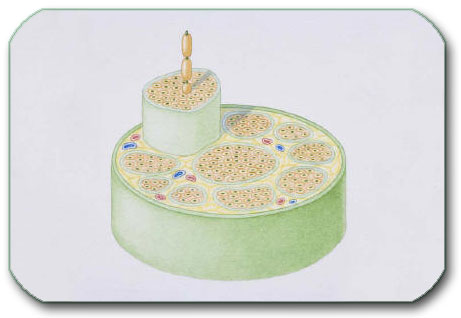
Image: Nerve cross-section — The green dots represent nerve fibers. They are surrounded by the myelin sheath (orange). Groups of fibers form bundles called fascicles, which are surrounded by the perineurium (light green). Blood vessels that nourish the nerve run between these bundles. Multiple fascicles together form a nerve, enclosed by the epineurium (dark green).
Why Nerve Structure Matters for Recovery
If only the nerve fiber (axon) is damaged, but the surrounding tube remains intact, the nerve often has a chance to recover without surgery.
However, if the protective tube is damaged or destroyed, recovery becomes difficult or impossible without surgical repair.
Even when the nerve structure is preserved, recovery may fail if the nerve becomes compressed later by:
- scar tissue
- inflammation
- bone, disc, or ligament pressure
This is why nerve compression can cause persistent or worsening symptoms.
How Nerves Originate in the Body
Spinal Nerve Roots
Nerves begin as nerve roots emerging from the spinal cord.
Each spinal nerve is formed by:
- a sensory root (dorsal) with dorsal root ganglion
- a motor root (ventral)
These roots join to form a spinal nerve.
Plexuses and Peripheral Nerves
In many regions, spinal nerves intertwine and form plexuses.
From these plexuses, peripheral nerves arise and travel to the limbs:
- Cervical plexus – neck and upper shoulder
- Brachial plexus – shoulder, arm, forearm, and hand
- Lumbar plexus – front of the thigh
- Sacral plexus – pelvis, buttock, leg, and foot
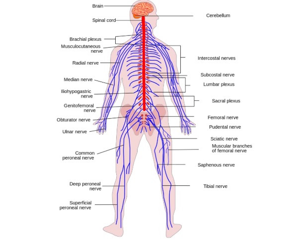
Image: Nerve plexuses in the body and the nerves that emerge from them.
Intercostal Nerves
Unlike limb nerves, intercostal nerves do not form plexuses.
They run directly:
- from the thoracic spine
- along each rib
- around the chest wall
They supply sensation and muscle control to the chest and upper abdominal wall.

Image: Anatomy of the Intercostal Nerve. Chronic irritation or damage to this nerve can lead to intercostal neuralgia.
Major Nerves of the Upper Limb (Basic Overview)
- Median nerve – sensation in the thumb, index, middle finger; controls fine hand movements
- Ulnar nerve – sensation in the little finger and part of the ring finger; controls grip strength
- Radial nerve – controls wrist and finger extension; sensation on the back of the hand
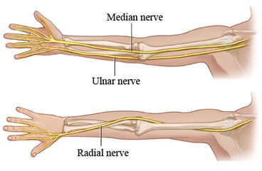
Image: Arm nerves- Median, Ulnar, Radial nerves
Major Nerves of the Lower Limb (Basic Overview)
- Femoral nerve – controls knee extension; sensation in the front of the thigh
- Obturator nerve – controls thigh adduction
- Sciatic nerve – the largest nerve in the body; supplies most of the leg
- Tibial nerve – controls foot push-off and plantar sensation
- Peroneal (fibular) nerve – controls foot lifting and toe extension

Image: Leg nerves — Femoral, Obturator, Sciatic, Tibial, and Peroneal nerves.
Peripheral nerves can be injured or damaged by pressure along their path (entrapment syndrome), such as carpal tunnel syndrome, cubital tunnel syndrome, Guyon canal syndrome, peroneal nerve compression, meralgia paresthetica, and thoracic outlet syndrome.