Dr Željko Kojadinović — NEUROHIRURGIJA I LEČENJE BOLA
Dr Zeljko Kojadinovic — Pain Treatment & Neurosurgery

Author:
Dr. Zeljko Kojadinovic, MD, PhD
— Neurosurgeon and Pain Management Specialist
Specialized Experience:
30 years of clinical expertise in neurosurgery
Last medically reviewed:
January 7, 2026
The brain is the central control organ of the nervous system.
It processes information, controls movement, sensation, thinking, emotions, and vital body functions.
Although very complex, the brain can be understood by dividing it into a few main parts, each with a clear role.
Main parts of the brain
Main parts of the brain are: cerebrum (the large brain), brainstem, cerebellum (the ‘little brain’).
Cerebrum (the large brain)
The cerebrum is the largest part of the brain.
It controls higher brain functions such as thinking, voluntary movement, sensation, language, and memory.
It is divided into two hemispheres and several regions called lobes, each with specialized roles.
Brainstem
The brainstem is a stalk-like part that connects the brain to the spinal cord and controls vital life functions, including:
- breathing
- heart rate
- blood pressure
- consciousness and wakefulness
Injury to the brainstem can be life-threatening even when other parts of the brain are intact. The brainstem is also the origin point for 10 of the 12 cranial nerves. These nerves control essential functions such as eye movement, facial sensation, hearing, swallowing, and the movement of the face and tongue.
Cerebellum
The cerebellum is located at the back of the brain and is responsible for:
- coordination of movement
- balance and posture
- fine motor control
Damage to the cerebellum typically causes unsteady movement and poor coordination rather than weakness.

Image: Main parts of the brain-The cerebrum with its four lobes (frontal, parietal, temporal, and occipital), the cerebellum, and the brainstem.
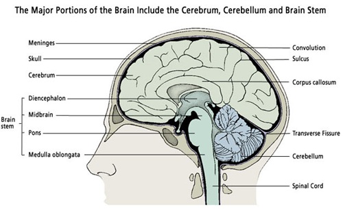
Image: Sagittal section of brain anatomy

Image: Coronal section of brain anatomy – showing the two cerebral hemispheres with their internal structures and the ventricles within them.
The Skull (Cranium)
The brain is housed within the skull, a rigid bony structure that provides vital protection. Because the skull is inelastic, any increase in volume inside—such as from a tumor, hemorrhage, or swelling—leads to increased intracranial pressure, which can affect brain function.
Cerebrum (the large brain)
The cerebrum is the largest and most complex part of the brain.
It is responsible for higher brain functions, including thinking, movement, sensation, language, and behavior.
Brain hemispheres
The cerebrum is divided into two hemispheres:
- the left hemisphere, which in most people controls language and logical functions
- the right hemisphere, which is more involved in spatial awareness and visual processing
Each hemisphere controls the opposite side of the body. The two hemispheres are not isolated; they are connected by a thick bundle of nerve fibers called the corpus callosum. This structure acts as a communication bridge, allowing the left and right sides of the brain to share information and coordinate complex tasks.
Brain lobes
Each hemisphere is further divided into regions called lobes, each with specific roles:
- Frontal lobe – planning, decision-making, behavior, personality, and voluntary movement
- Parietal lobe – sensation such as touch, pain, temperature, and body awareness
- Temporal lobe – hearing, memory, and understanding language
- Occipital lobe – vision and visual processing
Damage to different lobes produces distinct symptoms, depending on the function of the affected area.
Cerebral cortex
The outer layer of the cerebrum is called the cerebral cortex.
It is the most highly developed part of the brain and is responsible for:
- conscious thought
- voluntary movement
- sensory perception
- language and higher cognitive functions
The cortex has a folded surface made up of:
- gyri (ridges)
- sulci (grooves)
These folds increase the surface area of the brain, allowing more processing capacity.
Functional centers in the cortex
Specific regions of the cerebral cortex serve as functional centers, such as:
- motor areas controlling movement
- sensory areas processing touch and pain
- language areas responsible for speech and comprehension
- visual areas processing sight
Understanding which cortical area is involved helps explain symptoms seen on neurological examination or imaging.
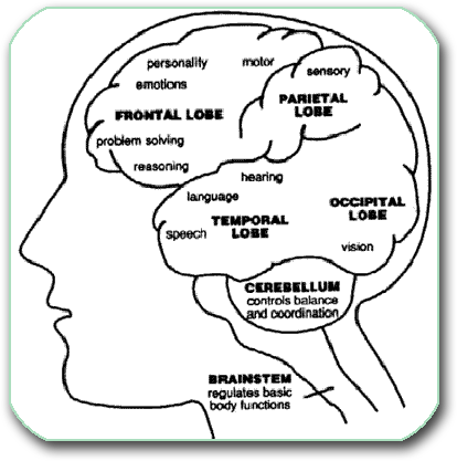
Image: Functional centers in the cortex
Brain ventricles and cerebrospinal fluid (CSF)
Inside the brain are interconnected cavities called ventricles.
They produce and circulate cerebrospinal fluid (CSF), a clear liquid that:
- cushions the brain
- protects it from injury
- helps maintain normal pressure
CSF flows from the ventricles into the subarachnoid space, where it surrounds the brain and spinal cord.
A detailed explanation of ventricles, CSF flow, and pressure regulation is available on separate pages.
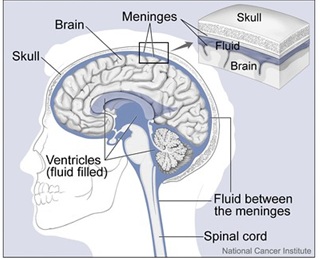
Image: Diagram of the brain ventricles (lateral, 3rd, and 4th) and the subarachnoid space surrounding the brain. Both are filled with cerebrospinal fluid (CSF). CSF is produced within the ventricles, circulates through the subarachnoid space around the brain, and is eventually absorbed into the veins. The entire volume of CSF is produced and completely replaced three times a day.
Brain coverings (meninges)
The brain is protected by three thin layers called meninges:
- Dura mater – the tough outer layer
- Arachnoid mater – the delicate middle layer
- Pia mater – the thin inner layer that lies directly on the brain surface
There is no space between the brain and the pia mater.
The space that contains cerebrospinal fluid — the subarachnoid space — is located between the arachnoid mater and the brain surface.
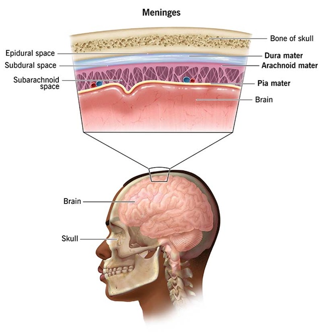
Image: meninges anatomy
Blood supply of the brain
The brain requires a constant supply of oxygen and nutrients.
This is provided by specialized arteries and drained by veins.
Even short interruptions of blood flow can cause serious neurological damage.
Arterial and venous circulation of the brain is explained in more detail on a dedicated page.
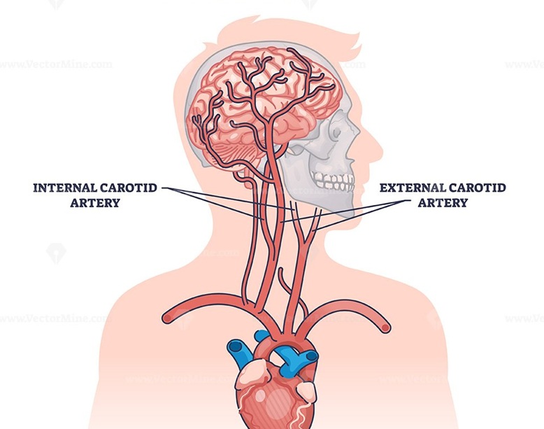
Image: Brain arteries – the two carotid arteries in the front and the two vertebral arteries in the back, which enter the skull from the neck to supply the brain with blood.
Why understanding brain anatomy matters
Understanding basic brain anatomy helps patients and families:
- interpret CT and MRI findings
- understand the location of injury or disease
- better follow explanations about conditions such as stroke, trauma, hemorrhage, or increased intracranial pressure
This page provides a simplified overview.
Each structure is explained in greater detail on linked pages for those who want to learn more.