Dr Željko Kojadinović — NEUROHIRURGIJA I LEČENJE BOLA
Dr Zeljko Kojadinovic — Pain Treatment & Neurosurgery

Primary and Secondary CNS Lymphoma
Author:
Dr. Zeljko Kojadinovic, MD, PhD
— Neurosurgeon and Pain Management Specialist
Specialized Experience:
30 years of clinical expertise in neurosurgery.
Last medically reviewed:
January 28, 2026
Who This CNS Lymphoma Page Is For
This page is intended for patients diagnosed with lymphoma involving the central nervous system (CNS) and for family members seeking a clear, medically accurate explanation.
It explains the difference between primary CNS lymphoma and secondary CNS involvement, why these tumors are usually not treated with surgical removal,
how diagnosis is established (MRI, biopsy, laboratory and systemic evaluation), and how modern chemotherapy-based treatment works.
The page also clarifies why some brain lesions that resemble glioblastoma on MRI are in fact lymphomas, why steroids can temporarily improve symptoms,
and why premature surgery can be harmful or misleading in this disease.
Readers may focus only on the sections relevant to their current situation, without reading the entire page.
If MRI findings are ambiguous, symptoms improve dramatically with steroids, surgery is being proposed without clear biopsy confirmation,
or there is uncertainty about whether a lesion represents lymphoma or glioma — an individualized
neurosurgical second opinion
can help clarify diagnosis, biopsy strategy, and optimal treatment planning.
When patients usually seek a second opinion for CNS lymphoma
- MRI shows a contrast-enhancing brain lesion, and it is unclear whether the diagnosis is glioblastoma or lymphoma
- Neurological symptoms improve rapidly after corticosteroid therapy, raising suspicion of lymphoma
- Surgical resection is being proposed despite imaging features suggestive of lymphoma
- A biopsy is recommended, but there is uncertainty about the safest trajectory or the most diagnostic target
- It is unclear whether the lymphoma is primary to the CNS or secondary from systemic disease
- There are conflicting opinions about treatment sequencing (biopsy, chemotherapy, radiotherapy)
- Imaging or clinical findings do not fully explain the patient’s neurological deterioration
In these situations, an expert neurosurgical review can help clarify the most likely diagnosis, determine whether biopsy is indicated, identify the safest and most informative biopsy target, and prevent unnecessary or harmful surgery: Request Consultation
CNS Lymphoma — Quick Summary (Read This First)
- CNS lymphoma is a malignant tumor of lymphoid cells affecting the brain, spinal cord, leptomeninges, or eyes. It may be primary (confined to the CNS) or secondary, spreading to the brain from systemic lymphoma.
- Primary CNS lymphoma (PCNSL) is rare but aggressive. It accounts for approximately 2–3% of all primary brain tumors and behaves very differently from glioblastoma.
- Surgery is usually not curative and is often avoided. Unlike glioblastoma, CNS lymphoma is treated primarily with chemotherapy, and extensive surgical resection does not improve outcome.
- MRI with contrast is the key initial diagnostic tool. CNS lymphoma often appears as a strongly enhancing lesion, sometimes mimicking glioblastoma or metastases, which makes correct interpretation critical.
- Rapid clinical or radiological improvement after corticosteroids strongly suggests lymphoma. Steroids can temporarily shrink the tumor, which may complicate diagnosis if biopsy is delayed.
- Definitive diagnosis requires tissue. Stereotactic brain biopsy is usually required to confirm primary CNS lymphoma and determine its subtype before treatment. In contrast, in secondary CNS lymphoma—which is present in approximately 30–40% of patients with systemic lymphoma—diagnosis can often be established through peripheral tissue biopsy (e.g., lymph node, bone marrow, or extranodal sites), avoiding direct brain biopsy. Primary CNS lymphoma typically does not metastasize outside the central nervous system, with extracranial spread being rare.
- Standard treatment is based on high-dose chemotherapy. High-dose methotrexate–based regimens form the backbone of therapy; radiotherapy is used selectively.
- Prognosis is generally better than glioblastoma but remains serious. Outcomes depend on age, functional status (KPS), response to chemotherapy, and whether disease is primary or secondary.
- This page is structured so you can focus only on what matters to you. Use the Contents box to jump to sections on diagnosis, treatment strategy, prognosis, or when a neurosurgical second opinion is important.
Most readers benefit from the Quick Summary plus the sections on Diagnosis, Why Surgery Is Usually Avoided, and Treatment Strategy. Other sections provide deeper clinical context.
Contents
- Who this page is for
- When to seek a second opinion
- Quick summary
- What is brain lymphoma?
- Symptoms
- Why it mimics glioblastoma
- The steroid pitfall
- Diagnosis
- Tissue diagnosis (biopsy)
- CSF examination
- Treatment
- Chemotherapy
- Radiotherapy
- Course and response
- Key Decisions in Brain Lymphoma
- Second opinion & biopsy strategy
- Prognosis
- Key clinical perspective
- Role of the neurosurgeon
- When a second opinion is critical
- Bottom line
- Final perspective
- FAQs
Brain lymphoma is a malignant tumor of the lymphatic system that involves the brain, spinal cord, or eyes. Unlike glioblastoma, brain lymphoma is not primarily a surgical disease, and its treatment, behavior, and prognosis are fundamentally different. Correct diagnosis is critical, because early management decisions can dramatically influence outcome. Central nervous system lymphomas account for approximately 2–4% of all brain tumors and about 4–6% of malignant brain tumors in adults. Primary CNS lymphoma representing roughly 60–70% of these cases and secondary CNS involvement making up the remainder.
The incidence of primary CNS lymphoma has increased over recent decades, particularly among immunocompromised patients. Individuals with HIV/AIDS, organ transplant recipients, and patients receiving long-term immunosuppressive therapy have a markedly higher risk. In the general population, primary CNS lymphoma accounts for approximately 2–3% of all primary brain tumors. In contrast, among patients with AIDS, CNS lymphoma may represent up to 10–15% of brain tumors. Although the widespread use of antiretroviral therapy has reduced its incidence, CNS lymphoma remains an important diagnostic consideration in immunocompromised patients presenting with contrast-enhancing brain lesions.
Age at Diagnosis: The median age at diagnosis for immunocompetent patients is typically between 65 and 67 years. However, in HIV-positive individuals, the disease occurs much earlier, with a median age at diagnosis usually between 30 and 40 years.
Read more about brain tumor classification and other types of brain tumors on this page.
What Is Brain Lymphoma?
Brain lymphoma refers to lymphoma affecting the central nervous system (CNS). It appears in two main forms:
- Primary CNS lymphoma (PCNSL) 60-70%— arises directly within the brain or spinal cord, without systemic disease at diagnosis. Primary CNS lymphoma typically does not metastasize outside the central nervous system, with extracranial spread being rare.
- Secondary CNS lymphoma 30-40%— spreads to the brain from lymphoma elsewhere in the body, most commonly from the lymph nodes, bone marrow, testes, breasts, lungs, or gastrointestinal tract.
While the overall risk is 5–10%, it escalates to 15–30% in specific scenarios. This includes aggressive biological subtypes (such as Burkitt lymphoma or ‘double-hit’ lymphomas) and cases where the primary disease originates in sanctuary sites like the testes or breasts, which act as reservoirs for cells that can bypass standard treatment and reach the brain.
Both forms often present in the same way and can look very similar to glioblastoma on MRI, which is why misdiagnosis is common early on.
Histological subtype
More than 90–95% of primary CNS lymphomas are Diffuse Large B-cell Lymphomas (DLBCL). T-cell lymphomas, Burkitt lymphoma, and follicular lymphomas are extremely rare in the CNS and represent only a small minority of cases. This biological profile, dominated by B-cells, explains the typical steroid responsiveness of CNS lymphoma.
Typical Locations of Lymphoma in the Brain
Unlike many other brain tumors, CNS lymphoma has a preference for specific regions, often appearing in deeper structures of the brain:
- Periventricular areas: Frequently found near the fluid-filled spaces (ventricles) of the brain.
- Deep Gray Matter: Often involves the thalamus or basal ganglia, which can impact motor control and sensory processing.
- Corpus Callosum: It has a tendency to cross the midline or involve this bridge between the two hemispheres.
- Multifocal lesions: In about 30–40% of cases, lymphoma appears in multiple locations simultaneously, which is a major differentiator from glioblastoma (which is usually a single mass).
- Eyes and Leptomeninges: In some patients, the disease may also involve the eyes (intraocular lymphoma) or the thin layers covering the brain (meninges).
Symptoms of Brain Lymphoma
Symptoms usually develop over weeks to a few months, often faster than low-grade tumors but sometimes less explosively than glioblastoma.
Common symptoms include:
- Progressive headache, often with nausea or vomiting
- New-onset seizures, especially in adults without prior epilepsy
- Cognitive decline — confusion, memory loss, slowed thinking
- Personality or behavioral changes
- Focal neurological deficits, depending on tumor location:
- weakness on one side of the body
- speech or language problems
- visual disturbances
- gait instability
Symptoms may temporarily improve dramatically after corticosteroids, which is an important diagnostic clue — but also a major pitfall.
General Symptoms of Secondary Lymphoma (Systemic Disease)
Secondary CNS lymphoma develops in patients who already have lymphoma elsewhere in the body. Because of this, symptoms are often both systemic and neurological.
Common general (systemic) symptoms include:
- Unexplained weight loss
- Persistent fatigue and weakness
- Night sweats (often drenching)
- Prolonged or recurrent fever
- Loss of appetite
- Generalized itching
- Painless lymph node enlargement (neck, armpits, groin)
These symptoms may precede brain involvement by months.
Organ-Specific Symptoms Outside the Brain
Depending on where the lymphoma originates or spreads, patients may also have symptoms related to other organs:
- Lymph nodes: painless swelling in the neck, chest, abdomen, or groin
- Bone marrow: anemia, frequent infections, easy bruising or bleeding
- Lungs: cough, shortness of breath, chest discomfort
- Liver and spleen: abdominal fullness, pain under the ribs, early satiety
- Gastrointestinal tract: abdominal pain, diarrhea, bleeding, weight loss
- Skin: nodules, plaques, or persistent skin lesions
- Testes (in men): painless testicular enlargement (a known risk factor for CNS spread)
Why Brain Lymphoma Is Often Confused With Glioblastoma on MRI
On MRI, brain lymphoma often appears as:
- a contrast-enhancing mass
- deep or periventricular location
- significant surrounding edema
These features overlap with glioblastoma. However, there are key differences:
- Lymphoma is usually more cellular and less necrotic
- It often shows restricted diffusion — On Diffusion-Weighted Imaging (DWI), lymphoma typically appears very „bright“ due to its high cellular density. This specific MRI sequence helps distinguish it from more necrotic or cystic tumors like glioblastoma.
- It may shrink rapidly after steroids
Because imaging alone cannot be fully reliable, histopathological confirmation is essential.
The Steroid Pitfall: Why Diagnosis Can Be Delayed or Missed
Corticosteroids (such as dexamethasone) can cause rapid tumor regression in lymphoma. While this may temporarily improve symptoms, it can also:
- make the lesion partially or completely disappear on MRI
- render biopsy non-diagnostic
- delay definitive treatment
For this reason, steroids should be avoided before biopsy whenever lymphoma is suspected, unless there is life-threatening brain swelling.
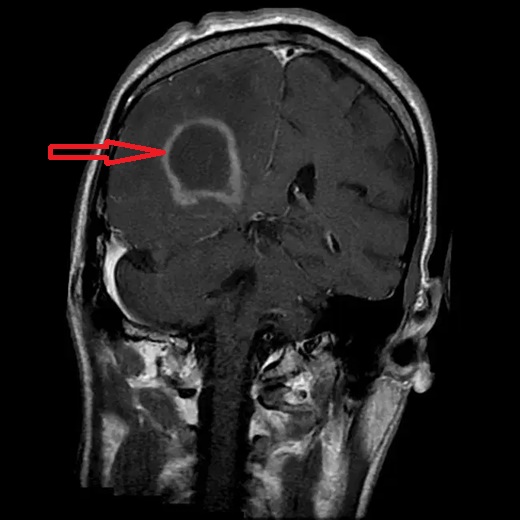
Image: MRI scan of the brain where a red arrow points to a primary CNS lymphoma in an immunocompromised individual, closely mimicking the appearance of a glioblastoma.
Diagnostic Workup After MRI Suggests CNS Lymphoma
Once MRI findings raise suspicion for a CNS lymphoma, imaging alone is never sufficient to establish the diagnosis or determine the disease type. Further steps are mandatory to confirm the pathology and define whether the lymphoma is primary or secondary, as treatment strategies differ fundamentally.
Tissue Diagnosis (Mandatory Step)
Definitive diagnosis requires histopathological confirmation.
- Stereotactic brain biopsy is the standard approach. If systemic lymphoma has already been histologically confirmed outside the brain and MRI findings are consistent with CNS involvement, a brain biopsy may not be necessary. However, if no systemic lymphoma has been proven, or if imaging findings are ambiguous, stereotactic brain biopsy is essential to establish the diagnosis and avoid inappropriate surgery or delayed chemotherapy.
- Advanced imaging (perfusion MRI, spectroscopy, PET) can support suspicion, but cannot replace biopsy.
- Surgical resection is usually avoided, as it does not improve outcome in lymphoma
- Immunohistochemistry and molecular analysis are performed to define lymphoma subtype (most commonly diffuse large B-cell lymphoma)
Important: Corticosteroids should be avoided before biopsy whenever possible, as they may cause temporary tumor regression and lead to false-negative results.
Systemic Staging: Excluding Secondary Lymphoma
At the time of diagnosis, it is not known whether the lymphoma is primary or secondary.
Therefore, systemic staging is required in all patients.
This typically includes:
- Whole-body PET-CT (preferred)
or CT of the chest, abdomen, and pelvis - Blood tests:
- complete blood count
- LDH
- liver and kidney function tests
- inflammatory markers
- Bone marrow biopsy, if systemic involvement is suspected
These investigations determine whether lymphoma is present outside the central nervous system.
Cerebrospinal Fluid (CSF) Examination
In selected patients and when safe to perform:
- Lumbar puncture with CSF cytology and flow cytometry
- Helps identify leptomeningeal involvement and supports staging
Final Classification
Only after completing biopsy and systemic staging can the diagnosis be finalized:
- Primary CNS lymphoma: disease confined to the brain, leptomeninges, spinal cord, or eyes, without systemic involvement
- Secondary CNS lymphoma: brain involvement as part of a systemic lymphoproliferative disease
Key Clinical Point
Primary CNS lymphoma does not normally spread outside the nervous system.
If lymphoma is detected elsewhere in the body, the disease is classified as secondary.
Treatment of Brain Lymphoma
Why Surgery Is Usually Not the Treatment
Unlike glioblastoma, surgical resection does not improve survival in brain lymphoma. The tumor is highly sensitive to chemotherapy, and surgery risks neurological damage without benefit.
Surgery is therefore limited to:
- biopsy
- emergency decompression in exceptional cases
Standard Treatment Protocols
Chemotherapy (Mainstay of Treatment)
The backbone of treatment is high-dose methotrexate–based chemotherapy, often combined with other agents. Treatment is typically delivered in specialized oncology centers and may require hospitalization.
Radiotherapy
Radiotherapy may be used:
- as consolidation therapy
- in relapse
- in patients unable to tolerate intensive chemotherapy
Because of potential long-term cognitive toxicity, especially in older patients, radiotherapy is used selectively.
Symptomatic Treatment
Supportive care plays an important role and includes:
- corticosteroids (after diagnosis)
- anti-epileptic drugs for seizures
- treatment of brain edema
- rehabilitation and cognitive support
Symptomatic treatment does not replace oncological therapy, but improves quality of life and functional status.
Course of Disease and Response to Treatment
Brain lymphoma is often highly treatment-sensitive, especially initially. Many patients experience:
- rapid radiological response
- significant clinical improvement
However:
- relapses are common
- long-term disease control remains challenging
The disease course varies widely depending on patient age, functional status, and response to therapy.
Key Decision Points in Suspected CNS Lymphoma
In suspected CNS lymphoma, the most important issue is not how to remove the tumor, but how to avoid incorrect initial decisions. Unlike most brain tumors, surgical resection is usually not beneficial, and the priority is to establish a correct diagnosis through biopsy before starting treatment.
When Biopsy Should Be Performed Instead of Surgery
The most important step is to recognize the possibility of lymphoma before any surgical decision is made. If this is not considered early, a lesion that mimics glioblastoma or metastasis on MRI may be unnecessarily operated on. The key step in management is obtaining a tissue diagnosis, most often through stereotactic biopsy. Performing surgical resection instead of biopsy may delay appropriate chemotherapy without improving outcome. The central question is not how much tumor to remove, but how to establish the correct diagnosis in the safest and fastest way.
Can Steroids Delay or Compromise Diagnosis
Corticosteroids can temporarily shrink the tumor, which may lead to a non-diagnostic biopsy or even make the lesion disappear on imaging. For this reason, steroids should be used cautiously and usually avoided before biopsy, unless there is a life-threatening situation.
When MRI Suggests Lymphoma but Diagnosis Is Uncertain
CNS lymphoma can closely mimic glioblastoma on MRI, which creates a high risk of misdiagnosis. In these situations, treatment decisions should be based on careful interpretation of imaging, clinical course, and response to steroids, not on imaging alone.
When Surgery May Be Unnecessary or Harmful
In many cases, proceeding directly to surgery may expose the patient to unnecessary neurological risk without therapeutic benefit. The most important decision is recognizing when surgery should be avoided, and when a targeted biopsy and oncological treatment represent the correct approach.
Request CNS Lymphoma Neurosurgery Consultation — 24-Hour Review or Priority Option (Usually Within 3 Hours)
When MRI suggests CNS lymphoma, families often face urgent, high-stakes questions:
what the scan most likely represents, whether steroids should be started (or stopped) before biopsy,
where the safest diagnostic target is, and whether surgery is appropriate (usually it is not, beyond biopsy).
An independent neurosurgical second opinion can help you interpret MRI features, avoid common pitfalls (especially the steroid–biopsy issue),
clarify a biopsy strategy, and understand what treatment typically involves after diagnosis is confirmed.
- ✔ Send a short message describing the suspected CNS lymphoma diagnosis, symptoms, and what the MRI report says
- ✔ You’ll receive a reply within 24 hours explaining if and how we can help in your specific CNS lymphoma situation
- ✔ Time-sensitive cases: if neurological status is worsening, there is reduced consciousness, significant mass effect/ICP concern, or doctors are urgently recommending biopsy — write PRIORITY in your first message
- ✔ MRI (DICOM) images and hospital documentation can be reviewed once initial contact is established
- ✔ During the consultation, we explain biopsy targeting and timing (including steroid considerations), expected next steps, and realistic goals — with up to 10 days of follow-up for brief questions
Consultation fees typically range from $180–250, depending on case complexity and imaging findings.
Secure payment by credit card, PayPal invoice (USD), or bank transfer.
This is within the usual range for international specialist telehealth second opinions in neurosurgery.
Prognosis of Brain Lymphoma
Overall prognosis of brain lymphoma is significantly better than glioblastoma, but the disease remains serious and potentially life-threatening. Outcomes depend strongly on early diagnosis, correct classification (primary vs secondary), and response to chemotherapy.
Overall Prognostic Factors
Prognosis is influenced by:
- Age (younger patients do better)
- Karnofsky Performance Status (KPS)
- Response to first-line chemotherapy
- Extent of systemic disease (in secondary CNS lymphoma)
- Immune status (immunocompetent vs immunocompromised)
Survival Expectations — Primary CNS Lymphoma (PCNSL)
In immunocompetent patients treated with modern high-dose methotrexate–based protocols:
- Median overall survival:
30–60 months (≈ 2.5–5 years) - 5-year survival:
30–40% - 10-year survival:
15–20% (in selected long-term responders)
Younger, immunocompetent patients (<60 years) with good functional status (KPS ≥70) and a complete response to chemotherapy represent the most favorable subgroup.
Survival Expectations — Secondary CNS Lymphoma
Secondary CNS lymphoma generally has a worse prognosis because it reflects advanced systemic disease.
- Median survival:
6–24 months, depending on systemic control - Prognosis is strongly linked to:
- activity of lymphoma outside the brain
- chemosensitivity of the systemic disease
- ability to tolerate intensive therapy
Relapse and Disease Control
- Relapse remains the main limiting factor
- Most recurrences occur within the first 2–3 years
- Salvage therapies can prolong survival but are rarely curative
Key Clinical Perspective
- Brain lymphoma is not surgically cured, but it is one of the few malignant brain tumors that is highly chemotherapy-sensitive
- Early and correct diagnosis is crucial — mistaking lymphoma for glioblastoma and proceeding directly to surgery or radiation can delay effective treatment
Bottom Line
Statistics describe populations, not individuals.
Some patients experience durable long-term remission, while others progress rapidly.
Functional status, biological behavior, and response to therapy matter more than averages.
A Critical Note on Surgery and Brain Lymphoma
Although brain lymphoma is not a surgical disease, in real-world practice there are centers with a disproportionately high number of operated lymphomas. This usually reflects diagnostic uncertainty before surgery, rather than true surgical indication.
The primary responsibility of the neurosurgeon is not to operate at any cost, but to recognize when a lesion is likely a lymphoma before committing the patient to an unnecessary resection.
The Role of the Neurosurgeon in Suspected Brain Lymphoma
The neurosurgeon plays a decisive role at several critical points:
1. Recognizing lymphoma before surgery
A neurosurgeon must actively consider lymphoma in the differential diagnosis when MRI features, clinical presentation, or steroid responsiveness suggest it. Proceeding directly to tumor resection without this consideration can delay appropriate chemotherapy and expose the patient to unnecessary risk.
2. Correct indication for biopsy
When tissue diagnosis is required, the correct decision is usually stereotactic biopsy, not resection. This decision must be based on imaging characteristics, clinical context, and expected therapeutic consequences.
3. Choosing the correct biopsy target
Not all parts of a lesion are diagnostically equivalent. Sampling necrotic, hemorrhagic, or steroid-altered tissue can lead to:
- inconclusive pathology
- false-negative or misleading results
- delayed or incorrect treatment
An experienced neurosurgeon selects the biopsy target that is most likely to yield definitive histopathological and molecular diagnosis.
4. Selecting the safest biopsy trajectory
Equally important is choosing the safest needle pathway — minimizing the risk of:
- hemorrhage
- neurological deficit
- ventricular breach
- sampling error
5. Integrating all results
The neurosurgeon must interpret imaging, pathology, molecular markers, steroid response, and clinical evolution together — not as isolated data points.
Frequently Asked Questions About CNS Lymphoma
What is brain lymphoma or CNS lymphoma?
Brain lymphoma, also called CNS lymphoma when it involves the central nervous system, is a malignant tumor made of lymphoid cells affecting the brain, spinal cord, leptomeninges, or eyes. It is different from glioblastoma and most other brain tumors because it is usually treated primarily with chemotherapy rather than surgical removal. CNS lymphoma may be primary, meaning it starts within the central nervous system, or secondary, meaning lymphoma from another part of the body spreads to the brain or other CNS structures. Correct diagnosis is essential because surgery, steroids, biopsy timing, and chemotherapy decisions are very different from other brain tumors.
What is the difference between primary CNS lymphoma and secondary CNS lymphoma?
Primary CNS lymphoma begins inside the brain, spinal cord, leptomeninges, or eyes, without evidence of lymphoma elsewhere in the body at the time of diagnosis. Secondary CNS lymphoma means that a systemic lymphoma, usually an aggressive non-Hodgkin lymphoma, has spread to the central nervous system. The two conditions can look similar on MRI and may cause similar neurological symptoms, but staging and treatment planning differ. Primary CNS lymphoma is usually treated with high-dose methotrexate–based chemotherapy directed at CNS disease. Secondary CNS lymphoma requires assessment of both brain involvement and systemic lymphoma activity, because prognosis and treatment depend strongly on disease outside the CNS.
Can lymphoma spread to the brain?
Yes. Lymphoma can spread to the brain, spinal cord, leptomeninges, or eyes, and this is called secondary CNS lymphoma. It is more likely in certain aggressive lymphoma subtypes and in patients with high-risk systemic disease. Some sites, such as the testes, breast, bone marrow, or certain extranodal locations, may carry a higher risk of CNS involvement. When lymphoma spreads to the brain, patients may develop headache, confusion, weakness, speech problems, seizures, visual symptoms, or gait difficulty. Diagnosis requires careful MRI interpretation and systemic staging. Treatment depends on both CNS disease and the status of lymphoma elsewhere in the body.
Is brain lymphoma a brain tumor?
Yes. Brain lymphoma is a type of brain tumor, but it behaves differently from glioma, glioblastoma, meningioma, or metastasis. It arises from lymphoid cells rather than from brain-supporting glial cells. This difference is clinically important because brain lymphoma is usually not treated by removing as much tumor as possible. Instead, the key first step is establishing the correct diagnosis, usually through stereotactic biopsy when systemic lymphoma has not already been confirmed. After diagnosis, treatment is primarily chemotherapy-based. For this reason, brain lymphoma should not be managed like a typical resectable brain mass unless there is an exceptional emergency situation.
What symptoms can brain lymphoma or CNS lymphoma cause?
Brain lymphoma symptoms depend on tumor location, number of lesions, brain swelling, and whether the disease is primary or secondary. Common symptoms include progressive headache, nausea, vomiting, confusion, memory problems, personality change, slowed thinking, weakness on one side of the body, speech problems, visual disturbance, balance problems, and seizures. Symptoms often develop over weeks to a few months. In secondary CNS lymphoma, patients may also have systemic lymphoma symptoms such as weight loss, night sweats, fever, fatigue, itching, enlarged lymph nodes, or organ-related symptoms. A striking but temporary improvement after corticosteroids can be an important clue that the lesion may be lymphoma.
What does brain lymphoma look like on MRI?
Brain lymphoma often appears on MRI as a strongly contrast-enhancing lesion, commonly located near the ventricles, deep gray matter, basal ganglia, thalamus, corpus callosum, or other deep brain structures. It may be single or multifocal. CNS lymphoma is often highly cellular, so it commonly shows restricted diffusion on diffusion-weighted MRI. Surrounding edema may be present. In some patients, the lesion can resemble glioblastoma, metastasis, infection, or inflammatory disease. MRI is essential for suspicion and biopsy planning, but imaging alone is not enough for a definitive diagnosis. Histopathological confirmation is usually required unless systemic lymphoma has already been proven and CNS involvement is clinically clear.
Can CNS lymphoma look like glioblastoma on MRI?
Yes. CNS lymphoma can closely mimic glioblastoma on MRI because both may appear as contrast-enhancing brain masses with surrounding edema and neurological symptoms. However, there are important differences. Lymphoma often has dense cellularity, restricted diffusion, deep or periventricular location, basal ganglia or thalamic involvement, corpus callosum involvement, and sometimes multifocal disease. Glioblastoma more often shows necrosis, irregular ring enhancement, and infiltrative features, although overlap is common. A major clue is rapid improvement after corticosteroids, which is more typical of lymphoma. Because MRI cannot always separate these diagnoses safely, stereotactic biopsy is often the correct step when lymphoma is suspected. Mistaking lymphoma for glioblastoma can lead to unnecessary resection and delayed chemotherapy.
Why do steroids sometimes improve brain lymphoma symptoms?
Brain lymphoma is often very sensitive to corticosteroids such as dexamethasone. Steroids can reduce swelling and may also temporarily shrink the lymphoma itself, leading to dramatic clinical or radiological improvement. This can be helpful in emergencies with dangerous brain swelling, but it creates a diagnostic problem. If steroids are given before biopsy, the lesion may partially disappear or become less diagnostic, and the biopsy may be falsely negative or inconclusive. For this reason, when CNS lymphoma is suspected, steroids are usually avoided before biopsy unless the patient has severe mass effect, reduced consciousness, or another urgent reason requiring immediate treatment.
How is brain lymphoma diagnosed?
Brain lymphoma diagnosis begins with MRI, but definitive diagnosis usually requires tissue confirmation. If systemic lymphoma has not already been proven, stereotactic brain biopsy is commonly performed to obtain a sample for histology, immunohistochemistry, and molecular testing. If the patient already has confirmed systemic lymphoma and MRI findings are typical for CNS involvement, biopsy from a peripheral site or existing pathology may sometimes be sufficient, but this depends on the clinical situation. Systemic staging is also important and may include PET-CT, CT chest/abdomen/pelvis, blood tests, bone marrow evaluation, eye examination, and CSF analysis when safe and clinically indicated.
When is brain biopsy needed for suspected CNS lymphoma?
Brain biopsy is needed when MRI suggests CNS lymphoma but no reliable tissue diagnosis exists. This is especially important when the alternative diagnosis could be glioblastoma, metastasis, infection, demyelination, or another inflammatory lesion. Stereotactic biopsy allows the doctor to confirm lymphoma type and guide chemotherapy. The biopsy target must be chosen carefully because necrotic, hemorrhagic, or steroid-altered tissue may produce inconclusive results. Steroids should usually be avoided before biopsy when possible. If systemic lymphoma is already proven and the brain findings are clearly compatible with secondary CNS involvement, a separate brain biopsy may not always be necessary, but this must be individualized.
Is brain surgery a treatment option for CNS lymphoma or brain lymphoma?
Brain surgery is usually not the main treatment for CNS lymphoma or brain lymphoma. In most patients, surgery is used to obtain tissue for diagnosis, usually by stereotactic biopsy, while treatment is based mainly on high-dose methotrexate–based chemotherapy. Extensive surgical removal is usually avoided because CNS lymphoma is a systemic-type malignant lymphoid disease within the nervous system and visible tumor removal does not usually improve survival. Surgery may be considered only in exceptional situations, such as life-threatening mass effect, diagnostic uncertainty that cannot be resolved by biopsy, or selected cases where urgent decompression is required. In most cases, biopsy rather than resection is the correct neurosurgical procedure.
What is the standard treatment for primary CNS lymphoma?
The standard treatment for primary CNS lymphoma is high-dose methotrexate–based chemotherapy, usually managed by neuro-oncology or hematology-oncology teams experienced with CNS disease. Other chemotherapy agents, rituximab-based combinations, targeted therapies, or consolidation strategies may be added depending on age, performance status, kidney function, immune status, and lymphoma subtype. In selected medically fit patients, autologous stem cell transplantation may be considered as consolidation after response to induction therapy. Treatment often requires careful monitoring and may involve hospitalization. Radiotherapy is used selectively because whole-brain radiation can cause significant cognitive toxicity, especially in older patients. Surgery is not the main treatment except for biopsy.
Is radiotherapy always part of CNS lymphoma treatment?
No. Radiotherapy is not always part of CNS lymphoma treatment. Whole-brain radiotherapy was used more often in the past, but it can cause long-term cognitive decline, especially in older adults. Modern treatment for primary CNS lymphoma is usually based first on high-dose methotrexate–based chemotherapy. Radiotherapy may be considered as consolidation in selected patients, in relapse, or when chemotherapy cannot be tolerated or does not provide adequate control. The decision depends on age, neurological status, response to chemotherapy, disease extent, and expected neurocognitive risk. This is one reason treatment planning should be individualized and coordinated between oncology, neurology, radiation oncology, and neurosurgery.
What is the prognosis for brain lymphoma or primary CNS lymphoma?
The prognosis for brain lymphoma depends on whether the disease is primary or secondary, the patient’s age, Karnofsky Performance Status, immune status, extent of disease, and response to chemotherapy. Primary CNS lymphoma generally has a better prognosis than glioblastoma but remains potentially life-threatening. In immunocompetent patients treated with modern high-dose methotrexate–based regimens, median survival is often measured in years rather than months, but results vary widely. Secondary CNS lymphoma usually has a worse prognosis because it reflects systemic disease with CNS involvement. Statistics describe groups, not individuals. Functional status, diagnosis timing, and treatment response are often more important than averages.
What is the prognosis for B-cell lymphoma in the brain?
The prognosis for B-cell lymphoma in the brain depends on whether it is primary CNS lymphoma or secondary CNS involvement from systemic B-cell lymphoma. More than 90–95% of primary CNS lymphomas are diffuse large B-cell lymphomas, and many respond to high-dose methotrexate–based treatment. Prognosis is more favorable in younger, immunocompetent patients with good functional status and strong response to chemotherapy. Secondary B-cell lymphoma involving the brain usually has a more difficult prognosis because it reflects aggressive systemic disease reaching the nervous system. MRI appearance alone cannot predict outcome. Tissue diagnosis, systemic staging, neurological status, immune status, and treatment response are the most important prognostic factors.
What is the life expectancy for lymphoma in the brain?
Life expectancy for lymphoma in the brain varies substantially. Primary CNS lymphoma may respond well to chemotherapy, and some patients achieve long remissions. In eligible patients treated with modern high-dose methotrexate–based regimens, median overall survival in recent series is often reported in the range of approximately 3–5 years, but this varies strongly with age, performance status, immune function, treatment tolerance, and response to chemotherapy. Secondary CNS lymphoma generally has a shorter expected survival because it usually occurs in the setting of aggressive or advanced systemic lymphoma. Individual prognosis cannot be determined from MRI alone. It requires tissue diagnosis, systemic staging, neurological status assessment, and evaluation of how the disease responds to treatment.
What is the survival rate for CNS lymphoma with treatment?
The survival rate for CNS lymphoma with treatment varies widely because primary CNS lymphoma and secondary CNS lymphoma are different clinical situations. In primary CNS lymphoma, high-dose methotrexate–based treatment can produce meaningful remission, and some patients live for years, especially when they are younger, medically fit, immunocompetent, and respond well to first-line therapy. Survival is worse when performance status is poor, disease is multifocal or advanced, immune suppression is present, or treatment cannot be tolerated. Secondary CNS lymphoma usually has lower survival rates because nervous system involvement occurs together with systemic lymphoma. Survival statistics should therefore be interpreted only in the context of the individual diagnosis and treatment response.
What is the prognosis when systemic lymphoma spreads to the brain?
When systemic lymphoma spreads to the brain, spinal cord, leptomeninges, eyes, or cerebrospinal fluid, the condition is called secondary CNS lymphoma. Prognosis is usually worse than in primary CNS lymphoma because the disease reflects both systemic lymphoma biology and nervous system involvement. Outcome depends on the lymphoma subtype, response of systemic disease to treatment, extent of CNS involvement, patient age, performance status, immune status, and ability to tolerate intensive therapy. Some patients respond to treatment, but secondary CNS lymphoma is often aggressive and relapse risk is high. Management usually requires coordinated hematology-oncology, neuro-oncology, neurology, and neurosurgical input when biopsy or diagnostic clarification is needed.
Is brain lymphoma curable?
Brain lymphoma can sometimes enter long-term remission, especially in younger or medically fit patients who respond well to high-dose methotrexate–based treatment. However, it remains a serious malignant disease, and relapse is common. Cure is more realistic in selected primary CNS lymphoma patients with good performance status and strong treatment response, but it cannot be assumed at diagnosis. Secondary CNS lymphoma usually has a more difficult prognosis because it reflects lymphoma involving both the nervous system and systemic disease. The most important early steps are correct diagnosis, avoiding unnecessary surgery, proper staging, and rapid initiation of appropriate chemotherapy once tissue diagnosis is secured.
Is CNS lymphoma prognosis better than glioblastoma?
In many patients, primary CNS lymphoma has a better prognosis than glioblastoma because it is more sensitive to chemotherapy. Some patients respond dramatically to high-dose methotrexate–based treatment and may achieve long remission. However, CNS lymphoma is still an aggressive malignant disease, relapse is common, and outcomes vary widely. The comparison also depends on age, immune status, performance status, lesion distribution, and treatment response. Glioblastoma is usually treated with maximal safe resection, radiation, and temozolomide, while CNS lymphoma is usually treated with biopsy followed by chemotherapy. This difference is why correct early diagnosis is so important when MRI could represent either lymphoma or glioblastoma.
Why is a neurosurgical second opinion important in suspected CNS lymphoma?
A neurosurgical second opinion is important when MRI findings are ambiguous, symptoms improve rapidly after steroids, biopsy is being planned, open surgery is being proposed, or steroids have already been given and the lesion has partially resolved on repeat MRI. In suspected CNS lymphoma, the key neurosurgical decision is often not how much tumor to remove, but whether the lesion should be biopsied instead of resected. The biopsy target and trajectory must be chosen carefully to avoid non-diagnostic tissue and unnecessary neurological risk. A second opinion can also help clarify whether the imaging pattern is more consistent with lymphoma, glioblastoma, metastasis, infection, or another condition. Correct early decisions can prevent harmful surgery and treatment delay.
Can patients receive an online neurosurgical second opinion for CNS lymphoma?
Yes. Patients can receive an online neurosurgical second opinion for suspected or confirmed CNS lymphoma when MRI images, reports, clinical history, medication history, and available pathology or systemic staging results are provided. The consultation can help clarify whether the lesion pattern suggests lymphoma, whether steroids may have affected diagnosis, whether biopsy is needed, and what biopsy strategy appears safest. It can also explain why surgical removal is usually not the main treatment and what questions should be discussed with oncology teams. Online review is especially useful when families are uncertain whether the proposed plan is biopsy, surgery, chemotherapy, radiotherapy, or additional diagnostic workup.
Trusted resources for patients and families (CNS Lymphoma focus)
- National Cancer Institute (NCI) — Primary CNS Lymphoma Patient Guide
- Macmillan Cancer Support — Primary CNS Lymphoma: Support & Information
- American Brain Tumor Association — Lymphoma of the Brain (Patient FAQ)
These resources provide general educational information and do not replace individualized medical assessment or expert consultation.
On this page, you can learn more about other types of brain metastases.