Dr Željko Kojadinović — NEUROHIRURGIJA I LEČENJE BOLA
Dr Zeljko Kojadinovic — Pain Treatment & Neurosurgery


Author: Dr. Zeljko Kojadinovic, MD, PhD – Neurosurgeon
Last medically reviewed: October 10, 2026
WHO THIS PAGE IS FOR
If you have tailbone pain that becomes worse while sitting, on hard surfaces, or when rising from a chair, this page explains the most common causes, how coccydynia is diagnosed, and which treatments may help.
If your pain has persisted despite cushions, medications, physical therapy, or previous injections, a telehealth pain consultation can help assess whether the pain is arising from the coccyx itself, abnormal coccygeal mobility, a joint, nerve, pelvic floor structure, or another source.
If an injection, ganglion impar block, radiofrequency procedure, or coccygectomy has been suggested, we can review your imaging and clinical history and clarify whether the proposed treatment is appropriate through an online second opinion.
The specialist who can identify the true pain generator does not necessarily need to be located in your country.
When patients usually seek a second opinion for persistent tailbone pain
- Tailbone pain persists despite cushions, medications, physical therapy, or other conservative treatment
- Pain is particularly severe while sitting, on hard surfaces, or when rising from a chair
- Standard X-rays or MRI are normal or do not clearly explain the symptoms
- There is uncertainty about whether abnormal coccygeal mobility, a joint, pelvic floor structure, or nerve is the true pain generator
- Previous injections or other procedures provided little, temporary, or unclear benefit
- A ganglion impar block, radiofrequency procedure, or coccygectomy has been suggested and you want to know whether it is truly indicated
- The pain has remained unresolved despite examinations and treatment in your country, and the appropriate specialist is not available locally
If this reflects your situation, a focused telehealth review can help clarify the most likely pain generator, whether additional imaging such as dynamic sitting and standing X-rays may be useful, which treatment options are reasonable, and whether an intervention or surgery is actually indicated in your case: Request Consultation
Coccydynia (Tailbone Pain) — Quick Summary (Read This First)
- Coccydynia is pain arising from the coccyx or the structures immediately around it. It is typically worse while sitting, especially on a hard surface, when leaning backward, or when rising from a chair. However, not every pain felt in the tailbone region is true coccydynia.
- The key question is: what is the true pain generator? Pain may originate from the coccyx itself, the sacrococcygeal or inter-coccygeal joints, surrounding ligaments and soft tissues, pelvic floor muscles, or neural structures. Similar pain may also come from the sacroiliac joint, lumbosacral spine, sacral nerve roots, or other pelvic structures.
- Common causes include trauma, abnormal coccygeal mobility, degenerative or inflammatory changes, and childbirth. A fall directly onto the buttocks may cause contusion, fracture, subluxation, or persistent mechanical instability. In some patients, no single clear cause can be identified and the condition is classified as idiopathic coccydynia.
- Pain when rising from a chair can be an important diagnostic clue. In patients with hypermobility, subluxation, or another form of mechanical instability, movement of the coccyx during the transition from sitting to standing may provoke a sudden increase in pain.
- A normal static X-ray or MRI does not exclude coccydynia. MRI is useful for assessing soft tissues, bone marrow edema, inflammation, inter-coccygeal discs, and other lesions, but it is usually performed while lying down and does not show how the coccyx behaves under load.
- Dynamic sitting and standing X-rays can reveal abnormalities that routine imaging misses. In selected patients, comparing the coccyx in standing and loaded sitting positions may demonstrate hypermobility, subluxation, or another abnormal movement pattern that is not visible on a conventional static image.
- Imaging findings must match the clinical picture. Coccygeal shape, curvature, a bony spicule, or another anatomical variation may be seen on imaging without causing symptoms. A finding becomes clinically meaningful when it corresponds with the location of pain, focal tenderness, and reproduction of the patient’s usual symptoms.
- Treatment is usually conservative at first. Reducing direct coccygeal pressure, modifying sitting posture, using an appropriate cushion or seat cut-out, medications, targeted physical therapy, pelvic floor treatment, and selected manual techniques may all be useful, depending on the underlying mechanism.
- Persistent pain may require a more targeted diagnostic and interventional approach. Injections can be both therapeutic and diagnostic. Depending on the suspected pain generator, options may include sacrococcygeal or inter-coccygeal injections, a ganglion impar block, ESWT, or selected radiofrequency procedures.
- Neuropathic symptoms change the diagnostic approach. Burning or shooting pain, tingling, sensory loss, weakness, reflex changes, or perineal symptoms may indicate involvement of sacral nerve roots, peripheral nerves, or another neural structure rather than an isolated coccygeal disorder.
- Tarlov cysts and other MRI findings do not automatically explain the pain. Most Tarlov or perineural cysts are incidental. Their clinical significance depends on whether the cyst location and neurological symptoms correspond with the patient’s pain pattern.
- Coccygectomy is not the next step simply because pain has lasted a long time. Surgery is generally reserved for carefully selected patients with chronic, clearly localized and functionally limiting pain, failure of adequate conservative and interventional treatment, and convincing evidence that the coccyx is the true pain generator.
- Some tailbone pain requires broader investigation. Progressive or constant pain unrelated to position, marked night pain, fever, unexplained weight loss, a known malignancy, neurological deficit, saddle numbness, or bladder or bowel dysfunction should prompt evaluation for causes other than ordinary mechanical coccydynia.
- How to use this page: If your pain is mainly provoked by sitting or rising from a chair, focus first on Abnormal Coccygeal Mobility, Dynamic Coccyx X-rays, and the treatment sections. If you also have neurological or perineal symptoms, read When Does Pain in the Tailbone Region Come From the Spine or Nerves? If surgery has been suggested, review When Is Coccygectomy Considered? and How Successful Is Coccygectomy?
Most patients only need the sections that match their pain pattern, followed by Diagnosis, Dynamic Coccyx X-rays, and the treatment options most relevant to their findings.
Contents
- What Is the Coccyx?
- Who Develops Coccydynia?
- Causes of Coccydynia
- Other Causes of Tailbone Pain
- Typical Tailbone Pain
- How Long Can Pain Last?
- Diagnosing Coccydynia
- Spine or Nerve Pain
- Treating Coccydynia
- When Is Coccygectomy Considered?
- Coccydynia Prognosis
- Final Clinical Principle
- Start Telehealth Consultation
- FAQ
Coccydynia is pain arising from the coccyx or the immediately surrounding structures of the sacrococcygeal region. It is typically aggravated by sitting, particularly on a hard surface, leaning backward, or rising from a chair. In some patients, the pain becomes persistent and can significantly limit daily activities, driving, desk work, and travel. Not every pain felt in the tailbone region originates from the coccyx itself, so identifying the true pain generator is an important part of diagnosis.
What Is the Coccyx and Why Can It Hurt?
The coccyx, or tailbone, is the lowest part of the spinal column. It consists of several small bony segments whose number and shape can vary considerably between individuals. It is connected to the sacrum through the sacrococcygeal joint, while additional joints with varying degrees of mobility may exist between individual coccygeal segments.
Ligaments, pelvic floor muscles, and other soft-tissue structures attach to the coccyx. Neural structures also pass nearby and may contribute to pain. The anatomy of this region is therefore not purely bony; pain may arise from a joint, ligament, muscle, nerve, or surrounding soft tissue.
Anatomical variations of the coccyx are common. The coccyx may be almost straight, mildly curved forward, markedly curved, or may show a sharp anterior angulation between segments, sometimes approaching 90 degrees. Some individuals also have partial displacement between coccygeal segments or a prominent bony spicule at the tip. These variations are not diseases by themselves, but they may alter pressure distribution and biomechanics during sitting and, in predisposed patients, contribute to coccydynia.
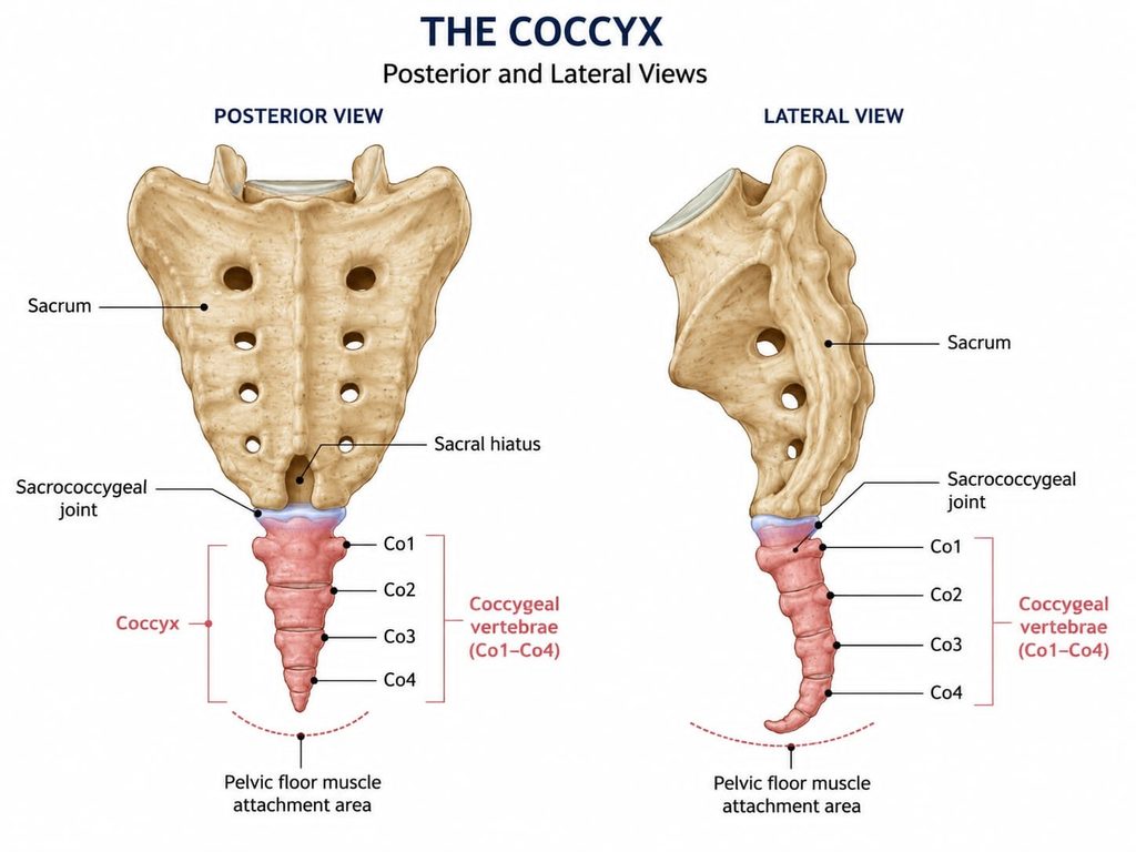
Figure: Illustration of the anatomy of the sacrum and tailbone (coccyx) with their joint
Who Is More Likely to Develop Coccydynia?
Coccydynia occurs considerably more often in women than in men, with some series reporting an approximate ratio of 5:1. The reason is probably not a single factor, but a combination of pelvic anatomy, sitting biomechanics, childbirth, and the distribution of soft tissues around the coccyx.
Body weight may also influence how the coccyx is loaded. In patients with obesity, posterior subluxation during sitting has been described more frequently, whereas thinner patients may more often show hypermobility or a bony spicule at the distal coccyx. This does not mean that body weight itself causes the disorder, but it may influence biomechanics and the type of lesion that develops.
In a significant proportion of patients, no single clear cause can be identified. These cases are generally classified as idiopathic coccydynia.
What Causes Coccydynia?
Trauma
One of the best-known causes is a direct fall onto the buttocks. This may result in a contusion, fracture, subluxation, or abnormal mobility of the coccyx.
Pain does not always begin after a severe injury. A relatively minor trauma may become clinically important if it leads to persistent mechanical instability or irritation of the joints and surrounding soft tissues.
Abnormal Coccygeal Mobility
In some patients, pain occurs because the coccyx moves more than expected during sitting or because subluxation or dislocation occurs.
Classic studies using dynamic radiography have shown that a substantial proportion of patients with coccydynia demonstrate abnormal mobility only when images obtained in standing and sitting positions are compared. A normal static image therefore does not exclude mechanical coccydynia.
Degenerative and Inflammatory Changes
Pain may arise from the sacrococcygeal joint, inter-coccygeal joints, or discs. In some patients, MRI shows changes resembling inflammation, endplate abnormalities, fluid within inter-coccygeal discs, or soft-tissue changes around the tip of the coccyx.
Coccygeal Shape and Bony Prominences
Certain morphological variants, marked curvature, or a prominent bony tip may concentrate pressure during sitting. However, an imaging variant alone is not a diagnosis. It becomes clinically relevant only when it corresponds with the location of pain, focal tenderness, and reproduction of the patient’s symptoms.
Prolonged Pressure During Sitting
Prolonged sitting, particularly on a hard surface or in a position that places direct pressure on the coccyx, can aggravate an existing mechanical or inflammatory disorder. Sitting is therefore often an important provoking factor, but it is not necessarily the original cause of the condition.
Childbirth
The coccyx is exposed to substantial biomechanical forces during childbirth. In some women, injury or abnormal mobility of the sacrococcygeal region may develop following delivery.
Neural Structures
Pain in this region does not necessarily originate only from bones or joints. The anococcygeal nerve, perforating cutaneous nerve, ganglion impar, and other neural structures within the pelvis may contribute to the pain.
This is clinically important because it helps explain why different patients with pain in a similar location may respond to different interventions.
Does Every Pain in the Tailbone Region Represent Coccydynia?
No. A number of different disorders can produce pain in the sacral and coccygeal region.
The differential diagnosis may include lumbosacral pathology, sacroiliac pain, pelvic muscle and fascial pain, pudendal and other neuropathies, Tarlov cysts, pilonidal cysts, infection, inflammatory disorders, tumors, and other lesions of the sacrococcygeal region. Radiological reviews particularly emphasize that infections, neoplasms, crystal deposition disorders, and cystic lesions can mimic ordinary mechanical coccydynia.
Persistent tailbone pain should therefore not automatically be attributed to sitting or to a nonspecific “inflammation of the coccyx.”
What Does Typical Tailbone Pain Feel Like?
The most characteristic symptom is localized midline pain over the coccyx. It is commonly aggravated by sitting, hard surfaces, leaning backward, prolonged sitting, and the transition from sitting to standing.
In some patients, pain may also be provoked during defecation, particularly when there is direct pressure or activation of the pelvic floor muscles. Less commonly, pain during sexual intercourse or variation in symptom intensity during the menstrual cycle has been described. These symptoms are not specific to coccydynia and should prompt consideration of other pelvic pain disorders when typical local coccygeal tenderness is absent.
For some patients, rising from a chair is particularly painful, which may be an important clue to abnormal mobility or subluxation of the coccyx. In Maigne’s study, this pattern was more frequent in patients with radiographically confirmed luxation.
The pain may be dull and deep, although some patients describe it as sharp, stabbing, or burning.
Why Can Pain Be Worse When Standing Up Than While Sitting?
During the transition from sitting to standing, both the direction and intensity of forces acting on the coccyx change. If hypermobility, subluxation, or another form of mechanical instability is present, movement of the coccyx during unloading may trigger sudden pain.
For this reason, the statement “sitting itself is not the worst part — standing up is” is not unusual and may have diagnostic value.
How Long Can Tailbone Pain Last?
In acute coccydynia, particularly after a minor injury or transient local inflammation, symptoms gradually improve over the following weeks or months in many patients with pressure relief and conservative treatment.
When pain persists, especially when it limits sitting and daily activities, it is important to consider whether there is hypermobility, subluxation, a bony spicule, chronic inflammation, or another underlying pain generator.
How Is Coccydynia Diagnosed?
Diagnosis begins with a detailed history and a focused clinical examination. Important questions include the exact location of the most severe pain, whether symptoms began after a fall, childbirth, or another event, how long the patient can sit, whether pain is worse when rising, whether it depends on seat hardness and pelvic position, and whether neurological, urinary, gastrointestinal, or other symptoms are present.
Clinical examination includes targeted palpation of the coccyx and an attempt to reproduce the patient’s characteristic pain. Localized tenderness that reproduces the patient’s usual symptoms increases the likelihood that the coccygeal region is the true pain generator.
Coccyx X-rays
Standard anteroposterior and lateral X-rays may show coccygeal shape, deformity, previous trauma, or other abnormalities, but a normal static X-ray does not exclude coccydynia. When pain is strongly related to loading during sitting, dynamic imaging may provide more relevant information.
Dynamic Coccyx X-rays — Why Are Sitting and Standing Images Used?
Dynamic radiography compares the position of the coccyx while the patient is standing with its position while sitting in a posture that loads the tailbone. It may demonstrate hypermobility, subluxation, or another abnormal movement pattern that is not visible on a static image.
This is particularly useful when pain clearly depends on sitting, when rising from a chair is painful, when static imaging has not explained the symptoms, or when mechanical coccydynia is suspected. Dynamic X-rays are not necessary for every patient, but in the appropriate clinical setting they may be considerably more informative than a conventional unloaded study.
Can a Coccyx MRI Be Normal Despite Persistent Pain?
Yes. MRI is usually performed with the patient lying down and therefore does not necessarily show what happens when the coccyx is loaded during sitting. However, MRI is very useful for assessing soft tissues, bone marrow edema, inflammatory changes, inter-coccygeal discs, and other processes that are not well visualized on plain radiographs.
MRI and dynamic radiography therefore do not answer exactly the same diagnostic question.
When Are CT or Additional Imaging Needed?
CT can be useful when more detailed assessment of bony anatomy is required, particularly after trauma or when a structural lesion is suspected. Contemporary reviews recommend MRI or CT selectively, especially in cases of trauma, unclear findings, or when there are features that raise concern for tumor, infection, or another more serious disorder.
When Does Pain in the Tailbone Region Come From the Spine or Nerves?
Pain that a patient describes as “tailbone pain” does not always originate from the coccyx itself. Lumbosacral radiculopathy, sacral nerve roots, peripheral neuropathies, and certain sacral lesions may produce pain that is perceived in the coccygeal, gluteal, or perineal region.
Tarlov, or perineural, cysts are a common source of diagnostic uncertainty. Most are incidental findings and do not cause symptoms, but when their location and the clinical picture are compatible, they may be associated with sacral or perineal pain, sensory symptoms, or disturbances of pelvic organ function. The presence of a cyst on MRI does not by itself prove that the cyst is the cause of pain.
In patients with atypical pain, lumbar disc herniation, foraminal or central stenosis, sacral radiculopathy, and other spinal causes should also be considered. Pain following a dermatomal distribution, tingling, objective sensory loss, weakness, or reflex changes is more suggestive of a neural source than of an isolated coccygeal pain generator.
Rarely, pain in the sacrococcygeal region may be the first manifestation of a tumor, including a sacral chordoma, or of infection. Progressive, constant, or night pain, a palpable mass, neurological deficit, or other red flags require further imaging and a broader diagnostic evaluation. Although these conditions are uncommon, they should be excluded when the clinical pattern is atypical.
How Is Coccydynia Treated?
Treatment depends on the cause, duration of symptoms, and mechanism of pain. There is no single treatment that is appropriate for every patient.
Reducing Coccygeal Pressure and Modifying Sitting
The first step is often to reduce direct pressure on the painful region. This may include changing position more frequently, avoiding prolonged uninterrupted sitting, adjusting pelvic angle, and using an appropriate cushion or seat cut-out.
The goal is not to avoid sitting completely, but to reduce direct loading of the painful structure. No single cushion is best for every patient. A cushion that unloads the coccyx may redistribute pressure to other areas of the pelvis, so the response should be assessed individually.
Medications
In appropriate patients, simple analgesics and nonsteroidal anti-inflammatory drugs (NSAIDs) may be used, particularly when an inflammatory or mechanical component is suspected. If the pain has neuropathic features, such as burning, shooting pain, tingling, or sensory symptoms, medications commonly used for neuropathic pain may sometimes be considered. Muscle relaxants may occasionally be useful when significant pelvic floor or surrounding muscle spasm is present. Medication will not correct coccygeal hypermobility, subluxation, or another structural abnormality, but it may reduce pain sufficiently to improve sitting tolerance, movement, and participation in rehabilitation.
Physical Therapy
Physical therapy may be useful, but it should be tailored to the specific problem. In some patients, attention may need to be directed toward the pelvic floor muscles, gluteal and surrounding muscles, pelvic biomechanics, protective muscle spasm, and sitting and movement patterns.
Contemporary systematic reviews suggest that certain conservative treatments may reduce pain, but the quality and consistency of evidence are not the same for all techniques.
Injections and Targeted Procedures
When conservative treatment is insufficient, targeted injection procedures may be considered. These may have both a therapeutic role, by temporarily reducing pain and inflammation, and a diagnostic role, because the response to a precisely targeted procedure may help confirm the true pain generator.
The injection site depends on the findings and the suspected source of symptoms and may include the sacrococcygeal joint, an inter-coccygeal segment, surrounding structures, or the ganglion impar.
Ganglion Impar Block for Tailbone Pain
The ganglion impar is a sympathetic neural structure located anterior to the distal sacrum and coccyx and is one of the interventional targets used in chronic coccygeal and perineal pain. A contemporary systematic analysis of ganglion impar block studies suggests that significant pain reduction may occur in appropriately selected patients, although the quality of evidence and duration of response vary between studies.
A ganglion impar block is therefore not a universal solution for every patient with tailbone pain. It is most appropriate when the clinical pain pattern corresponds with the neural structure being targeted.
Manual Therapy and Coccygeal Mobilization
Manual techniques may include stretching and massage of the pelvic floor muscles and, in selected patients, internal transrectal mobilization of the coccyx. This approach may be used to assess or modify coccygeal mobility and surrounding pelvic floor tension. Manual therapy is not a universal treatment and should be tailored to the individual findings related to coccygeal mobility, tenderness, and pelvic floor muscle function.
Extracorporeal Shock Wave Therapy — ESWT
Extracorporeal shock wave therapy has been investigated as a noninvasive treatment option for chronic coccydynia. More recent studies and systematic reviews suggest possible reductions in pain and improvement in function, but the number of high-quality studies remains limited.
ESWT therefore cannot currently be regarded as a standard treatment for every patient with coccydynia.
Radiofrequency Procedures for Tailbone Pain
In some patients with chronic and refractory pain, radiofrequency procedures may be considered. Contemporary treatment reviews include them among the options that may be used after failure of basic conservative measures.
The decision depends on the response to previous diagnostic procedures and on accurate identification of the pain generator.
When Is Coccyx Surgery — Coccygectomy — Considered?
Coccygectomy is the surgical removal of part or all of the coccyx. It should not be recommended simply because pain has been present for a long time.
It is most appropriate in patients with chronic and clearly localized pain, substantial limitation of daily function, failure of adequate conservative and interventional treatment, findings that support the coccyx as the true pain generator, and good correlation between the clinical picture, imaging findings, and, when performed, diagnostic blocks.
Either partial or total coccygectomy may be performed depending on the location of the pathological segment and the surgeon’s assessment. The literature does not provide sufficiently strong evidence to consider one technique universally superior for all patients. If a symptomatic segment is incompletely removed, revision surgery may occasionally be required.
How Successful Is Coccygectomy?
A systematic review and meta-analysis of 21 studies included 826 surgically treated patients with refractory coccydynia. The average reduction in pain was approximately 5 points on a 0–10 scale, and improvement was maintained during longer-term follow-up.
This indicates that surgery may be highly beneficial in properly selected patients. However, most available evidence comes from observational studies, so the outcome cannot be guaranteed in advance for every patient.
Improvement after surgery does not have to be immediate. Pain and function may continue to improve over several months, and the final result should therefore not be judged too early. Long-term series indicate that improvement may persist for years.
Wound healing is a particular concern after coccygectomy because of the anatomical location of the incision, making patient selection and surgical technique especially important. Surgical-site infection is the most important specific complication of coccygectomy. In larger systematic reviews and contemporary series, it has been reported in approximately 8–10% of operated patients, although higher rates have been described in older and smaller series. The risk is related to the location of the incision near perineal bacterial flora.
What Is the Prognosis of Coccydynia?
Prognosis depends largely on the true cause of the pain. A patient with transient inflammation after a minor injury does not have the same clinical course as a patient with persistent subluxation, hypermobility, marked degenerative change, or chronic neuropathic pain.
Conservative treatment is sufficient for many patients. In others, symptoms become chronic and more precise identification of the pain generator is necessary before interventional treatments are considered in a stepwise manner.
When an anatomical and mechanical cause is clearly confirmed, the prognosis after targeted treatment can be good, including in carefully selected patients who ultimately require surgery.
When Does Tailbone Pain Require More Urgent Medical Assessment?
Typical mechanical coccydynia changes with position and loading. More prompt evaluation is needed when symptoms do not follow the usual mechanical pattern.
Important warning features include severe progressive or constant pain unrelated to position, marked night pain, fever or other signs of infection, unexplained weight loss, known malignancy, significant trauma, new neurological weakness, new perineal or saddle-area numbness, and new disturbance of bladder or bowel control.
These symptoms require consideration of causes other than ordinary coccydynia. Radiological literature particularly emphasizes tumors, infections, and other lesions of the sacrococcygeal region in the differential diagnosis.
Final Clinical Principle
Tailbone pain is not a diagnosis by itself. In one patient, the problem may be local inflammation following trauma; in another, hypermobility or subluxation that becomes visible only during sitting; in another, soft-tissue or inter-coccygeal disc abnormalities on MRI; and in another, pain may arise from neural or other structures in the region.
For this reason, it is not enough simply to obtain an MRI, buy a cushion, or repeatedly undergo the same physical therapy. The key is to determine where the true pain generator is, what provokes and maintains the symptoms, and by what mechanism the pain is produced.
Only then can a rational decision be made about whether modification of sitting and targeted rehabilitation are sufficient, whether an injection or ganglion impar block is appropriate, or whether coccygectomy should be considered in the small number of carefully selected patients who may benefit from surgery.
Tailbone Pain — Start Your Telehealth Consultation
If your tailbone pain has persisted despite cushions, medications, physical therapy, manual treatment, or previous injections, a detailed telehealth consultation can help identify the most likely pain generator. The pain may arise from the coccyx itself, abnormal coccygeal mobility, the sacrococcygeal or inter-coccygeal joints, surrounding soft tissues, pelvic floor structures, nerves, or another source in the lumbosacral and pelvic region. Your symptoms are assessed together with available X-rays, MRI, CT, and previous treatment results to determine what should be investigated or treated next.
If the appropriate pain specialist is not available within your healthcare system, an international consultation can provide the missing expertise. Recommendations are adapted to the diagnostic tests, procedures, and treatments realistically available in your country, so your local doctor can use them when planning further care.
- ✔ Send a short message describing your problem
- ✔ You’ll receive a reply within 24 hours with details about whether and how we can help, including consultation cost and scheduling
- ✔ Only then, you can send your medical documentation
- ✔ During the consultation, you will receive clear guidance. A written summary is provided when appropriate, and you may send free follow-up questions for 10 days.
- ✔ Secure payment by credit card, PayPal invoice (USD), or bank transfer.
Consultation fees typically range from $180–250, depending on case complexity.
Based on our medical report, reimbursement can often be obtained (if your insurance plan allows it).
This is within the usual international range for specialist telehealth consultations. A consultation may also help clarify whether further imaging, a targeted injection, ganglion impar block, another interventional procedure, or coccygectomy should be considered — and whether the coccyx is truly the source of the pain.
FAQ — Coccydynia (Tailbone Pain): Causes, Diagnosis and Treatment
What is coccydynia?
Coccydynia is pain arising from the coccyx, or tailbone, and the structures immediately around it. Typical coccydynia is usually felt in the midline at the lowest part of the spine and is often aggravated by sitting, hard surfaces, leaning backward, or rising from a chair. However, not every pain felt in the tailbone region is true coccydynia. Similar symptoms may come from the sacroiliac joint, lumbosacral spine, sacral nerve roots, pelvic floor muscles, peripheral nerves, or other pelvic structures. The most important diagnostic task is therefore to identify the true pain generator rather than assuming that every pain near the coccyx originates from the coccyx itself.
What are the most common causes of coccydynia?
Common causes include direct trauma, abnormal coccygeal mobility, degenerative or inflammatory changes, childbirth, and anatomical variations that increase pressure on the coccyx during sitting. A fall onto the buttocks may cause bruising, fracture, subluxation, or persistent mechanical instability. In other patients, the sacrococcygeal or inter-coccygeal joints may become painful without a major injury. Some people have hypermobility, a prominent bony spicule, or marked coccygeal angulation. These findings are important only when they correspond with the location and behavior of the pain. In a significant proportion of patients, no single definite cause is identified, and the condition is classified as idiopathic coccydynia.
Why does tailbone pain become worse when I sit?
Sitting places direct mechanical load on the coccyx and surrounding soft tissues. This may compress an inflamed joint, ligament, bony prominence, or sensitive soft tissue and may also provoke abnormal movement between coccygeal segments. Hard surfaces and positions that tilt the pelvis backward usually increase pressure on the tailbone. Sitting itself is not always the original cause of the disorder; it may simply expose an underlying problem such as hypermobility, subluxation, inflammation, previous trauma, or abnormal coccygeal shape. This is why treatment should not consist only of avoiding sitting. The underlying pain mechanism should be identified whenever symptoms persist.
Why can coccydynia be worse when I stand up from a chair?
Pain during the transition from sitting to standing can be an important diagnostic clue. When a person rises from a chair, the direction and intensity of forces acting on the coccyx change rapidly. If the coccyx is excessively mobile, partially displaced, or mechanically unstable, movement during unloading can reproduce sharp pain. Some patients therefore report that sitting is uncomfortable but that the strongest pain occurs during the first seconds after standing up. This pattern can support suspicion of abnormal coccygeal mobility, although it is not diagnostic by itself. Dynamic sitting and standing X-rays may be particularly useful when this mechanical pattern is prominent.
Can tailbone pain persist even when the X-ray or MRI is normal?
Yes. A normal static X-ray or MRI does not exclude a coccygeal pain disorder. Standard imaging is usually performed without the coccyx being loaded in the same way as during sitting. MRI is useful for assessing soft tissues, bone marrow edema, inflammatory changes, inter-coccygeal discs, and other lesions, but it may not show abnormal movement that occurs only under pressure. A static X-ray may also appear normal despite hypermobility or subluxation during sitting. Imaging must therefore be interpreted together with the pain pattern, focal tenderness, clinical examination, and, in selected patients, dynamic radiographs obtained in both standing and sitting positions.
When are dynamic X-rays useful for coccydynia?
Dynamic coccygeal radiography compares the position of the coccyx while the patient is standing with its position during loaded sitting. The purpose is to determine whether the coccyx moves abnormally when pressure is applied. These images may demonstrate hypermobility, excessive movement, subluxation, or another abnormal mechanical pattern that is not visible on a conventional static X-ray. Dynamic imaging is particularly useful when symptoms clearly depend on sitting, when pain is pronounced while rising from a chair, or when routine imaging has failed to explain an otherwise typical mechanical pattern. It is not required for every patient and should be used when the clinical picture suggests abnormal coccygeal mobility.
Can tailbone pain actually come from the spine or nerves?
Yes. Pain perceived around the coccyx may sometimes originate from the lumbosacral spine, sacral nerve roots, peripheral nerves, or other structures within the pelvis. Neural pain is more likely when symptoms include burning or shooting pain, tingling, numbness, a dermatomal distribution, weakness, reflex changes, or perineal sensory symptoms. Lumbar disc disease, foraminal or central stenosis, sacral radiculopathy, pudendal or other neuropathies, and certain sacral lesions may imitate coccygeal pain. This distinction matters because treatment directed only at the coccyx will not help if the dominant pain generator is neural. Symptoms, neurological findings, and imaging must therefore be assessed together.
Can a Tarlov cyst cause tailbone pain?
Tarlov, or perineural, cysts are frequently discovered incidentally on MRI and most do not cause symptoms. Their presence alone therefore does not prove that they are responsible for pain. In selected patients, however, a cyst located at an appropriate sacral level may be clinically relevant when symptoms include sacral or perineal pain, sensory disturbance, neurological features, or pelvic-organ symptoms that correspond anatomically with the involved nerve roots. The important principle is correlation. A cyst should not automatically be blamed simply because it appears on MRI, and other causes of coccygeal and pelvic pain should still be considered.
Can childbirth cause coccydynia?
Yes. During childbirth, the coccyx is exposed to considerable mechanical forces as the pelvic outlet expands. In some women, this can result in injury to the sacrococcygeal region, abnormal mobility, subluxation, joint irritation, or surrounding soft-tissue pain. Symptoms may begin immediately after delivery or become more obvious when prolonged sitting resumes. The diagnosis still requires careful assessment because pelvic floor dysfunction, sacroiliac pain, pudendal or other neuropathic pain, and musculoskeletal changes associated with pregnancy may produce similar symptoms. Treatment should therefore be directed toward the structure most likely to be generating the pain rather than assuming that all postpartum pain comes directly from the coccyx.
What should be done if tailbone pain persists despite treatment?
Persistent pain despite cushions, medications, physical therapy, manual treatment, or previous injections does not automatically mean that surgery is required. The first step is to reconsider the diagnosis and determine whether the true pain generator was correctly identified. Persistent symptoms may reflect abnormal coccygeal mobility, a painful sacrococcygeal or inter-coccygeal joint, a bony prominence, pelvic floor dysfunction, neural pain, or another structure outside the coccyx. Static imaging may also have missed a mechanical problem. In selected patients, dynamic X-rays, targeted diagnostic injections, neurological assessment, or additional imaging can clarify the mechanism. Further treatment should then be chosen according to the most likely pain generator rather than simply repeating therapies that have already failed.
What treatments are available for coccydynia?
Treatment is usually stepwise and depends on the underlying mechanism. Initial measures may include modification of sitting, reducing direct coccygeal pressure, an appropriate cushion or seat cut-out, analgesics or anti-inflammatory medication, and targeted physical therapy. When neuropathic features are present, medications commonly used for neuropathic pain may sometimes be considered, while significant pelvic floor spasm may require a different approach. Other options include manual therapy, targeted injections, ganglion impar block, extracorporeal shock wave therapy, and selected radiofrequency procedures. Coccygectomy is reserved for carefully selected patients with persistent, clearly localized and functionally limiting pain when adequate non-surgical and interventional treatment has failed.
Which medications may be used for tailbone pain?
Medication depends on the suspected pain mechanism. Simple analgesics and nonsteroidal anti-inflammatory drugs may be useful when mechanical irritation or inflammation is prominent. When pain has burning, shooting, tingling, or other neuropathic features, medications commonly used for neuropathic pain may sometimes be considered. Muscle relaxants may occasionally help when pelvic floor or surrounding muscle spasm contributes substantially to symptoms. Medication does not correct hypermobility, subluxation, a bony prominence, or another structural lesion. Its role is primarily to reduce pain sufficiently to improve sitting tolerance, movement, sleep, and participation in rehabilitation while the underlying pain generator is addressed.
What type of physical therapy is used for coccydynia?
Physical therapy should be individualized rather than based on one standard exercise program. Depending on the findings, treatment may address pelvic floor muscle tension, gluteal and surrounding muscle dysfunction, pelvic biomechanics, protective muscle spasm, posture, and sitting mechanics. Some patients benefit from reducing mechanical overload and gradually restoring movement, while others require more specific pelvic floor treatment. Physical therapy is less likely to succeed when the dominant pain generator is an unstable coccygeal segment, significant subluxation, or another structural problem that has not been recognized. Persistent symptoms after generic therapy should therefore prompt reassessment rather than simply repeating the same exercises indefinitely.
Can manual therapy or transrectal mobilization help coccydynia?
Manual therapy may help selected patients, particularly when pelvic floor muscle tension, restricted coccygeal movement, or surrounding soft-tissue dysfunction contributes to pain. Techniques may include stretching and massage of the pelvic floor and, in selected cases, internal transrectal mobilization of the coccyx. This allows the clinician to assess and sometimes modify coccygeal mobility and surrounding muscle tension. It is not appropriate for every patient, and the expected benefit depends on the underlying mechanism. Manual treatment should not be repeated indefinitely when symptoms do not improve, particularly if imaging or examination suggests instability, significant subluxation, fracture, another structural lesion, or a pain generator outside the coccyx.
What are the risks of manual treatment for tailbone pain?
Manual and transrectal techniques are less invasive than injection or surgery, but they are not completely free of adverse effects. Temporary soreness, increased local pain, pelvic floor discomfort, or a symptom flare may occur after treatment. Internal techniques may also be uncomfortable and should only be performed when clinically indicated, with appropriate consent and technique. Forceful manipulation is not appropriate when fracture, marked instability, infection, tumor, or another contraindication is suspected. The procedure should therefore follow a clinical assessment rather than being used routinely. If pain becomes progressively worse after treatment or develops a different pattern, the diagnosis should be reconsidered.
When are injections used for coccydynia?
Targeted injections may be considered when conservative treatment has not provided sufficient relief and when examination or imaging suggests a specific pain generator. Depending on the suspected source, the target may be the sacrococcygeal joint, an inter-coccygeal segment, surrounding tissues, or another anatomically relevant structure. Injections can serve two purposes. They may reduce local inflammation and pain, but they may also provide diagnostic information. Meaningful temporary improvement after a precisely targeted injection supports the possibility that the injected structure is contributing to symptoms. This information may help guide further treatment and can be particularly valuable before considering more invasive procedures.
What are the possible complications of injections for tailbone pain?
Complications are uncommon when targeted injections are appropriately performed, but they can occur. Possible problems include temporary pain flare, local bleeding or bruising, infection, vasovagal reactions, allergic reactions, and adverse effects related to the injected medication. Procedures performed near neural or pelvic structures also require accurate anatomical targeting to reduce the risk of unintended needle placement. Repeated corticosteroid injections may have additional local or systemic effects and should not be performed without a clear indication. The expected benefit and risk depend on the exact target, technique, imaging guidance when used, and the patient’s medical condition.
When is a ganglion impar block used for coccydynia?
The ganglion impar is a sympathetic neural structure located anterior to the distal sacrum and coccyx. A ganglion impar block is used in selected patients with chronic coccygeal or perineal pain. Local anesthetic, sometimes combined with another medication, is placed near this structure to interrupt pain transmission. A successful block may provide therapeutic relief and can also support the conclusion that pain pathways involving the ganglion impar contribute to symptoms. It is not a universal treatment for every patient. Its greatest value comes from careful patient selection and from using it when the clinical pain pattern is compatible with the structure being targeted.
What are the complications of a ganglion impar block for tailbone pain?
Ganglion impar block is generally considered minimally invasive, but complications are possible. These include temporary worsening of pain, bleeding, infection, allergic or medication-related reactions, and vasovagal symptoms. Because the ganglion impar lies in the presacral region close to the rectum and other pelvic structures, accurate needle placement is important. Unintended injury to adjacent structures is uncommon but is one reason why appropriate technique and anatomical guidance matter. Relief may also be incomplete or temporary, which is not technically a complication but is clinically important when discussing expectations. A block should therefore be performed for a clear indication rather than simply because pain has persisted.
Can ESWT help chronic coccydynia?
Extracorporeal shock wave therapy, or ESWT, has been investigated as a noninvasive treatment for chronic coccygeal pain. Some studies report reductions in pain and improvement in function, but the evidence remains less extensive and less consistent than for established conservative measures. ESWT may therefore be considered in selected patients, particularly when a noninvasive option is preferred, but it should not be presented as a universally effective treatment. Its usefulness depends on the underlying pain mechanism, and a patient with marked coccygeal instability, neural pain, or another specific structural lesion may require a different approach. Failure of ESWT should prompt reconsideration of the pain generator rather than automatic repetition of treatment.
What are the possible side effects of ESWT for tailbone pain?
ESWT is noninvasive, but temporary adverse effects may occur. Patients may experience local soreness, tenderness, temporary pain flare, mild swelling, or bruising after treatment. The intensity of treatment and sensitivity of the coccygeal region influence tolerability. Significant complications are uncommon, but ESWT should still be used selectively and avoided when there is concern for conditions that require another form of evaluation or treatment. Progressive pain, suspected fracture, infection, tumor, or neurological disease should not simply be treated with repeated shock-wave sessions. The main limitation is often not toxicity but uncertainty about which patients are most likely to benefit.
When is radiofrequency treatment considered for coccydynia?
Radiofrequency procedures may be considered in selected patients with chronic pain that has not responded adequately to simpler conservative and interventional treatment. The procedure is intended to modify pain transmission from a carefully selected neural target rather than to correct a structural abnormality of the coccyx. Patient selection is therefore essential. A previous diagnostic block or another convincing response may help determine whether the planned target is actually involved in the pain. Radiofrequency treatment should not replace evaluation for hypermobility, subluxation, tumor, infection, neural compression, or another correctable cause. Its role is greatest when the pain generator has been reasonably well defined.
What are the complications of radiofrequency treatment for tailbone pain?
Possible adverse effects include temporary local pain, soreness, numbness, neuritic pain, bleeding, infection, and incomplete or short-lived relief. Because radiofrequency changes neural pain transmission, inaccurate targeting could affect structures that were not intended to be treated. The exact risk profile depends on the anatomical target and technique used. Some patients obtain substantial relief, while others improve only temporarily or not at all. This is why diagnostic reasoning remains important before treatment. Radiofrequency should not be performed simply because symptoms are chronic; it should be used when the clinical pattern and previous diagnostic procedures support the selected target.
When is coccygectomy considered for coccydynia?
Coccygectomy is considered only in carefully selected patients with chronic, clearly localized, functionally limiting pain that has not responded to adequate conservative and interventional treatment. The decision should be supported by a convincing relationship between the patient’s symptoms, focal examination findings, imaging, and, when relevant, response to diagnostic injections. Surgery should not be recommended simply because symptoms have lasted for many months or years. Before coccygectomy, alternative pain generators such as sacroiliac pain, lumbar or sacral nerve pathology, pelvic floor dysfunction, and other pelvic lesions should be considered. Both partial and total coccygectomy are used depending on the location of the symptomatic segment and surgical assessment.
What are the complications of coccygectomy for tailbone pain?
The most important specific complication of coccygectomy is surgical-site infection because the incision is located close to the perineal region. Other potential surgical complications include wound-healing problems, hematoma, persistent postoperative pain, incomplete relief, painful scarring, and the general risks associated with anesthesia and surgery. Injury to deeper presacral structures is uncommon but anatomically possible, which is why careful dissection close to the coccyx is important. Some patients may also have persistent symptoms if the original pain generator was incorrectly identified or if another source of pelvic pain remains active. Careful patient selection is therefore as important as the surgical technique itself.
How successful is coccygectomy for coccydynia?
Published series and systematic reviews indicate that coccygectomy can provide substantial improvement in carefully selected patients with refractory disease. Outcomes are generally better when pain is clearly localized to the coccyx, symptoms are mechanically reproducible, imaging or examination supports the diagnosis, and appropriate conservative and interventional treatments have already failed. However, most available evidence comes from observational studies rather than large randomized trials, so surgery cannot guarantee complete relief for every patient. Improvement may also continue gradually for several months after the operation. The decision should therefore be based on the strength of the diagnosis and the expected balance between potential benefit and surgical risk.
How long can recovery take after treatment for tailbone pain?
Recovery depends on the underlying cause and the treatment used. Pain after a minor injury or transient inflammation may gradually improve over weeks or months with pressure reduction and conservative care. Recovery after an injection or other intervention varies according to the pain generator and whether the procedure successfully targeted it. After coccygectomy, improvement is not necessarily immediate. Pain and function may continue to improve over several months, so the final surgical result should not be judged too early. Persistent symptoms should prompt reassessment of whether the original pain generator was correctly identified and whether another coccygeal, pelvic, spinal, or neuropathic source remains active.
When can tailbone pain indicate a more serious condition?
Typical mechanical pain changes with sitting, position, and loading. More urgent medical assessment is appropriate when symptoms are progressive, constant, or unrelated to position; when severe night pain is present; or when pain is associated with fever, unexplained weight loss, known malignancy, significant trauma, a palpable mass, new neurological weakness, saddle-area numbness, or new bladder or bowel dysfunction. These features may indicate infection, tumor, significant neural compression, or another disorder rather than an ordinary mechanical coccygeal problem. Such symptoms should not be managed only with cushions, physical therapy, injections, or telehealth. Direct medical evaluation and appropriate imaging may be required.
Can an online consultation help when coccydynia has not improved?
Yes. A focused online consultation can be useful when symptoms persist despite cushions, medications, physical therapy, manual treatment, or previous injections, or when the cause remains uncertain. The consultation can review the exact pain location and behavior, previous trauma or childbirth, sitting tolerance, neurological or perineal symptoms, MRI, X-rays, CT, and previous treatments. It can also identify whether additional testing such as dynamic sitting and standing X-rays or a targeted diagnostic procedure may be appropriate. The purpose is not simply to provide another general opinion, but to determine whether the dominant pain generator is the coccyx, a joint, pelvic floor structure, nerve, spine, or another source and then plan the next rational step.
Do I need surgery if treatment for tailbone pain has failed?
Not necessarily. Failure of cushions, medications, physical therapy, or injections does not automatically mean that coccygectomy is the next step. Treatment may have failed because the true pain generator was not identified, because therapy was not directed toward the correct mechanism, or because more than one structure contributes to symptoms. Some patients require dynamic imaging, targeted injections, ganglion impar block, pelvic floor treatment, or evaluation for neural or spinal causes before surgery is considered. Coccygectomy is most appropriate when pain remains clearly localized to the coccyx, significantly limits function, and persists despite appropriate non-surgical and interventional treatment with convincing evidence that the coccyx is truly responsible.
Why can coccydynia persist after several different treatments?
Treatment often fails when several therapies are applied without first establishing the dominant pain generator. A cushion may reduce pressure but cannot correct hypermobility. Medication may reduce inflammation but cannot stabilize an abnormal coccygeal segment. Physical therapy may help pelvic floor or muscle dysfunction but may have limited effect on a painful unstable joint. Likewise, a ganglion impar block is unlikely to solve pain arising predominantly from another anatomical structure. Persistent symptoms therefore require a mechanism-based approach. The important question is not simply which treatments have already been tried, but whether each treatment was directed at the structure actually responsible for the patient’s symptoms.